Introduction
One of the most important stages in clinical infertility treatment today is frozen embryo transfer. With assisted reproductive techniques (ART), a greater number of oocytes is obtained compared with natural cycles with controlled ovary stimulation. The cryopreservation of human embryos allows us to increase the potential of conception for any in vitro fertilization (IVF) cycle to its highest level and to prevent the waste of embryos. The risk of multiple pregnancy is reduced with the transfer of one or two embryos alone, and embryos can be preserved for later use. Cryopreservation offers an alternative option to fresh embryo transfer in patients with endometrial pathologies, unsuitable endometrial receptivity, or excessive ovary response, thereby preventing ovarian hyperstimulation syndrome (OHSS). It allows sufficient time for preimplantation genetic diagnosis (PGD) and can ensure the preservation of fertilization by forming an oocyte/embryo pool in patients with low ovary reserves.
Vitrification, which is currently used to freeze human oocytes and embryos at the pronucleus, cleavage, morula, and blastocyst stages, is a simple, low-cost, and effective method (Liebermann and Tucker, Reference Liebermann and Tucker2006; Al-Hasani et al., Reference Al-Hasani, Ozmen, Koutlaki, Schoepper, Diedrich and Schultze-Mosgau2007; Desai et al., Reference Desai, Blackmon, Szeptycki and Goldfarb2007; Kuwayama, Reference Kuwayama2007). However, there is no consensus regarding which stage of embryonic development is most suitable for vitrification with maximum development after the embryo is dissolved (Ghandy and Karimpur Malekshah, Reference Ghandy and Karimpur Malekshah2017). Vitrification can be used in all embryo development stages. The extant literature focuses mostly on day 3 (cleavage stage) and day 5 (blastocyst stage) vitrification. These studies have reported that vitrification has similar survival and clinical pregnancy rates for embryos on days 3 and 5 (Cobo et al., Reference Cobo, de los Santos, Castello, Gamiz, Campos and Remohi2012; Fernandez-Shaw et al., Reference Fernandez-Shaw, Cercas, Braña, Villas and Pons2015). However, as day 3 vitrification requires early embryo quality assessment, it cannot correctly project the embryos’ further development potential. It is maintained that, even though some blastomeres lose vitality after they are dissolved (as an embryo, in the blastocyte stage, has a greater number of cells compared with an embryo in the early division stage), they are still able to preserve their general vitality at a high level. As the blastocyte stage is the last embryonic stage before implantation, it allows us to make a more accurate estimate about implantation success. However, as only limited numbers of embryos are able to reach the blastocyte stage, transfers must be made from among the limited number of embryos remaining after the freezing and dissolving stages. Yet, the embryos frozen on day 4 have the chance to be monitored in the incubator for an extra day after being dissolved. This may allow us the opportunity to choose embryos of better quality from those dissolved. Therefore, in our study, we aimed to utilize the day 4 embryo freezing method alongside the day 3 and day 5 embryo freezing procedures.
Materials and methods
We included 128 patients, and 148 cycles belonging to these patients, in our study. All patients applied to the Gülhane Training and Research Hospital Department of Gynecology and Obstetrics Reproductive Medical Center of the University of Health Sciences, Ankara, Turkey between January 2016 and December 2018 for treatment. Male majority factor cases and the transfers of four patients whose embryos were frozen on day 3 were cancelled and excluded from the study, as their embryo quality was poor after being dissolved. The ethics committee’s consent for the study was received from the ethics committee presidency at Gülhane Training and Research Hospital of the University of Health Sciences (Ethics Committee Date: 26 February 2019 No. 19/48). Patients’ data were obtained retrospectively from hospital archive records. The patients’ ages, infertility reasons, number of oocytes collected, embryo transfer dates, embryo quality, days of embryo freezing, clinical pregnancy rates, and ongoing pregnancy rates were evaluated. The results of the embryos frozen were compared in three groups based on the day they were frozen: days 3, 4 or 5. Demographic data were also compared among the embryos frozen on days 3, 4, and 5. The period between freezing and thawing of embryos is between 3 and 24 months. Those patients who have frozen embryo transfer (FET) cycles also had fresh embryo transfer before. Our embryo quality were graded according to Alpha Istanbul consensus at 2011 (Alpha Scientists In Reproductive Medicine, 2011). Embryos were frozen using the closed vitrification method and dissolved using the quick dissolve method. We vitrified grade 1 or 2 embryos on day 3, compact embryos on day 4 and grade A blastocysts on day 5. For cases in which the number of embryos to be vitrified during patient monitoring was low, embryos were vitrified on day 3 or day 4 and were monitored morphologically after being dissolved. Then, the best embryos were chosen and transferred on day 4 or day 5. For cases in which the number of embryos to be frozen was high, embryos that were able to reach the blastocyte stage were vitrified on day 5. We transfer five grade 1 of the 29 embryos frozen and dissolved on day 3. Thirteen grade 1 and 2 embryos on day 4, and grade A and B seven embryos were transferred on day 5. The transfers of four patients’ embryos were cancelled due to poor embryo quality. Of the 80 day 4 embryos dissolved, 21 were transferred on day 4, and 59 were transferred on day 5. 39 day 5 embryos frozen and dissolved were transferred within 4 to 5 h on the same day they were dissolved.
Here, 18 patients were transferred embryos that had been frozen and dissolved twice, and one patient was transferred embryos that had been frozen and dissolved three times. Those 18 patients were counted separately for statistical analysis. While some patients were transferred a high number of frozen embryos dissolved in one cycle without waiting, in another cycle the embryos were transferred after being monitored for 1 to 2 days. Repetitive transfers were obtained by dissolving large numbers of vitrified embryos that were grouped in the previous ART cycle. The clinical pregnancy was defined according to the presence of gestational sac.
The SPSS 22 program (SPSS Inc., Chicago, II., USA) was used for statistical analysis. Student’s t-test and one-way analysis of variance (ANOVA) were applied for categorical data, and Shapiro–Wilks test was conducted to see whether the data complied with the normal distribution. Data that did not comply with the normal distribution were compared dyadically using the Mann–Whitney U-test.
Embryo freezing and thawing
We used closed vitrification system ‘Rapid-i’ for embryo vitrification and thawing. The embryo freezing and thawing procedure was based on the method propounded by Nina N. Desai et al. (Desai et al., Reference Desai, Goldberg, Austin and Falcone2013). A three-step vitrification protocol was used for cleavage, morula, and blastocyst-stage embryos. We used a three-step kit (RapidVit Omni, VitroLife, Sweden AB) for vitrification. Embryos were loaded singly, doubly, or, rarely, in triple groups onto the Rapid-i carrier and vitrified. During the warming procedure, we used a four-step kit: RapidWarm Omni (VitroLife, Sweden AB).
After the embryos were thawed, if they were to be transferred on the same day they were frozen, they were transferred after being left in incubation for at least 4 h. Embryos frozen on days 3 and 4 were transferred after a 1 or 2 day incubation period based on the number and quality of embryos.
Results
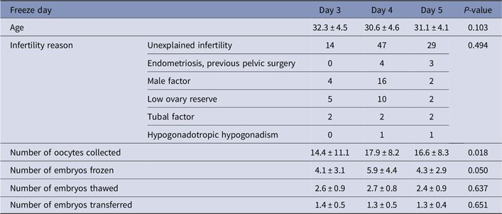
No significant differences were observed between the three groups based on age, the number of oocytes collected, and infertility reasons. There was also no significant difference between the three groups in terms of the number of embryos thawed, the transfer days, and the number of embryos transferred (Table 1).
Patients’ demographic data and cycle specifications based on which embryos used in frozen–thawed embryo transfer were obtained

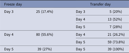
There was no significant difference in terms of clinical pregnancy and ongoing pregnancy rates according to the day the embryos were frozen (Table 2). Here, 17.4% of the patients’ embryos were frozen on day 3, 55.6% on day 4, and 27% on day 5; 20% of the embryos frozen on day 3 were also transferred on day 3; 52% were transferred on day 4; and 28% were transferred on day 5; 26.2% of the embryos frozen on day 4 were transferred on day 4, and 73.8% were transferred on day 5. All embryos frozen on day 5 were transferred the same day (Table 3).
Clinical pregnancy and ongoing pregnancy rates according to freeze day

Transfer day number and percentages according to freeze day

The clinical pregnancy and ongoing pregnancy results were examined, after thawing, in all groups according to their infertility reasons, with no significant difference observed in terms of clinical pregnancy and ongoing pregnancy rates.
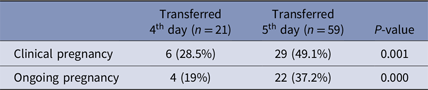
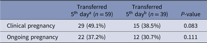
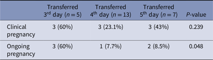
Clinical pregnancy and ongoing pregnancy rates were observed to be significantly higher in the results of embryos frozen on day 4 and transferred on day 5, according to the transfer day (P = 0.000) (Table 4). There were no statistical difference between the embryos that were frozen on the fourth and fifth days (Table 5). Although the numbers of embryos were limited, clinical pregnancy and ongoing pregnancy rates were observed to be significantly higher from the results of embryos frozen on day 3 and transferred on days 3, 4 or 5 according to the transfer day (P = 0.048) (Table 6).
Pregnancy rates of embryos frozen on the fourth day

Pregnancy rates of embryos frozen on the fourth and fifth days

a Embryos frozen and thawed on the fourth day (waited 1 day for transfer).
b Embryos frozen and thawed on the fifth day.
Pregnancy rates of embryos frozen on the third day

Discussion
Our study is the first one in the extant literature in which pregnancy results are compared following freezing and thawing of human embryos in the cleavage, morula, and blastocyte stages. Although there are certain case statements on this topic (Tao et al., Reference Tao, Tamis and Fink2001), there is no other study comparing a high number of cases.
One of the most important problems in IVF cycles is choosing the embryo with the highest life capacity for the freezing process. There is currently no technology able to determine this with complete accuracy. However, current developments in embryo laboratories allow us to take embryo cultures to the blastocyte phase and, hence, increase our chances of choosing embryos with higher implantation and life capacities. It is hypothesized that the chance of success will increase when blastocytes are used in freezing–thawing cycles because an embryo that has survived in extended culture has a higher probability of remaining alive after the thawing process (Jones et al., Reference Jones, Trounson, Lolatgis and Wood1998; Noyes et al., Reference Noyes, Reh, McCaffrey, Tan and Krey2009). One of the interesting results of our study is that a higher pregnancy rate has been found in embryos frozen in the morula stage and transferred in the blastocyte stage. However, as no morphological and genetic changes could be projected in the thawed embryos, shortcomings continue to remain on this topic in the literature. It is possible to better monitor the morphological development of embryos by freezing the embryos in the day 4 morula stage and transferring them on day 5. This would provide the opportunity to choose the correct embryo with the highest possibility of implantation, therefore increasing the chance of conception. Our literature scan contains numerous studies on embryos frozen on day 3 and day 5. However, no comparative human embryo study was found concerning embryos frozen on day 4.
A study conducted by Kong et al. suggested that it is necessary for embryos frozen in the cleavage stage to be frozen at the 7-cell or 8-cell stages (Kong et al., Reference Kong, Yang, Gong, Lu, Zhang, Lu and Lin2016). Chromosomal anomalies have been found to be higher in embryos that have less than six or more than nine cells. In our study, embryos in the cleavage stage on day 3 were frozen during the 7-cell or 8-cell stages.
In a study of the transfer of frozen–thawed (FT) embryos in the cleavage stage, the pregnancy rates of embryos kept in a 2-h culture following thawing and those held in a 24-h culture were compared after transfer. The cleavage and the pregnancy rates for embryos kept in the overnight culture environment were found to be higher (Joshi et al., Reference Joshi, Banker, Patel and Shah2010). Similarly, in our study, embryos frozen on day 4 were kept in the culture environment for a day after thawing and were transferred on day 5; higher pregnancy rates were achieved with the embryos in this group.
Brugnon et al. (Reference Brugnon, Bouraoui, Ouchchane, Gremeau, Peikrishvili, Pouly and Janny2010) assessed FT embryos in the blastocyte and cleavage stages, observing that the survival rates of embryos in the cleavage stage following thawing were significantly higher. However, the implantation rate is higher in embryos frozen in the blastocyte stage, with no difference seen in terms of pregnancy rates between the two groups.
In another study that compared transfers in the blastocyte and cleavage stages, fresh blastocyte transfers were reported to provide significantly higher clinical pregnancy and live birth rates compared with the cleavage stage. No difference was found in fresh and FET comparisons between the rates of clinical pregnancy and live birth in embryos in the cleavage and blastocyte stages. However, the cancellation rate between transfers was found to be higher in FT embryos in the blastocyte stage (Glujovsky et al., Reference Glujovsky, Farquhar, Quinteiro Retamar, Alvarez Sedo and Blake2016).
In a 2012 study, viability and pregnancy rates were shown to be better in embryos thawed in the cleavage stage and transferred in the blastocyte stage (Eftekhar et al., Reference Eftekhar, Aflatoonian, Mohammadian and Tabibnejad2012). Another study compared cleavage stage and blastocyte stage embryos in fresh and frozen cycles. The cumulative pregnancy rates and live birth rates in both fresh and frozen cycles were found to be significantly higher in the blastocyst group (Glujovsky and Farquhar, Reference Glujovsky and Farquhar2016). In yet another study, the implantation rates and clinical pregnancy rates for embryos transferred in the blastocyst stage was also observed to be higher compared with those of embryos transferred in the cleavage stage (Kaur et al., Reference Kaur, Swarankar, Maheshwari and Acharya2014).
Meanwhile, a study by Fernandez-Shaw et al. (Reference Fernandez-Shaw, Cercas, Braña, Villas and Pons2015) compared embryos frozen on day 3 and day 5 in patients aged under 35 years and patients aged 35 years and over. Therefore, in our research, we aimed to utilize the day 4 embryo freezing method. While the pregnancy rates in blastocyst-stage embryo transfers in the 35 and older age group were observed to be higher, no difference was seen in the under 35 age group.
Yet, some studies have suggested that embryos be frozen in the blastocyst stage. There are, however, reasons why this is not recommended, one being that a limited number of embryos are able to reach the expanded blastocyst stage, and the other being the concern of failure to transfer any embryos per couple that was significantly higher in the blastocyst group after thawing (Blake et al., Reference Blake, Farquhar, Johnson and Proctor2007). In an article published by Tao et al. (Reference Tao, Tamis and Fink2001) two factors were presented in which the embryos were frozen in the morula/compact stage; accordingly, it has been reported that, when compared with early stage embryos (i.e., the period from the pronuclear stage to the 8-cell stage) the morula stage provides the best opportunity for selecting better embryos. Even though embryos in the blastocyst stage allowed for better embryo selection compared with those in the morula stage, no transfer was aimed in the blastocyst stage, because it has been emphasized that the morula stage is a better option for transfer than the blastocyst stage. The reasons the morula/compact stage was chosen rather than the blastocyst stage include: preference of the assisted hatching method in all the embryos transferred, the idea that embryos are more defenceless during the transfer process in the blastocyst stage, and the expanded culture environment of blastocyst-stage embryos, which requires greater effort and incubator space.
Vanderzwalmen et al. (Reference Vanderzwalmen, Bertin, Debauche, Standaert, van Roosendaal, Vandervorst, Bollen, Zech, Mukaida, Takahashi and Schoysman2002) conducted a study in which the transfer of blastocyst embryos whose blastocoele liquid was drained was compared with blastocyst embryos whose blastocoele liquid was not drained after freezing–thawing, the degeneration rate was higher in blastocysts whose blastocoele liquid was not drained, accompanied by a lower transfer rate. This has shown that higher viability and pregnancy rates can be achieved in morula whose blastocoele cavities have not yet formed, as well as in early blastocysts. It has also indicated that, for blastocysts in which the blastocoele cavity has expanded prominently, the viability rate after freezing–thawing may be increased through artificially reducing the blastocoele cavity before vitrification. In this way, concerns over the viability rate of blastocyst-stage embryos after freezing–thawing have been eliminated.
In a study similar to ours, morula and blastocysts vitrified in mouse embryos were compared with the blastocyst group not vitrified with blastocysts. Trophectoderm (TE) and inner cell mass (ICM) were evaluated in embryos that reached the blastocyst stage after FT. No significant difference was found between the thawed morula and the control group. When comparing the thawed blastocysts and the control group, TE and ICM were found to be significantly lower in the thawed blastocyst group (Ling et al., Reference Ling, Zhang, Cao, Chen, Peng, Guo, Heng, Tong and Wang2009).
In another study conducted on rats, embryos vitrified in the cleavage, morula, and blastocyst stages were compared with the implantation rates of fresh embryos. Here, 100% of embryos frozen in the morula stage reached the blastocyst stage after thawing. Implantation rates in embryos frozen at the morula stage were also observed to be significantly higher (Han et al., Reference Han, Niwa and Kasai2003).
In a human study on fresh cycles, no significant difference was observed in terms of pregnancy rates after embryo transfers at the day 4 morula and day 5 blastocyst stages (Holschbach et al., Reference Holschbach, Weigert, Dietrich, Roesner, Montag, Strowitzki and Toth2017).
In another study, embryos frozen on day 4 were transferred on the same day, before 24 h had elapsed after thawing. Here, embryos in the morula stage were grouped and compared among themselves as the early morula, compact morula, and late morula stages. The viability and transfer rates after thawing in good quality embryos in the late morula stage were found to be higher (Tao et al., Reference Tao, Craig, Johnson, Williams, Lewis, White and Buehler2004).
We preferred to freeze cleavage or morula stage embryos in groups of two or three instead of one by one. After thawing and transferring frozen embryos in a group, the remaining good quality embryos can be frozen again, giving patients a second chance to transfer. In our study, in one of our patients, one embryo left over after transfer on day 5, from the embryos similarly frozen and thawed on day 4, was frozen again.
In conclusion, the extant literature does not contain enough data relating to the transfer results of embryos frozen and thawed in the morula stage. Our study has concluded that embryos at the morula stage can also be safely frozen and thawed, leading to similar pregnancy results. Therefore, we believe that higher pregnancy and live birth rates can be achieved by transferring, in the blastocyst-stage, embryos that were frozen and thawed in the morula stage. There is a need for comparative studies on this with a larger patient population.
Acknowledgements
We would like to thank KOC Merve for her considerable support regarding statistical analysis in this manuscript.
Financial support
This research received no specific grant from any funding agency, commercial or not-for-profit sectors.
Conflicts of interest
The authors declare there are no conflicts of interest.
Ethical standards
All procedures followed were in accordance with the ethical standards.