I. INTRODUCTION
Fused six- and five-membered cyclic rings are multifunctional building blocks found in many natural products and compounds of medicinal interest (Hussain et al., Reference Hussain, Hussain, Carroll and Walsh2013; Bautista et al., Reference Bautista, Maldonado and Ortega2012). Representative examples include terpenoids, steroids, dictyoxetane, faurinone, pathylactone, and canangone, which contain a hydrindane (6, 5 carbocyclic system) or oxaspirodecane (6, 5 carbooxacyclic system) skeleton as part of a fused polycyclic compound. Synthesis and characterization of fused 6, 5 cyclic systems have attracted considerable attention because of their intriguing molecular architecture along with a range of biological activities (Hog et al., Reference Hog, Huber, Mayer and Trauner2014; Defaut et al., Reference Defaut, Parsons, Spencer, Male, Kariuki and Grainger2012; Findley et al., Reference Findley, Sucunza, Miller, Davies and Procter2008). The relative configuration of several natural products and their synthetic analogues was established by extensive nuclear magnetic resonance (NMR) studies and single crystal X-ray structure analysis (Ebner and Carreira, Reference Ebner and Carreira2015; Wagner and Kratky, Reference Wagner and Kratky2015; Seco et al., Reference Seco, Quinoa and Riguera2004).
With the recent advances in direct space approaches for structure solution (David and Shankland, Reference David and Shankland2008; Favre-Nicolin and Cerny, Reference Favre-Nicolin and Cerny2004; Pagola et al., Reference Pagola, Stephens, Bohle, Kosar and Madsen2000; Harris and Cheung, Reference Harris and Cheung2004), ab-initio crystal structure analysis from powder X-ray diffraction (PXRD) has been reported for organic systems with considerable molecular flexibility (Dey et al., Reference Dey, Chatterjee, Bhattacharya, Pal and Mukherjee2016; Das et al., Reference Das, Naskar and Mukherjee2015; Williams et al., Reference Williams, Hughes and Harris2015; Arlin et al., Reference Arlin, Bhardwaj, Johnston, Miller, Bardin, MacDouqall, Fernandes, Shankland, David and Florence2014). It should, however, be noted that structural crystallography with PXRD is much more challenging than that of its single-crystal counterpart (Harris et al., Reference Harris, Tremayne and Kariuki2001) because, first, the information content of a powder diffractogram is markedly lower and second, this information is far more difficult to extract because of the systematic as well as random overlapping of peaks in the powder diffraction pattern. This is reflected in the Cambridge Structure Database (CSD, version 5.37, November 2015 release) (Groom et al., Reference Groom, Bruno, Lightfoot and Ward2016) search, which revealed that out of the total 350 196 entries for organic compounds only 1842 (0.53%) structures have been solved from PXRD data (including both laboratory and synchrotron X-ray data) without referring to an isotypic single-crystal structure.
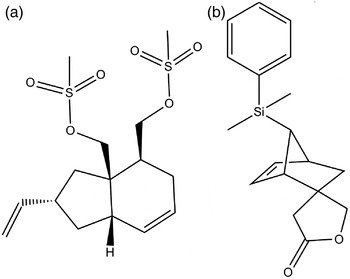
The structural study of two fused 6, 5 carbocyclic and 6, 5 carbooxacyclic systems, 4-(methyl(sulfonyl)methoxy-2-vinyl)-2S*,3aR*,4S*,5,7aS*-(hexahydro-1H-indan-3a-yl)methylmethanesulfonate (1) and (1S*,2S*,4S*,7R*)-7-(dimethyl(phenyl)silyl)-4′,5′-dihydro-2′H-spiro[bicyclo[2.2.1]hept[5]ene-2,3′-furan]-2′-one (2) (Scheme 1) are described in the present paper along with the density functional theory (DFT) calculations to analyze their molecular geometry. Since our attempts to grow single crystals suitable for X-ray analysis resulted in microcrystalline powders, structure determination of 1 and 2 was accomplished from PXRD. An investigation of close intermolecular interactions in 1, 2 and a few related 6, 5 carbocyclic and 6, 5 carbooxacyclic systems via Hirshfeld surface analysis is also presented.
Chemical diagram of (a) C15H24O6S2 (1), (b) C18H22O2Si (2).

II. EXPERIMENTAL
A. Materials
Synthesis of 4-(methyl(sulfonyl)methoxy-2-vinyl)-2S*,3aR*,4S*,5,7aS*-(hexahydro-1H-indan-3a-yl)methylmethanesulfonate (1) and (1S*,2S*,4S*,7R*)-7-(dimethyl(phenyl)silyl)-4′,5′-dihydro-2′H-spiro[bicyclo[2.2.1]hept[5]ene-2,3′-furan]-2′-one (2) has been reported earlier (Maity and Ghosh, Reference Maity and Ghosh2009; Maity, Reference Maity2010). Despite the presence of multiple chiral centers in the molecules of 1 and 2, both compounds were synthesized as racemates and crystallized in centrosymmetric space groups. The NMR and IR data reported previously are in agreement with the chemical formulas of 1 and 2 refined by PXRD.
B. X-ray powder diffraction
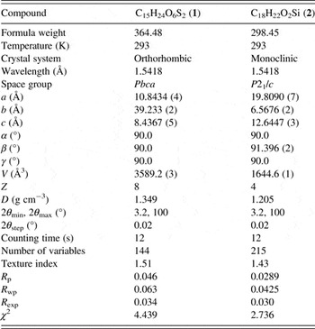
PXRD data of compounds 1 and 2 were recorded at ambient temperature [293(2) K] using a Bruker D8 Advance diffractometer operating in the Bragg–Brentano geometry with CuK α radiation (λ = 1.5418 Å). Indexing of PXRD patterns using EXPO 2014 (Altomare et al., Reference Altomare, Cuocci, Giacovazzo, Moliterni, Rizzi, Corriero and Falcicchio2013) showed an orthorhombic (in 1) and a monoclinic (in 2) unit cells. Given the volume of unit cells and a consideration of expected density of these compounds, the number of formula units in the unit cell of 1 and 2 turned out as 8 and 4, respectively. Although the correct assignment of space group from the systematic absences of reflections in the PXRD patterns is not often obvious, a statistical analysis of PXRD data using the FINDSPACE module of EXPO 2014 (Altomare et al., Reference Altomare, Cuocci, Giacovazzo, Moliterni, Rizzi, Corriero and Falcicchio2013) indicated Pbca and P21/c as the most likely space groups for 1 and 2, respectively, which were chosen for structure solution. The structures of 1 and 2 were solved by global optimization of structural models in direct space based on a Monte Carlo search using the simulated annealing technique (in parallel tempering mode) as implemented in the program FOX (Favre-Nicolin and Cerny, Reference Favre-Nicolin and Cerny2002). The optimization of isolated molecules was performed using the energy gradient method as incorporated in MOPAC 9.0 (Stewart, Reference Stewart2007).
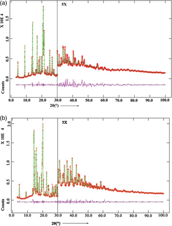
The best solution (i.e., structure with the lowest R wp) was used as the initial structural model of 1 and 2 for Rietveld refinement (Rietveld, Reference Rietveld1967), which was carried out using the GSAS program (Larson and Von Dreele, Reference Larson and Von Dreele2000). A pseudo-Voigt peak profile function was used during refinement and the background of the PXRD patterns was modeled by a Shifted Chebyshev function of first kind with 16 points in 1 and 2, regularly distributed over the entire 2θ range. Standard restraints were applied to bond lengths and bond angles, and planar restraint was used for the phenyl ring in 2. The standard restraints on the bond distances and bond angles of 1 and 2 are listed in Table SI. Common isotropic displacement parameters were refined for carbon, oxygen, sulfur atoms (in 1), and silicon, oxygen, carbon atoms of methyl and phenyl groups, and spiro-fused rings (in 2). Hydrogen atoms in the molecules of 1 and 2 were placed in calculated positions with a fixed U iso (0.07 Å2 in 1 and 0.1 Å2 in 2) value. In the final stage of refinement, a preferred orientation correction using the generalized spherical harmonic model was applied. The final Rietveld plots of 1 and 2 (Figure 1) showed good agreement between the observed PXRD profiles and calculated powder patterns. A summary of crystal data and relevant refinement parameters for 1 and 2 is listed in Table I.
Final Rietveld plot for (a) C15H24O6S2 (1), (b) C18H22O2Si (2). The intensity in the high-angle region has been multiplied by a factor of 5.

Crystal data and structure refinement parameters of C15H24O6S2 (1) and C18H22O2Si (2).

C. Hirshfeld surface analysis
The Hirshfeld surfaces (Hirshfeld, Reference Hirshfeld1977; Spackman and Jayatilaka, Reference Spackman and Jayatilaka2009) and associated two-dimensional (2D) fingerprint plots (Spackman and Mckinnon, Reference Spackman and McKinnon2002; Parkin et al., Reference Parkin, Barr, Dong, Gilmore, Jayatilaka, McKinnon, Spackman and Wilson2007) were generated using the Crystal Explorer 3.1 program (Wolff et al., Reference Wolff, Grimwood, McKinnon, Turner, Jayatilaka and Spackman2012). For each point on the Hirshfeld isosurface, two distances d e, the distance from the point to the nearest nucleus external to the surface, and d i, the distance to the nearest nucleus internal to the surface, are defined. The normalized contact distance (d norm) based on d e and d i is given by
 $$d_{{\rm norm}} = \displaystyle{{d_i - r_i^{{\rm vdw}}} \over {r_i^{{\rm vdw}}}} + \displaystyle{{d_{\rm e} - r_{\rm e}^{{\rm vdw}}} \over {r_{\rm e}^{{\rm vdw}}}}, $$
where r i vdW and r e vdW are the van der Waals radii of the particular atoms involved in the close contact to the surface. The value of d norm is negative or positive depending on the intermolecular contacts being shorter or longer than the van der Waals separations. The parameter d norm displays a surface with a red–white–blue color scheme, where the bright red spots highlight shorter contacts, the white areas represent contacts around the van der Waals separation, and the blue regions are devoid of close contacts.
D. Computational study
Density functional theory (DFT) calculations were performed in the solid state (periodic) for 1 and 2 with the DMol3 code (Delley, Reference Delley1990) in the framework of a generalized gradient approximation (Perdew et al., Reference Perdew, Burke and Ernzerhof1996). The geometry optimization was carried out using the BLYP correlation functional (Becke, Reference Becke1988; Lee et al., Reference Lee, Yang and Parr1988) with a double numeric plus polarization basis set. The starting atomic coordinates were taken from the final X-ray refinement cycle and geometry optimizations were carried out without any structural constraints.
III. RESULT AND DISCUSSION
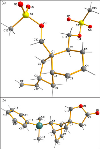
The molecular views of 1 and 2 with atom labeling scheme are shown in Figure 2. The molecule 1 consists of a cis-fused 6, 5 bicyclic ring (C1–C9 atoms) with the C12–C1–C2–H2 torsion angle of 41°, while that in 2 comprises a 5, 6, 5 tricyclic ring skeleton with the norbornene fragment (C1–C7 atoms) spiro-fused with the tetrahydrofuran (THF) moiety (C7–C10, O1 atoms). The ring puckering parameters (Cremer and Pople, Reference Cremer and Pople1975) for the five-membered ring containing atom C9 in 1 (Table II) indicate a half-chair conformation. The six-membered cyclohexene ring (C1–C6 atoms) in 1 adopts a half-chair conformation with the methylmethanesulfonate group at C6 in an equatorial position. The torsion angle C12–C1–C6–C14 of –58.6(6)° in 1 indicates a syn-clinal orientation of two methanesulfonate groups about the C1–C6 bond. Similarly, the ring puckering parameters (Table II) of the 6,5 bridged norbornene system (C1–C7 atoms) in 2 show conformation close to a boat for the cyclohexene moiety (C1–C4, C6 and C7 atoms) and an envelope for the THF ring (C7–C10 and O1 atoms). An extended molecular conformation in 2 is established by torsion angles C7–C4–C5–Si of 176.6(2)° and C4–C5–Si–C13 of −175.9(2)°.
Molecular views of (a) C15H24O6S2 (1), (b) C18H22O2Si (2) with the atom-labeling scheme.

Ring-puckering parameters (Å, °) of C15H24O6S2 (1) and C18H22O2Si (2).

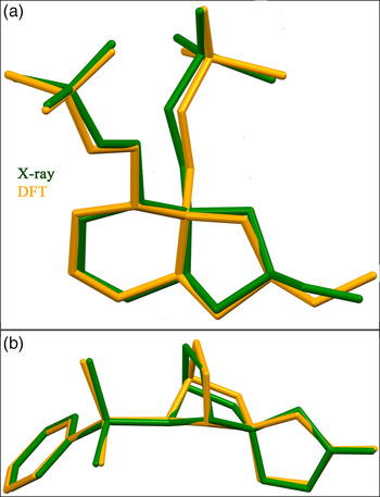
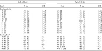
The geometrical parameters (Table III) of molecules with S = O bond distances of 1.414(13)–1.432(12) Å in 1 and Si–C bond lengths of 1.863(3)–1.885(4) Å in 2 agree well with the corresponding mean values (S = O: 1.426 Å for 791 structures and Si–C: 1.864 Å for 277 structures) obtained with MOGUL (Bruno et al., Reference Bruno, Cole, Kessler, Luo, Motherwell, Purkis, Smith, Taylor, Cooper, Harris and Orpen2004) from the CSD search (version 5.37, November 2015 release). An overlay of molecular conformations as determined by the X-ray analysis and solid state DFT calculations of 1 and 2 is shown in Figure 3. The rms deviations between the geometrically optimized bond lengths, bond angles (Table III), and the corresponding crystallographically determined values are 0.04 Å, 2.9° in 1 and 0.03 Å, 2.6° in 2; the corresponding rms Cartesian displacements for 1 and 2 are 0.33 and 0.20 Å, respectively. Close agreement between the X-ray analyzed structure and that obtained via quantum mechanical calculations probably indicates that the compounds 1 and 2 are stable conformers.
Superposition of molecular conformations as obtained from the X-ray structure analysis (green) and solid state DFT calculation (yellow) for (a) C15H24O6S2 (1) and (b) C18H22O2Si (2).

Selected bond lengths (Å) and bond angles (°) of C15H24O6S2 (1) and C18H22O2Si (2) as determined from X-ray structure analysis and DFT calculations.

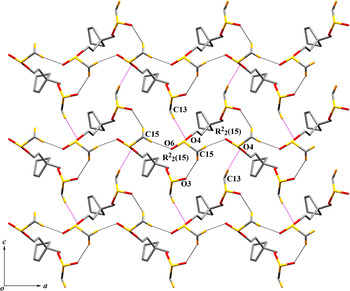
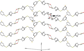
In the absence of any strong hydrogen bond donating group such as –OH or –NH, weak intermolecular C–H ⋯ O hydrogen bonds influence the packing of molecules of 1 and 2 in the crystalline state. Intermolecular C15–H15C ⋯ O6 [C ⋯ O 3.122(12) Å, H ⋯ O 2.17 Å, C–H ⋯ O 169°] hydrogen bonds connect molecules in 1 forming a polymeric C2 2(8) chain propagating along the [100]-direction. Further linking of molecules via C15–H15A ⋯ O3 [C ⋯ O 3.363(14) Å, H ⋯ O 2.50 Å, C–H ⋯ O 150°] hydrogen bonds generate a 1D strip built with edge fused R 2 2(15) rings (Figure 4). The parallel strips in 1 are joined by C13–H13B ⋯ O4 [C ⋯ O 3.518(13) Å, H ⋯ O 2.56 Å, C–H ⋯ O 169°] hydrogen bonds to produce a 2D framework in the (010) plane (Figure 4). In 2, pairs of C8–H8B ⋯ O2 [C ⋯ O 3.578(11) Å, H ⋯ O 2.63 Å, C–H ⋯ O 169°] hydrogen bonds join centrosymmetric molecules into dimers with an R 2 2(8) synthon. The propagation of R 2 2(8) rings via C17–H17 ⋯ π (C13–C18) interactions results in a 2D architecture parallel to (102) plane (Figure 5).
Two-dimensional framework in the (010) plane formed by the C–H ⋯ O hydrogen bonds in C15H24O6S2 (1).

Two-dimensional architecture in the (102) plane formed by the C–H ⋯ O hydrogen bonds and C–H ⋯ π interactions in C18H22O2Si (2).

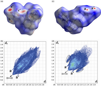
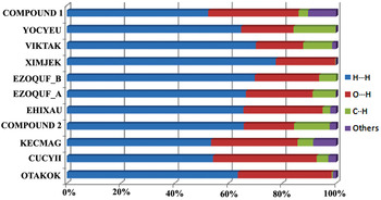
The Hirshfeld surfaces of 1 and 2 mapped over a d norm range of −0.5 to 1.5 Å are illustrated in Figure 6(a) and (c). The red spots labeled as ‘a/a′’, ‘b’ and ‘c′’ in Figure 6(a) and (c) are because of the C–H ⋯ O interactions in 1 and 2. Other red patches in Figure 6(a) correspond to the H ⋯ H interactions in 1. In the 2D fingerprint plot, two sharp spikes labeled as a/a′ in Figure 6(b) in the region of 2.1 < d e + d i < 2.4 Å are characteristics of C15–H15C ⋯ O6 bonded C2 2(8) chain in 1. Spikes corresponding to relatively weaker hydrogen bond (C15–H15A ⋯ O3) are masked within (a/a′). Spikes labeled as a/a′ in Figure 6(d) in the region of 2.5 < d e + d i < 2.85 Å are characteristics of C8–H8B ⋯ O2 bonded R 2 2(8) ring in 2. The wings marked with black circles in Figure 6(d) are because of C–H ⋯ π interactions in 2. The central spikes [Figure 6(b) and (d)] extending upto (d i, d e) region of (0.9 Å, 0.9 Å) in 1 and (1.2 Å, 1.2 Å) in 2 reveal relatively high percentage of H ⋯ H contacts in both cases. The relative contribution of different interactions to the Hirshfeld surfaces of 1, 2 and a few fused 6, 5 cyclic systems retrieved from the CSD are shown in Figure 7. It is evident from Figure 7 that H ⋯ H and O ⋯ H interactions can account for about 85% of the Hirshfeld surface area, 85.7% in 1 and 83.7% in 2, whereas the corresponding values for 3′-(2-hydroxypropyl)-5-methyldihydro-4′H-spiro[furan-3,2′-[5]oxatricyclo[4.2.1.03,7]nonane]-2,4′-dione (OTAKOK) (Kotha et al., Reference Kotha, Dipak and Mobin2011) and 3′-isopropyl-7a′-methylhexahydrospiro[[1,3]dioxolane-2,5′-inden]-1′(6′H)-one (XIMJEK) (Hog et al., Reference Hog, Mayer and Trauner2012) are 98.4 and 99.7%, respectively. An approximately twofold decrease in the contribution of O ⋯ H contacts from 33.3% in 1 to 18.7% in 2 is a consequence of reduced percentage of oxygen atoms in the molecular formula of 2 (4.4%) in comparison with that in 1 (12.8%).
Hirshfeld surface and 2D fingerprint plot of (a) C15H24O6S2 (1), (b) C18H22O2Si (2).

Relative contribution of different interactions to the Hirshfeld surfaces of C15H24O6S2 (1), C18H22O2Si (2) and a few related structures from the CSD.

IV. CONCLUSION
The present study highlights PXRD as a viable alternative for elucidating crystal structures of fused cyclic systems. The molecular conformation as established by the PXRD analysis can be supplemented using solid state DFT calculations. In the absence of conventional hydrogen bonds such as N–H ⋯ O and O–H ⋯ O, the crystal packing in 1 and 2 is influenced by intermolecular C–H ⋯ O hydrogen bonds and C–H ⋯ π (arene) interactions. Hirshfeld surface analysis of 1, 2 and a few related 6, 5 fused cyclic systems retrieved from the CSD reveals that H ⋯ H and O ⋯ H contacts can account for about 85% of the Hirshfeld surface area in these compounds.
SUPPLEMENTARY MATERIAL
The supplementary material for this article can be found at https://doi.org/10.1017/S0885715617000173.
ACKNOWLEDGEMENTS
The authors thank Dr. Soumitra Maity, IACS, Kolkata, for providing the compounds used in the present study. Financial support from the Council for Scientific and Industrial Research (CSIR), New Delhi, India, for a senior research fellowship (SRF) to T.D. is gratefully acknowledged.