Introduction
Junctional ectopic tachycardia is an arrhythmia that originates from the His bundle, with the precise pathogenesis remaining incompletely understood. Some evidence suggests it may be related to abnormalities in the cardiac conduction system during embryonic development. Based on aetiology, junctional ectopic tachycardia is classified into two types: congenital junctional ectopic tachycardia and postoperative junctional ectopic tachycardia. Congenital junctional ectopic tachycardia, a rare arrhythmia in children, usually presents with fetal tachycardia, which affects up to one-third of patients before birth. In 1990, Kylat et al. reported that congenital junctional ectopic tachycardia was associated with a mortality rate as high as 35%. Reference Kylat and Samson1 However, with advancements in pharmacological therapy and radiofrequency catheter ablation techniques, recent studies have demonstrated a significant reduction in mortality rates to approximately 4–9% particularly in the specialised cardiac electrophysiology centres. Reference Sasikumar, Kumar and Balaji2–Reference Catton and Peterson5
Ivabradine, a selective inhibitor of hyperpolarisation-activated cyclic nucleotide-gated (HCN4) channels, reduces heart rate without affecting cardiac function or blood pressure. Reference Tengqi and Xiaofei6 Ivabradine is primarily used to treat chronic heart failure, but its potential for managing cardiac arrhythmias has gained attention in recent years. Reference Dieks, Klehs, Muller, Paul and Krause7 In the current study, we report the clinical data from a case of ivabradine used to treat congenital junctional ectopic tachycardia. We also review the relevant literature to explore the potential applications of ivabradine for neonatal junctional tachycardia.
Case report
A male neonate was delivered via caesarean section at 37 weeks gestation due to intrauterine tachycardia. He presented with persistent tachycardia after birth, and electrocardiography suggested junctional tachycardia. The local hospital initiated treatment with intravenous cedilanid (10 µg/kg for 3 consecutive doses, followed by oral digoxin). However, this regimen failed to achieve conversion to sinus rhythm, and the heart rate remained elevated (198–210 beats per minute) after one week. Subsequently, intravenous amiodarone (5 mg/kg) was administered, followed by 15 µg/kg/min continuous intravenous infusion for 72 h, but no improvement in heart rate was observed. Amiodarone combined with propafenone (10 µg/kg/min continuous intravenous infusion) was continued for 2 weeks, but conversion to sinus rhythm was not achieved. Whole-exome genetic testing identified two variants in the DSG2 gene (c.3044T > C and c.1468G > A). However, based on the American College of Medical Genetics and Genomics (ACMG) guidelines, these variants were classified as “variants of uncertain significance.” Furthermore, functional prediction using the REVEL (Rare Exome Variant Ensemble Learner) supported this classification, indicating uncertain or benign outcomes. On postnatal day 25, the patient was transferred to our hospital.
Upon admission to our hospital, the neonate presented with marked tachycardia (190–220 bpm) with stable blood pressure (68/43 mmHg). Physical examination revealed no hepatosplenomegaly or peripheral oedema. Junctional ectopic tachycardia with a heart rate of 198 beats per minute was confirmed by electrocardiogram (Figure 1). Echocardiography examination demonstrated left ventricular enlargement (left ventricular end-diastolic dimension of 23 mm, z-score + 2.47), mild-to-moderate tricuspid regurgitation, and normal cardiac systolic function (ejection fraction 72%).
Electrocardiogram on admission.

Following admission, intravenous propafenone (1.5 mg/kg) was administered. The heart rate of the neonate decreased to 180–190 beats/min, but sinus rhythm was not restored. Oral propafenone (5 mg/kg, q8h) and amiodarone (5 mg/kg q8h) were given. After 24 h of this regimen, the heart rate remained elevated (180–190 beats per minute), and conversion to sinus rhythm was not achieved. Amiodarone was discontinued, and propafenone therapy was maintained. Oral ivabradine (0.1 mg/kg/dose q12h) was initiated. After 20 min of the first dose of ivabradine, the heart rate of the neonate decreased to 120–130 beats per minute with intermittent sinus rhythm. Within 3 h of ivabradine administration, sinus rhythm was restored (Figure 2). Ivabradine therapy was continued at the same dose, and the infant remained asymptomatic during follow-up. 24-hour Holter monitoring after one month of treatment showed an average heart rate of 114 beats per minute and occasional episodes of junctional ectopic tachycardia (accounting for 3.8% of total heartbeats). Echocardiography examination demonstrated a left ventricular end diastolic dimension of 20 mm (Z-score + 0.71), and the tricuspid regurgitation had resolved. The electrocardiogram results indicate a heart rate of 198 beats per minute with narrow QRS tachycardia, and atrioventricular (AV) dissociation is observed. It was diagnosed as junctional ectopic tachycardia.
Electrocardiogram taken 3 h after the first oral administration of ivabradine. The electrocardiogram result showed sinus rhythm with a heart rate of 125 beats per minute.

Literature review
Congenital junctional ectopic tachycardia is a rare arrhythmia characterised by narrow QRS complex tachycardia, often accompanied by AV dissociation or retrograde P-waves with ventriculoatrial conduction. Reference Drago, Battipaglia and Di Mambro8,Reference Liu, Ip, Markowitz, Lerman, Zipes, Jalife and Stevenson9 The rhythm may be regular or irregular, occasionally complicating diagnosis. Unlike other supraventricular tachycardias, congenital junctional ectopic tachycardia is typically refractory to conventional therapies, including adenosine administration and direct current (DC) cardioversion.
Amiodarone is the recommended first-line treatment for congenital junctional ectopic tachycardia in infants, used alone or with other antiarrhythmic drugs. Beta-blockers, digoxin, propafenone, and flecainide are among the alternatives. However, drug therapy often proves ineffective for congenital junctional ectopic tachycardia, which typically manifests as recurrent episodes. Radiofrequency ablation is widely used for various arrhythmias, Reference Kean, Hazle, LaPage, Bromberg and Dick10–Reference Thajudeen, Namboodiri, Choudhary, Valaparambil and Tharakan12 but its application in congenital junctional ectopic tachycardia is limited. The risk of complete AV block after radiofrequency ablation is high because of the proximity of the congenital junctional ectopic tachycardia origin to the His bundle. In addition, radiofrequency ablation in small infants is considerably limited by their low body weight and thin blood vessels. Reference Dieks, Klehs, Muller, Paul and Krause7
The pathophysiology of congenital junctional ectopic tachycardia remains incompletely understood. Some evidence suggests that it may be associated with the activation of HCN4 channels, leading to enhanced automaticity in the AV node and adjacent regions, including the bundle of His. Reference Dieks, Klehs, Muller, Paul and Krause7,Reference DiFrancesco, Ferroni, Mazzanti and Tromba13–Reference Yang, Tadros and Sun15 Ivabradine, a specific inhibitor of HCN4, reduces heart rate by inhibiting spontaneous diastolic depolarisation in the AV nodal area, making it a promising therapeutic option for congenital junctional ectopic tachycardia. Reference Al-Ghamdi, Al-Fayyadh and Hamilton16–Reference Di Marco, De Nigris, Pepe, Pagano, Di Nardo and Tipo20
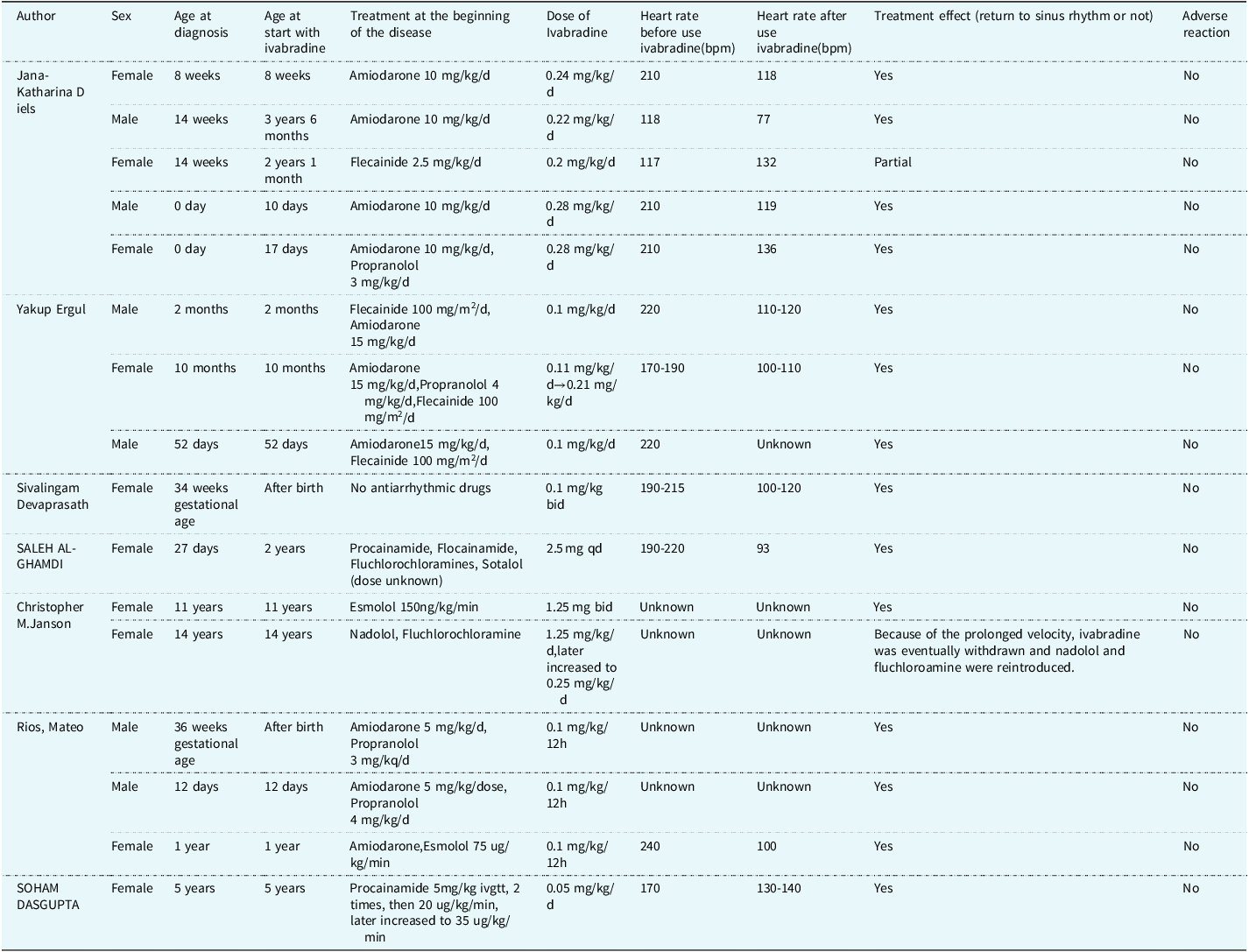
However, the use of ivabradine for supraventricular tachycardia in children remains unestablished. We searched 16 papers in PubMed for “Junctional Ectopic Tachycardia,” “children,” and “Ivabradine.” Excluding guidelines and reviews, we identified 7 case reports for analysis (Table 1). These reports described 16 cases of junctional ectopic tachycardia in children, consisting of 6 males and 10 females (age, 0 d –14 y; mean age, 2.5 y), including 15 congenital junctional ectopic tachycardia and 1 postoperative junctional ectopic tachycardia patient. All patients were initially treated with other antiarrhythmic drugs (amiodarone, digoxin, flecainide, propranolol), alone or in combination. After treatment failed, the patients were switched to ivabradine. Reference Dieks, Klehs, Muller, Paul and Krause7,Reference Al-Ghamdi, Al-Fayyadh and Hamilton16,Reference Dasgupta and Johnsrude21–Reference Rios, Chiesa, Arhcilles, Cuesta and Moltedo25
Summary of the literature on ivabradine for the treatment of junctional ectopic tachycardia in children

As described in 7 reports, sinus rhythm conversion was achieved in 15 of 16 cases treated with ivabradine (0.1–0.28 mg/kg/d). One report described congenital junctional ectopic tachycardia in a 14-year-old female patient in which ivabradine failed to restore sinus rhythm, leading to the development of sustained atrial tachycardia. Ivabradine was discontinued, whereas nadolol and flucytosine were maintained. Reference Janson, Tan, Iyer, Vogel, Vetter and Shah24 No adverse drug reactions were reported in any of the cases. Reference Dasgupta and Johnsrude21
Ivabradine has also demonstrated efficacy in congenital junctional ectopic tachycardia therapy in preterm infants. Suzan delivered a male infant at 31 weeks of gestation via emergency caesarean section. The infant developed unexplained tachycardia on the 19th postnatal day. Ambulatory electrocardiography suggested the presence of junctional ectopic tachycardia. The patient was started on propranolol (0.25 mg/kg/dose q12h), which failed to control the episode. Despite intravenous amiodarone (10 mg/kg/min) and increasing propranolol to 1 mg/kg/dose q6h, the heart rate remained 250–290 beats per minute. Ivabradine (0.05 mg/kg/dose q12h) was initiated on day 26 while propranolol and amiodarone therapy continued. A 24-hour ambulatory electrocardiogram showed restoration of sinus rhythm. No recurrence of the arrhythmia was observed during 6 months of follow-up. Reference Asfour, Al-Omran, Alodhaidan, Asfour, Khalil and Al-Mouqdad26
This case we reported presented with fetal tachycardia and was postnatally diagnosed as congenital junctional ectopic tachycardia. Initial treatment with digoxin, propafenone, and amiodarone proved unsuccessful. Following the administration of ivabradine (0.1 mg/kg/dose), the cardiac rhythm converted to a sinusoidal pattern within three hours after the first oral dose, demonstrating its efficacy in this case of junctional ectopic tachycardia. And this experience provided valuable clinical evidence supporting the use of ivabradine in treating neonatal congenital junctional tachycardia.
The most common adverse event associated with ivabradine is sinus bradycardia, which is dose-dependent. Reference Doctor, Scott, Tindel and Nguyen27 Therefore, the dosage should be adjusted based on heart rate during treatment, especially when used concomitantly with other heart rate-lowering medications such as amiodarone, sotalol, and verapamil. Although ivabradine does not directly prolong the QT interval, heart rate reduction may lead to secondary QT interval prolongation. Thus, it should be used cautiously in patients with congenital long QT syndrome. When combined with other QT-prolonging drugs, electrocardiographic monitoring of the QT interval is essential to prevent serious arrhythmias. Reference Duro Da Costa28,Reference Frommeyer, Weller and Ellermann29
Studies have demonstrated that the pharmacokinetic-pharmacodynamic (PK-PD) relationship of ivabradine is consistent between children and adults, with the magnitude of heart rate reduction showing a positive correlation with plasma drug concentration. However, due to developmental physiological differences, individualised dosing regimens are required for children of different age groups. In infants aged 6–12 months, the CYP3A4 enzyme system is not yet fully developed and the glomerular filtration rate is relatively low. These factors collectively lead to a significantly reduced clearance of ivabradine, resulting in an approximately 20% increase in plasma drug exposure compared to adults. Therefore, a lower initial dose (0.02 mg/kg) is clinically recommended for infants. For children over 12 months of age, as CYP3A4 enzyme activity gradually increases and renal function matures, drug clearance improves significantly. Consequently, this age group requires a higher initial dose (0.05 mg/kg) to achieve a similar area under the concentration-time curve as observed in adults. Reference Peigné, Bouzom, Brendel, Gesson, Fouliard and Chenel30,Reference Peigné, Fouliard, Decourcelle and Chenel31 However, these dosage recommendations are specifically targeted at the treatment of heart failure. The therapeutic goal of using ivabradine in heart failure is to reduce heart rate without inducing bradycardia. Previous case reports and our study all suggest that for the treatment of arrhythmias, the initial dose of ivabradine should be higher than that used for heart failure.
Summary
Further research and clinical validation are needed to support the use of ivabradine in children, especially for neonatal. Nonetheless, its potential for treating refractory arrhythmias should not be overlooked. The present case, along with existing literature, demonstrates the promising efficacy of ivabradine in paediatric junctional ectopic tachycardia, highlighting its ability to rapidly restore sinus rhythm and its potential as a preferred alternative for refractory cases.
Acknowledgements
I would like to thank Dr Sun for all her help in writing this article, as well as her meticulous care and support.
Author contribution
Conceptualisation: Peiwen Gong. Data curation: Huimin Su. Formal analysis: Ping Xiang. Methodology: Huichao Sun. Writing-original draft: Peiwen Gong. Writing-review& editing: Huichao Sun, Mi Li. This research received no specific grant from any funding agency, commercial or not-for-profit sectors.
Financial support
No funding was received for conducting this study.
Competing interests
All authors have no conflict of interest.
Ethics standard
This case report and review article does not require ethics approval.
Patient consent statement
We have already get the approval from patient’s parents.