INTRODUCTION
Human immunodeficiency virus (HIV) is the causative agent of acquired immunodeficiency syndrome (AIDS), which results in susceptibility to life-threatening opportunistic infections and cancer. Approximately 35 million people are estimated to live with HIV-1, with 1·6 million deaths annually, mainly in sub-Saharan Africa (UNAIDS, 2013).
HIV-1 is transmitted horizontally, mostly by sexual intercourse, and vertically by mother to child transmission. The acute phase of HIV-1 infection can be asymptomatic or be characterized by fever, lymphoadenopathy, headache and fatigue, not dissimilar from other acute viral infections. However a rapid loss of CD4+ T cells in the gastrointestinal tract is typical of early HIV-1 infection (Brenchley et al. Reference Brenchley, Schacker, Ruff, Price, Taylor, Beilman, Nguyen, Khoruts, Larson, Haase and Douek2004). T cell numbers recover only partially and a progressive decline ensues for several years, until they fall below 200 μL−1 (Lang et al. Reference Lang, Perkins and Anderson1989). At this stage, there is a clear ‘inverted ratio’ of CD8 and CD4T cells in blood and the T cell helper function is severely compromised. Hence severe immunodeficiency is established and opportunistic infections are frequent. Importantly, AIDS is often associated with malaria and tuberculosis (TB) in many countries, aggravating the clinical picture and promoting the emergence of drug-resistant strains of malaria and mycobacterium tuberculosis (Flateau et al. Reference Flateau, Le Loup and Pialoux2011; World Health Organization, 2012).
Currently there is no vaccine against HIV-1; prevention and antiviral drugs are the main tools to reduce the impact of the pandemic. In particular antiretroviral drugs have demonstrated efficacy in reducing viral load in infected individuals, restoring a functional immune system, reducing overall mortality and preventing transmission (Palella et al. Reference Palella, Delaney, Moorman, Loveless, Fuhrer, Satten, Aschman and Holmberg1998). However, problems of cost, distribution, adherence, monitoring and multiple drug resistance remain (Wainberg, Reference Wainberg2009). Significant gaps in our knowledge of how HIV-1 can evade acquired and innate immunity, the host-virus interactions critical for replication, how latency is established, and the determinants of transmission limit our ability to stop the epidemic.
HIV STRUCTURE
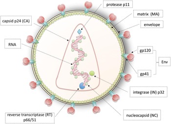
HIV is a lentivirus, member of the retrovirus family, capable of infecting mitotically active and non-dividing cells. Each virion is about 0·1 μm in diameter. The envelope forms the outermost layer of the viral particle and is adopted from the host cell membrane during budding. Embedded throughout the envelope are host proteins and protruding ‘spikes’ of HIV protein called Env. Each spike contains 3 molecules of glycoprotein gp120 on the extracellular face, anchored to the virion by 3 stems of transmembrane glycoprotein gp41. The matrix is between the envelope and the viral capsid. The viral core is cone-shaped, and is made of capsid protein p24 (CA). Inside this core there are 2 identical single-stranded HIV RNAs and enzymes crucial in viral replication: reverse transcriptase (RT) p66/51, protease p11 and integrase (IN) p32 (Fig. 1). Each of the single-stranded RNA encodes for 3 structural genes (gag, pol and env) important in the formation of structural proteins and for 6 regulatory genes (tat, rev, nef, vif, vpr and vpu) crucial in regulating replication of HIV (Freed and Martin, Reference Freed, Martin, Knipe and Howley2001).
Schematic representation of the HIV-1 particle structure.

HIV life cycle
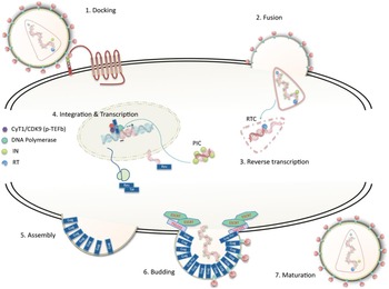
The HIV-1 life cycle has been extensively studied. It involves step-wise processes of binding, fusion, reverse transcription and integration, followed by transcription, assembly, viral budding and finally maturation (Fig. 2). It begins when the glycoprotein (gp120) on the envelope of HIV binds to the CD4 receptors present on helper T cells, macrophages and microglial cells, causing a conformational change and exposing the co-receptor binding domain. This allows binding to the chemokine co-receptors CCR5 or CXCR4, and a further conformational change that exposes the 6-helix bundle of gp41 containing the fusion domain (Kwong et al. Reference Kwong, Wyatt, Robinson, Sweet, Sodroski and Hendrickson1998; Wyatt, Reference Wyatt1998; Berger et al. Reference Berger, Murphy and Farber1999). Fusion between host cell and viral membranes enables entry of viral capsid and enzymes. Once in the host cytoplasm, the viral capsid must undergo uncoating. The capsid p24 itself is critical in maintaining optimal viral core stability, as well as facilitating the subsequent events of reverse transcription, nuclear import and integration (Fassati, Reference Fassati2012). Host proteins such as cyclophilin A participate in these early events (Fassati, Reference Fassati2012). The reverse transcription complex (RTC) is thus formed in the cytoplasm and the viral RNA genome is reverse transcribed into a double-stranded DNA form (Freed and Martin, Reference Freed, Martin, Knipe and Howley2001). Most of the p24 capsid proteins are lost once reverse transcription is completed, leading to the formation of the preintegration complex (PIC) (Fassati, Reference Fassati2012).
Schematic representation of the HIV-1 life cycle. Step 1, docking: the HIV-1 particle binds to the CD4 receptor, inducing a conformational change in the gp120 env protein. This exposes the co-receptor binding domain and another conformational change occurs in the TM domain of env (gp41) upon co-receptor binding. Step 2, fusion: co-receptor binding exposes a 6-helix bundle fgp41, which anchors to the cell membrane, inducing fusion and release of the viral core inside the cytoplasm. Step 3, reverse transcription: the viral core starts disassembly (uncoating) and the viral RNA is reverse transcribed into a double-stranded DNA molecule. The host cell factor cyclophilin A promotes these steps. This process takes place in the so-called reverse transcription complex (RTC). The pre-integration complex (PIC) is formed upon completion of reverse transcription and contains oligomerized integrase proteins. The PIC is transported into the nucleus. Step 4, integration and transcription: the PIC reaches the host chromatin and integrase catalyses the strand transfer reaction, which results in covalent integration of the viral DNA into cellular chromosomes. The host factor LEDGF/p75 promotes tethering of the PIC onto host chromatin. Low levels of transcription produce spliced mRNAs, including Tat and Rev, which promote further transcription and nuclear export of genomic viral RNA. 5, Assembly: gag and gag-pol polyproteins are synthesized and migrate towards the cell membrane, where they organize into a lattice together with env proteins. The NC domain of gag specifically binds viral RNA and recruits it into the assembly site. Step 6, budding: the host factor Tsg101 binds to the p6 domain of gag and recruits the ESCRT complex, which induce ‘vesiculation’ of the assembled particle, then the viral particle is cleaved off and released. Step 7, maturation: protease is activated, inducing a step-wise cleavage of the gag and gag-pol polyproteins, which results in a dramatic conformational change of the viral core from spherical to cone-shaped. The viral particle is now infectious.

The PIC then undergoes active nuclear import into the host cell nucleus (Fassati, Reference Fassati2006; Matreyek and Engelman, Reference Matreyek and Engelman2013). Once in the nucleus, the viral enzyme integrase permanently inserts the viral DNA into the host genome, producing the provirus (Freed and Martin, Reference Freed, Martin, Knipe and Howley2001). Both ends of the proviral DNA are flanked by identical sequences called long terminal repeats (LTR). The HIV-1 LTR is approximately 640 bp in length and is segmented into 3 regions: U3-R-U5. The U3 (unique 3′ sequence) is located at the 5′ end of each LTR, and is subdivided into 3 sections: modulatory, enhancer and core. This region contains the binding sites for cellular transcription factors, both constitutive and inducible, including Nuclear Factor-κB (NF-κB), CCAAT/enhancer binding protein (C/EBP), activating transcription factor/cyclic AMP response element binding (ATF/CREB), lymphocyte enhancer factor (LEF-1), nuclear factor of activated T cells (NF-AT) and activator protein 1 (AP-1) (Pereira et al. Reference Pereira, Bentley, Peeters, Churchill and Deacon2000). The R (repeated sequence) region is located between the U3 and U5 sequence and is where transcription begins. Importantly, this region contains the binding site for viral regulatory protein Tat (Krebs et al. Reference Krebs, Hogan, Quiterio, Gartner and Wigdahl2001). Next, the provirus utilizes its 5′ LTR domain as a promoter region to recruit host RNA polymerase II to make copies of its genomic RNA (Pollard and Malim, Reference Pollard and Malim1998). During this early stage, shorter, spliced viral mRNAs are made and exported to the cytoplasm for translation of Tat and Rev. Subsequently, Tat and Rev promote transcription and nuclear export of full length, unspliced viral mRNA for translation and packaging into new particles (Tazi et al. Reference Tazi, Bakkour, Marchand, Ayadi, Aboufirassi and Branlant2010).
Tat and Rev
The viral protein Tat is a critical transactivator that promotes transcriptional elongation. It binds to the TAR (trans-activation-responsive) element, an RNA stem-loop structure located in the R region of the LTR and recruits cellular co-factors Cyclin T1 and CDK9 in a complex called P-TEFb (Roy et al. Reference Roy, Delling, Chen, Rosen and Sonenberg1990; Wei et al. Reference Wei, Garber, Fang, Fischer and Jones1998; Bieniasz et al. Reference Bieniasz, Grdina, Bogerd and Cullen1999). This complex, also known as TAK (Tat-associated kinase), is crucial in phosphorylating the COOH terminal domain of RNA polymerase II, enhancing transcriptional elongation (Emerman, Reference Emerman1998).
The Rev protein binds a short RNA segment called Rev responsive element (RRE) located in the 3′ region of the viral mRNA. Rev recruits the nuclear export receptor CRM1, which, upon multimerization at the RRE, facilitates nuclear export of unspliced and incompletely spliced mRNAs (Pollard and Malim, Reference Pollard and Malim1998; Tazi et al. Reference Tazi, Bakkour, Marchand, Ayadi, Aboufirassi and Branlant2010). Thus Rev, by recruiting a host cell factor, overcomes the block to nuclear export of unspliced mRNAs in human cells.
Assembly, budding and maturation
The Gag polyprotein (which encodes for p17 matrix (MA), p24 capsid (CA), p7 nucleocapsid (NC) and p6) is the main driving force for the assembly of immature HIV (Ivanchenko et al. Reference Ivanchenko, Godinez, Lampe, Kräusslich, Eils, Rohr, Bräuchle, Müller and Lamb2009). Upon translation, most of the Gag proteins accumulate and multimerize at the plasma membrane, creating an assembly site. The N-terminus MA domain is tightly associated with the inner surface of the lipid bilayer while the C-terminus p6 projects away from the phospholipid membrane. Contacts between the nucleocapsid p7 domain and full length viral RNA act as a scaffold that recruits viral RNA towards the cell membrane for assembly. At this stage, Env is incorporated into the host membrane in the form of gp120 and gp41 (cleaved by host furine protease) (Freed and Martin, Reference Freed, Martin, Knipe and Howley2001). Subsequently, the Endosomal Sorting Complexes Required for Transport (ESCRT) is recruited to the assembly site to facilitate the budding of nascent immature virion (Garrus et al. Reference Garrus, von Schwedler, Pornillos, Morham, Zavitz, Wang, Wettstein, Stray, Côté, Rich, Myszka and Sundquist2001; Martin-Serrano et al. Reference Martin-Serrano, Zang and Bieniasz2001). The early interaction between the P(T/S)AP motif of the p6 protein and TSG101 (Tumor Susceptibility Gene 101) of the ESCRT-I complex initiates the formation of budding viruses This then activates ESCRT-II and consequently triggers the formation of ESCRT-III (Sundquist and Kräusslich, Reference Sundquist and Kräusslich2012). Another p6 domain, LXPXnL, serves as a docking site for the ESCRT-III binding partner: Alix [ALG-2 (apoptosis-linked gene 2)-interacting protein X] (Stuchell et al. Reference Stuchell, Garrus, Müller, Stray, Ghaffarian, McKinnon, Kräusslich, Morham and Sundquist2004; Usami et al. Reference Usami, Popov, Popova, Inoue, Weissenhorn and Göttlinger2009; Briant, Reference Briant2011). Lastly, the ESCRT complex recruits CHMPs (charged multivesicular body proteins) to promote closure of the membrane neck and VPS4 (vacuolar protein sorting) ATPase to complete the process of membrane fission (Sundquist and Kräusslich, Reference Sundquist and Kräusslich2012). As a result, an immature and non-infectious virion buds off from the host cell. Finally, the immature virion undergoes maturation by activating viral protease p11 (PR) through autocleavage. Together with major conformational changes and structural rearrangements, PR cleaves Gag and Gag-Pol polyproteins into mature proteins: MA, CA, NC, p6, IN and RT, producing a fully infectious virion (Briant, Reference Briant2011; Sundquist and Kräusslich, Reference Sundquist and Kräusslich2012).
Latency
Combined antiretroviral therapy (cART) is the standard treatment for HIV infection and targets different steps in the HIV replication cycle. Typically, there will be three or more antiviral drugs taken in combination (e.g. a nucleoside RT inhibitor, a protease inhibitor and a non-nucleoside RT inhibitor) to reduce the likelihood of developing viral resistance. cART is very effective (Palella et al. Reference Palella, Delaney, Moorman, Loveless, Fuhrer, Satten, Aschman and Holmberg1998) however, upon therapy suspension, the latent viral reservoir is activated and viral load rebounds to pre-treatment levels in a few weeks (Finzi et al. Reference Finzi, Hermankova, Pierson, Carruth, Buck, Chaisson, Quinn, Chadwick, Margolick, Brookmeyer, Gallant, Markowitz, Ho, Richman and Siliciano1997).
How the latent reservoir is established and maintained is an issue not fully resolved. It is accepted that there is very low viral production even under cART (Palmer et al. Reference Palmer, Maldarelli, Wiegand, Bernstein, Hanna, Brun, Kempf, Mellors, Coffin and King2008), but it is unclear if this residual viraemia is due to ongoing replication in specific sites (mainly the gastrointestinal lymphatic system – GALT) where drugs may diffuse poorly, or to a long-lived reservoir that is randomly activated, such as resting memory T cells (Hakre et al. Reference Hakre, Chavez, Shirakawa and Verdin2012). In latently infected cells, the virus remains transcriptionally silent. Latently infected cells are typically few, estimated at 0·03–3 per million resting CD4+ T cells, and are often undetectable by even the most sensitive clinical assays (Siliciano et al. Reference Siliciano, Kajdas, Finzi, Quinn, Chadwick, Margolick, Kovacs, Gange and Siliciano2003). These memory T cells can also survive for several years (Michie et al. Reference Michie, McLean, Alcock and Beverley1992; Chun et al. Reference Chun, Carruth, Finzi and Shen1997) and can be activated under appropriate stimulation, causing viral rebound. Therefore, the long-lived latent viral reservoir prevents HIV-1 eradication and a cure.
HIV-1 latency is a multifactorial process involving chromatin modifications, low levels of specific transcription factors, integration site selection and cell activation (Chan and Greene, Reference Chan and Greene2011; Hakre et al. Reference Hakre, Chavez, Shirakawa and Verdin2011; Van Lint et al. Reference Van Lint, Bouchat and Marcello2013) and it can be broadly divided in pre-integration latency and post-integration latency (Chun et al. Reference Chun, Carruth, Finzi and Shen1997; Donahue and Wainberg, Reference Donahue and Wainberg2013). Pre-integration latency occurs when the viral DNA failed to be integrated into the host genome. This could happen either because reverse transcription was impaired or PIC nuclear import was blocked (Zack et al. Reference Zack, Arrigo, Weitsman, Go, Haislip and Chen1990; Bukrinsky et al. Reference Bukrinsky, Sharova, Dempsey, Stanwick, Bukrinskaya, Haggerty and Stevenson1992). Inefficient reverse transcription and PIC nuclear import and integration in resting T cells is in part determined by the limited nucleotide pool, possibly due to host restriction factor SAMHD1 (Baldauf et al. Reference Baldauf, Pan, Erikson, Schmidt, Daddacha, Burggraf, Schenkova, Ambiel, Wabnitz, Gramberg, Panitz, Flory, Landau, Sertel, Rutsch, Lasitschka, Kim, König, Fackler and Keppler2012; Descours et al. Reference Descours, Cribier, Chable-Bessia, Ayinde, Rice, Crow, Yatim, Schwartz, Laguette and Benkirane2012) and in part by other host co-factors (Zack et al. Reference Zack, Arrigo, Weitsman, Go, Haislip and Chen1990). On the other hand, post-integration latency represents a more stable form of latency (Chun et al. Reference Chun, Finzi and Margolick1995). This post-integration latency may occur when infected, activated T cells return to resting G0 state (Chun et al. Reference Chun, Finzi and Margolick1995), or due to integration of viral DNA into repressed chromatin regions of the host genome, or due to interference with a flanking cellular promoter (Colin and Van Lint, Reference Colin and van Lint2009). Nonetheless, these latently infected cells can be activated and start producing virus again, but the signal transduction pathways that regulate this switch from latent to productive infection are understood only in part. It is generally believed that if the host cell is exposed to the appropriate antigens or activating stimuli, latency may be reversed and the cell can start producing new viruses (Siliciano and Greene, Reference Siliciano and Greene2011).
HEAT SHOCK PROTEIN 90 (HSP90)
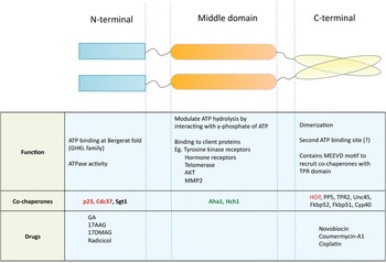
Heat shock proteins are a class of highly conserved proteins that are up-regulated as a response to elevated heat or other types or stress (Schlesinger, Reference Schlesinger1990). In general, these molecular chaperones bind to and stabilize another protein molecule or protein complex; and through a series of controlled binding and release, facilitate the correct folding or stabilization of the substrate molecule (Hartl, Reference Hartl1996). The Hsp90 N-terminal domain contains an ATP-binding site that shares tertiary structure similarity (the ‘Bergerat fold’) with other GHKL family members (Gyrase B, Hsp90, histidine kinase and MutL) (Dutta and Inouye, Reference Dutta and Inouye2000; Pearl and Prodromou, Reference Pearl and Prodromou2006). It is also the binding site for its inhibitor, geldanamycin (GA) (Stebbins et al. Reference Stebbins, Russo, Schneider, Rosen, Hartl and Pavletich1997). The middle domain of ∼280 residues contains critical sites for client protein binding. The highly charged linker region is also the site where the nuclear localization signal is located (Csermely and Schnaider, Reference Csermely and Schnaider1998); the C-terminal domain is important for dimerization and harbours the binding site for calmodulin (Csermely and Schnaider, Reference Csermely and Schnaider1998; Pearl and Prodromou, Reference Pearl and Prodromou2006) (Fig. 3). Hsp90 requires specific co-chaperones for its activity, for example Cdc37 is a kinase-specific co-chaperone whereas Hop and Tpr2 modulate interactions between Hsp90 and Hsp70 (Li et al. Reference Li, Soroka and Buchner2012) (Fig. 3). Crucially, because genetic knockout of Hsp90 is lethal in eukaryotes (Solit and Chiosis, Reference Solit and Chiosis2008), most studies on Hsp90 are mainly done utilizing selective inhibitors such as GA and 17-allylamino-17-demethoxygeldanamycin (17-AAG).
Schematic depiction of the Hsp90 domains, their function, their co-chaperones binding and their drug targeting. ATPase activity inhibits co-chaperones shown in red, and stimulates co-chaperones shown in green.

Hsp90 and HIV transcriptional control
Surprisingly, despite its abundance and critical functions in human cells, Hsp90 has been little studied in the context of HIV-1 infection, and its relevance has started to become apparent only recently.
Proteomics and microarray analyses on HIV-1 infected cells detected an increased expression of Hsp90 upon HIV-1 infection (Ringrose et al. Reference Ringrose, Jeeninga, Berkhout and Speijer2008; Boukli et al. Reference Boukli, Shetty, Cubano, Ricaurte, Coelho-Dos-Reis, Nickens, Shah, Talal, Philip and Jain2012), although it remained unclear if Hsp90 up-regulation was specifically induced by HIV-1 or it was simply a cellular response to infection-induced stress. Early studies showed that latently infected cells can reactivate HIV-1 when incubated in heat shock condition (42 °C) without any added cytokines (Stanley et al. Reference Stanley, Bressler, Poli and Fauci1990; Hashimoto et al. Reference Hashimoto, Baba, Gohnai, Sato and Shigeta1996) and this hyperthermia-induced activation has been exploited for rapid detection of HIV in clinical samples (Re et al. Reference Re, Furlini and La Placa1989). However, it remained unclear if HIV-1 reactivation upon heat shock actually depended on Hsp90.
More recently, hyperthermia (39·5 °C) was reported to enhance HIV-1 replication by stimulating viral gene expression (Roesch et al. Reference Roesch, Meziane, Kula, Nisole, Porrot, Anderson, Mammano, Fassati, Marcello, Benkirane and Schwartz2012). While both Hsp70 and Hsp90 were up-regulated in CD4+ T cells grown at 39·5 °C, selective inhibition of Hsp90 with small chemical compounds was sufficient to negate this enhanced HIV-1 replication (Roesch et al. Reference Roesch, Meziane, Kula, Nisole, Porrot, Anderson, Mammano, Fassati, Marcello, Benkirane and Schwartz2012). Furthermore, confocal microscopy experiments showed that hyperthermia promoted localization of Hsp90 at the viral transcriptional site (Roesch et al. Reference Roesch, Meziane, Kula, Nisole, Porrot, Anderson, Mammano, Fassati, Marcello, Benkirane and Schwartz2012). Therefore, the study demonstrated a direct link between enhanced replication of HIV-1 in conditions of hyperthermia and Hsp90. These findings have implications for the management of HIV-1-positive individuals, who often suffer from co-infection by other pathogens (tuberculosis, malaria) that cause high fever episodes.
An interesting question prompted by this study is why Hsp90 localizes at the HIV-1 transcriptional site. Indeed chromatin immunoprecipitation (ChIP) experiments showed that Hsp90 localizes on chromatin at the HIV-1 promoter (Vozzolo et al. Reference Vozzolo, Loh, Gane, Tribak, Zhou, Anderson, Nyakatura, Jenner, Selwood and Fassati2010). The functional significance of this observation was supported by the fact that selective Hsp90 inhibitors repressed HIV-1 gene expression in acutely infected cells (Vozzolo et al. Reference Vozzolo, Loh, Gane, Tribak, Zhou, Anderson, Nyakatura, Jenner, Selwood and Fassati2010). Interestingly, Hsp90 was required for efficient HIV-1 gene expression in acutely infected but not in chronically infected cells, suggesting that the chaperone is important for some specific early event regulating viral transcription.
One hypothesis to explain the role of Hsp90 in the early events post-HIV-1 integration is that the chaperone promotes chromatin remodelling to facilitate viral gene expression. The evidence supporting this hypothesis is growing. During the process of LTR activation, Tat recruits the SWI/SNF chromatin remodelling complex to the HIV promoter, facilitating nucleosomal reorganization (Mahmoudi et al. Reference Mahmoudi, Parra, Vries, Kauder, Verrijzer, Ott and Verdin2006; Tréand et al. Reference Tréand, Du Chéné, Brès, Kiernan, Benarous, Benkirane and Emiliani2006). Moreover, Hsp90, along with its co-chaperone p23, promotes disassembly of transcriptional regulatory complexes (Freeman and Yamamoto, Reference Freeman and Yamamoto2002). In yeast, Hsp90 has been shown to interact with components of the chromatin remodelling machinery such as The2 proteins and Rvb1 and Rvb2 helicases to promote transcription (Wong and Houry, Reference Wong and Houry2006). Hsp90 cofactors such as Tah1 and Pih1 are also suggested to mediate the folding and assembly of Rvb1/2 into the INO80 and SWR-C chromatin remodelling complexes (Zhao et al. Reference Zhao, Davey, Hsu, Kaplanek, Tong, Parsons, Krogan, Cagney, Mai, Greenblatt, Boone, Emili and Houry2005; Wong and Houry, Reference Wong and Houry2006).
In yeast, deletion of Hsp90 was shown to delay rapid transcriptional induction in response to galactose by inhibiting the removal of nucleosomes from the promoter DNA and recruitment of the transcriptional machinery (Floer et al. Reference Floer, Bryant and Ptashne2008). In Drosophilia, Hsp90 was shown to cooperate with Trithorax at Polycomb Response Element (PRE) to recruit Polycomb group proteins (PcG) for chromatin remodelling (Tariq et al. Reference Tariq, Nussbaumer, Chen, Beisel and Paro2009). After integration, the HIV-1 provirus can actually be considered like a cellular gene that must be rapidly activated. Therefore, the role of Hsp90 in regulating rapid chromatin remodelling to stimulate transcription in response to external stimuli (Floer et al. Reference Floer, Bryant and Ptashne2008) may be of particular relevance to HIV-1 replication. This is also important in HIV-1 reactivation from latency, where a degree of chromatin remodelling is required for induction of viral gene expression (Van Lint et al. Reference Van Lint, Bouchat and Marcello2013). Remarkably, a global ChIP analysis in Drosophila showed that Hsp90 maintains RNA polymerase II pausing, and is required for maximal and rapid activation of paused genes in response to environmental stimuli (Sawarkar et al. Reference Sawarkar, Sievers and Paro2012).
An alternative and complementary hypothesis to explain the role of Hsp90 in HIV-1 gene expression is that the chaperone is necessary for the function of specific transcription factors. For example Hsp90 is involved in the regulation of the NF-kB pathway, which is critical for early HIV-1 gene expression (Nabel and Baltimore, Reference Nabel and Baltimore1987; Bouwmeester et al. Reference Bouwmeester, Bauch, Ruffner, Angrand, Bergamini, Croughton, Cruciat, Eberhard, Gagneur, Ghidelli, Hopf, Huhse, Mangano, Michon, Schirle, Schlegl, Schwab, Stein, Bauer, Casari, Drewes, Gavin, Jackson, Joberty, Neubauer, Rick, Kuster and Superti-Furga2004; Pittet et al. Reference Pittet, Lee, Pespeni, O'Mahony, Roux and Welch2005; Chan and Greene, Reference Chan and Greene2011). Furthermore, Hsp90 was shown to aid the assembly of an active P-TEFb complex, which is recruited by Tat to phosphorylate the C-terminal domain of RNA Pol II and promote transcriptional elongation (O'Keeffe et al. Reference O'Keeffe, Fong, Chen, Zhou and Zhou2000).
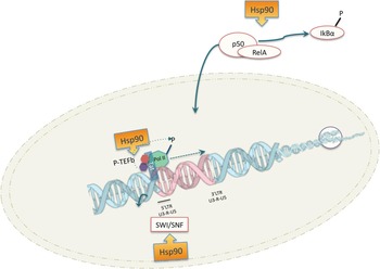
In summary, evidence is accumulating that Hsp90 may regulate HIV-1 gene expression at multiple levels and may play a significant role in HIV-1 reactivation from latency (Fig. 4).
Multiple roles of Hsp90 in regulating HIV-1 gene expression. Hsp90 is shown promoting activation of the NF-kB pathway, activation of the P-TEFb complex and the SWI/SNF chromatin remodelling complex.

Hsp90 and the stability of the viral capsid core
As a result of its protein-stabilizing role, Hsp90 has also been implicated in buffering genetic variations (Rutherford and Lindquist, Reference Rutherford and Lindquist1998). In other words, Hsp90 significantly contributes to the robustness of an organism phenotype in the presence of genetic variations caused by environmental insults or stochastic changes (Queitsch et al. Reference Queitsch, Sangster and Lindquist2002). If Hsp90 is inactivated, accumulated genetic variations become manifest, promoting stepwise evolution by adaptation (Queitsch et al. Reference Queitsch, Sangster and Lindquist2002). Importantly, evidence indicates that, at least in Drosophila, Hsp90 acts by buffering epigenetic variations as well (Pigliucci, Reference Pigliucci2003; Sangster et al. Reference Sangster, Queitsch and Lindquist2003). If Hsp90 is inactivated, changes in chromatin state become phenotypically expressed and heritable (Sollars et al. Reference Sollars, Lu, Xiao, Wang, Garfinkel and Ruden2003). Hsp90 may also prevent phenotypic variation by suppressing the mutagenic activity of transposons. Transposons are small DNA sequences that are able to ‘jump’ from one chromatin position to another, potentially creating mutations or genetic variability. Hsp90 regulates mobilization of these transposable elements by affecting Piwi-interacting RNA (piRNA) (Specchia et al. Reference Specchia, Piacentini, Tritto, Fanti, D'Alessandro, Palumbo, Pimpinelli and Bozzetti2010), a molecule associated with gene silencing (Grimson et al. Reference Grimson, Srivastava, Fahey, Woodcroft, Chiang, King, Degnan, Rokhsar and Bartel2008). Reduction in Hsp90 influences piRNA silencing activity, which causes stress-response-like activation of transposons, generating new variation (Specchia et al. Reference Specchia, Piacentini, Tritto, Fanti, D'Alessandro, Palumbo, Pimpinelli and Bozzetti2010).
Within this conceptual framework, one might propose that Hsp90 plays an important role in buffering genetic variations of viruses that have a high mutation rate. It is well-established that the mutation rate of HIV-1 is very high at 3·4×10−5 per base pair per cycle in vivo (Mansky and Temin, Reference Mansky and Temin1995), compared with human DNA polymerase with an error rate of 1×10⁻⁹ per nucleotide. HIV-1 genetic variability is under additional pressure in the presence of cART, because viral strains that become drug resistant often carry mutations that reduce viral fitness (Götte, Reference Götte2012; Kouyos et al. Reference Kouyos, Leventhal, Hinkley, Haddad, Whitcomb, Petropoulos and Bonhoeffer2012; Quashie PK et al. Reference Quashie, Mesplède and Wainberg2013). Hence the ability to ‘buffer’ such fitness-reducing mutations would be advantageous to HIV-1.
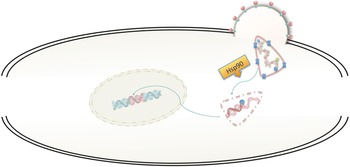
Support for this idea has come from two recent publications showing that Hsp90 rescues a defect of the viral core of mutated, protease-resistant HIV-1 strains, improving viral fitness and efficient replication (Joshi and Stoddart, Reference Joshi and Stoddart2011). Furthermore, Hsp90 was shown to rescue core stability defects of several HIV-1 mutants (Joshi et al. Reference Joshi, Sloan, Torbett and Stoddart2013). Hsp90 was found to help folding and stabilization of the capsids of hepatitis B virus (HBV) and poliovirus (Geller et al. Reference Geller, Vignuzzi, Andino and Frydman2007; Shim et al. Reference Shim, Quan, Yi and Jung2011). Not only did inhibition of Hsp90 impair viral replication, it also prevented the emergence of drug-resistant viruses (Geller et al. Reference Geller, Vignuzzi, Andino and Frydman2007), in agreement with the potential role of Hsp90 in buffering genetic variation (Fig. 5).
Role of Hsp90 in buffering genetic changes. Mutations in the capsid p24 protein (shown as an ‘X’) allow HIV-1 to escape protease inhibitors but reduce fitness, possibly by making the viral core too stable. Hsp90 restore, in part, viral fitness presumably by chaperoning the core.

Therefore, there is evidence supporting the exciting possibility that Hsp90 may be a capacitor for the evolution of HIV-1 and other viruses, by buffering genetic variations that reduce fitness. If this is the case, then Hsp90 may also be implicated in the evolution of drug-resistant variants, which represent a major problem of cART.
Immunological function
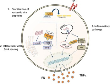
HIV-1 infection induces a broad range of immune responses, including innate responses leading to IFN and TNFα production. Indeed, during the acute phase of infection there is significant up-regulation of inflammatory cytokines (Borrow, Reference Borrow2011). While such responses may be beneficial in the short term, chronic immune activation is a feature of HIV-1 infection that best correlates with progression to AIDS (Giorgi et al. Reference Giorgi, Hultin, McKeating, Johnson, Owens, Jacobson, Shih, Lewis, Wiley, Phair, Wolinsky and Detels1999). Inhibition of Hsp90 has been shown to result in anti-inflammatory effects through several pathways, including the phosphatidylinositol-3 kinase (PI3K)/Akt and the NF-kB pathway (Shimp et al. Reference Shimp, Chafin, Regna, Hammond, Read, Caudell, Rylander and Reilly2012a , Reference Shimp, Parson, Regna, Thomas, Chafin, Reilly and Nichole Rylander b ). Interestingly, Hsp90 with its co-chaperone Cdc37 have also been implicated in intracellular sensing of retroviral DNA by stabilizing TBK1 and conversely chemical inhibition of Hsp90 abrogated retroviral DNA sensing in mouse embryonic cells lacking the TREX nuclease (Lee et al. Reference Lee, Roy, Ong, Mertins, Villani, Li, Dotiwala, Sen, Doench, Orzalli, Kramnik, Knipe, Lieberman, Carr and Hacohen2013).
HIV-1 infection also induces robust, albeit ultimately ineffective, CD8T cell responses. HIV-1-derived peptides have variable half-lives and some HLA-associated HIV-1 peptides obtained from clinical isolates have a shorter half-life relative to consensus sequence. The shorter half-life of the HLA-associate peptides impairs proper presentation and CTL recognition. Hsp90 in complex with histone deacetylase HDAC6 enhanced the stability of the peptides whereas chemical inhibition of Hsp90 dramatically reduced peptide stability (Lazaro et al. Reference Lazaro, Kadie and Stamegna2011). These results suggest that the chaperone may be important in vivo to mount an immune response to HIV-1 (Fig. 6).
Multiple roles of Hsp90 in regulating the immune response against HIV-1. Hsp90 is shown to promote processing of antigenic peptides for loading onto HLA-I, to facilitate sensing of viral DNA and to induce inflammation in response to HIV-1 infection.

CONCLUSIONS/FUTURE DIRECTIONS
There is a growing interest in understanding the different functions of Hsp90 in HIV-1 replication. Hsp90 seems critical for HIV-1 gene expression, reactivation from latency, stimulating replication in conditions of hyperthermia, core stability and anti-viral immune responses. Moreover, Hsp90 may influence HIV-1 ability to adapt to cART. Therefore Hsp90 may be an antiretroviral target with several distinct layers of activity. Several Hsp90 inhibitors are in Phase II clinical trials (www.clinicaltrials.gov) to treat solid malignancies and lymphomas. It is currently unknown if the safety profile of such inhibitors is good enough to justify their use in the clinical setting to repress HIV-1 reactivation from latency. Nonetheless, one may envisage two possible therapeutic approaches for Hsp90 inhibitors. First, Hsp90 inhibitors may be added to cART from the beginning, possibly resulting in therapeutic ‘intensification’ (Ramratnam et al. Reference Ramratnam, Mittler, Zhang, Boden, Hurley, Fang, Macken, Perelson, Markowitz and Ho2000; Buzon et al. Reference Buzon, Massanella, Llibre, Esteve, Dahl, Puertas, Gatell, Domingo, Paredes, Sharkey, Palmer, Stevenson, Clotet, Blanco and Martinez-Picado2010), with the advantage that reactivation from latency will be targeted in addition to viral replication. Second, Hsp90 inhibitors may allow temporary and structured interruptions of cART once viral loads become undetectable. Usually, interruption of cART results in rebound of viral loads in a few weeks, which is likely caused by reactivation of the latent reservoirs (Finzi et al. Reference Finzi, Hermankova, Pierson, Carruth, Buck, Chaisson, Quinn, Chadwick, Margolick, Brookmeyer, Gallant, Markowitz, Ho, Richman and Siliciano1997). However, if the latent reservoir is repressed by Hsp90 inhibitors after cART interruption, viral loads may not rebound or may rebound much later. Thus alternate, rather than concomitant use of cART and Hsp90 inhibitors may be feasible, reducing the risk of toxicity. Repressing HIV-1 reactivation in acutely infected individuals or at least in the early stages post-infection may lead to disappearance of the reservoir in a few years due to the natural turnover of latently infected cells, as observed in some individuals of the ANRS VISCONTI cohort (Sáez-Cirión et al. Reference Sáez-Cirión, Bacchus, Hocqueloux, Avettand-Fenoel, Girault, Lecuroux, Potard, Versmisse, Melard, Prazuck, Descours, Guergnon, Viard, Boufassa, Lambotte, Goujard, Meyer, Costagliola, Venet, Pancino, Autran and Rouzioux2013). Conversely, stimulation of Hsp90 by, for example, physiological hyperthermia (fever) might promote HIV-1 reactivation by mechanisms that are complementary to current strategies to purge latently infected cells based on histone deacetylase (HDAC) inhibitors (Archin et al. Reference Archin, Liberty, Kashuba, Choudhary, Kuruc, Crooks, Parker, Anderson, Kearney, Strain, Richman, Hudgens, Bosch, Coffin, Eron, Hazuda and Margolis2012; Durand et al. Reference Durand, Blankson and Siliciano2012). Therefore Hsp90 may be an important target to achieve a functional cure for HIV-1 infection.
In addition to the potential therapeutic implications, elucidating the interactions between Hsp90 and HIV-1 has revealed, and is likely to reveal in the future, novel functions of Hsp90 and novel cellular pathways regulated by the chaperone.
ACKNOWLEDGEMENTS
We thank Mehdi Baratchian, Mary Collins, Richard Jenner and David Selwood for helpful discussions.
FINANCIAL SUPPORT
AF is supported by an MRC Centre grant and the EU FP7 HIVINNOV grant (ref. 305137).