Introduction
Atherosclerotic cardiovascular diseases are the leading cause of death worldwide.Reference Gülel1 Modifiable risk factors such as smoking, obesity, and physical inactivity play a crucial role in the early development of coronary artery disease.Reference Mallika, Goswami and Rajappa2 Passive smoking is a major global health concern and has been associated with unhealthy lifestyle behaviours, including sedentary activity, insufficient sleep, and adverse cardiometabolic outcomes in children.Reference Li, Wang and Wang3,Reference Raghuveer, White and Hayman4
Previous studies have demonstrated that parental smoking increases the risk of childhood obesity, diabetes, and cardiovascular disease and is associated with components of the metabolic syndrome such as dyslipidaemia, elevated blood pressure, and impaired glucose regulation.Reference Li, Wang and Wang3,Reference Jaddoe, de Ridder, van den Elzen, Hofman, Uiterwaal and Witteman5–Reference Pan, Wang, Talaei, Hu and Wu8 In addition, parental smoking has been linked to alterations in adipokines, including increased leptin and retinol-binding protein-4 levels and reduced adiponectin and fibroblast growth factor-21 levels, contributing to metabolic and inflammatory disturbances in children.Reference Li, Wang and Wang3,Reference Somm, Schwitzgebel and Vauthay9–Reference Pardo, Geloneze, Tambascia and Barros11 Longitudinal data suggest that the vascular effects of environmental tobacco smoke exposure during childhood may persist into adulthood.Reference West, Juonala and Gall12,Reference Gall, Huynh and Magnussen13
Adipose tissue functions as an active endocrine organ involved in metabolic and inflammatory processes relevant to atherosclerosis.Reference Li, Wang and Wang3,Reference Lau, Ohashi and Wang14,Reference Scheja and Heeren15 Exposure to secondhand smoke in children has been associated with low-grade inflammation and disturbances in lipid metabolism, contributing to early atherosclerotic changes.Reference Juonala, Magnussen and Venn16 Atherosclerosis begins in childhood, and increased carotid intima-media thickness represents one of the earliest detectable indicators of this process.Reference Geerts, Bots, Grobbee and Uiterwaal6 Epicardial adipose tissue, located around the heart, exerts paracrine and inflammatory effects on the myocardium and coronary arteries. Both carotid intima-media thickness and epicardial adipose tissue thickness are recognised as non-invasive markers of subclinical atherosclerosis in paediatric populations.
Therefore, the present study aimed to evaluate the independent effect of parental smoking on early atherosclerotic changes in children by assessing carotid intima-media thickness and epicardial adipose tissue thickness.
Materials and methods
This prospective study was conducted at the paediatric cardiology outpatient clinics of Süleyman Yalçın City Hospital between August 2020 and January 2022. Ethical approval was obtained from the institutional review board (No: 2020/04). The study was carried out in accordance with the Declaration of Helsinki, and written informed consent was obtained from the parents of all participants prior to enrolment.
Study population
A total of 106 healthy children aged 6–16 years were prospectively enrolled. The exposed group consisted of 53 children with documented parental smoking exposure obtained during medical history, while the control group included 53 age- and sex-matched children without household smoking exposure. All participants were referred for evaluation of innocent cardiac murmurs and demonstrated normal findings on physical examination, electrocardiography, and echocardiography.
Exclusion criteria included congenital or acquired heart disease (except patent foramen ovale), chronic systemic diseases (chronic lung, renal, or hepatic disease), rheumatologic, genetic, or metabolic disorders, primary immunodeficiencies, hypertension (blood pressure ≥95th percentile for age, sex, and height), diabetes mellitus, insulin resistance, dyslipidaemia, and obesity defined as body mass index ≥95th percentile for age and sex.
Parents completed a structured questionnaire including information on birth weight, gestational age, breastfeeding duration, maternal smoking during pregnancy (number of cigarettes per day), gestational diabetes, and maternal anthropometric characteristics during pregnancy. Additional sociodemographic data were collected, including parental education, occupation, household income, family history of cardiovascular disease, and the child’s total daily sleep duration. Detailed information regarding parental smoking habits, including frequency of smoking in the presence of the child and smoking locations within the household, was also obtained.
Anthropometric measurements
Height and weight were measured using a digital scale and stadiometer, and body mass index was calculated as weight (kg)/height (m2). Body mass index percentiles for age and sex were assessed using nationally standardised growth charts for Turkish children.Reference Neyzi, Bundak and Gökçay17 Obesity was defined as a body mass index at or above the ninety-fifth percentile, and children meeting this criterion were excluded from the study.Reference Neyzi, Bundak and Gökçay17,Reference Sabuncu, Bayram and Kıyıcı18
Waist, mid-arm, and wrist circumferences were measured using a non-elastic tape measure according to standardised protocols. Waist circumference was measured at the level of the umbilicus at the end of expiration, mid-arm circumference at the midpoint between the acromion and olecranon, and wrist circumference at the distal ends of the radius and ulna. All measurements were recorded in centimetres. Reference values were obtained from national anthropometric studies.Reference Hatipoglu, Mazicioglu, Poyrazoglu, Borlu, Horoz and Kurtoglu19,Reference Öztürk, Çiçek, Mazıcıoğlu, Zararsız and Kurtoğlu20
Blood pressure and biochemical markers
Blood pressure was measured in the right brachial artery using a digital monitor (Omron M2) with the child in the seated position after a 15-minute rest. Systolic and diastolic blood pressure values were interpreted according to age-, sex-, and height-specific percentile curves, and values ≥95th percentile were defined as hypertension.Reference Falkner, Daniels and Flynn21
Laboratory evaluations included complete blood count, iron parameters, vitamin B12, 25-hydroxy vitamin D, fasting glucose, insulin, lipid profile components (high-density lipoprotein, low-density lipoprotein, total cholesterol, and triglycerides), and thyroid function tests. Insulin resistance was defined as a homeostatic model assessment of insulin resistance value >2.5. All blood samples were obtained after a 12-hour fasting period. Dyslipidaemia was defined according to established paediatric thresholds.Reference Bayram, Satman and Sabuncu22
Echocardiographic assessment
Echocardiographic examinations were performed in a single-blind manner using a Philips EPIQ 7C ultrasound system equipped with S5–1 and S8–3 probes, and all assessments were conducted by the same paediatric cardiologist. Standard M-mode measurements included left ventricular end-diastolic internal diameter, interventricular septal systolic and diastolic thickness, left ventricular posterior wall systolic and diastolic thickness, ejection fraction, shortening fraction, and left ventricular mass.
Aortic pulse wave velocity was assessed using Doppler ultrasound by calculating the ratio of the distance between the ascending and descending aorta to the transit time of blood flow between these sites.
Epicardial adipose tissue thickness was measured perpendicular to the free wall of the right ventricle at end-systole in the parasternal long-axis view by identifying the echo-free space between the outer myocardial surface and the visceral pericardium.
Carotid intima-media thickness was measured on the far wall of the common carotid artery approximately 1 cm proximal to the carotid bifurcation by averaging three cardiac cycles, in accordance with the recommendations of the American Society of Echocardiography.Reference Stein, Korcarz and Hurst23
Statistical analysis
Statistical analyses were performed using the Number Cruncher Statistical System (NCSS) 2007 software (Utah, USA). Continuous variables were expressed as mean ± standard deviation, and categorical variables as number and percentage. Normality of distribution was assessed using the Shapiro–Wilk test. Group comparisons were conducted using the independent-samples t test or the Mann–Whitney U test for continuous variables and the chi-square test for categorical variables, as appropriate. Correlation analyses were performed using Pearson’s or Spearman’s correlation coefficients.
To address the issue of multiple comparisons, epicardial adipose tissue thickness and carotid intima-media thickness were predefined as primary outcomes, and a Bonferroni-adjusted significance threshold was applied for the primary outcome analyses (α = 0.05/2).
To evaluate the independent association between parental smoking and subclinical atherosclerotic markers, analysis of covariance, and multivariable linear regression analyses were performed. Age, sex, sleep duration, maternal educational level, and lipid parameters (triglycerides, low-density lipoprotein cholesterol, and total cholesterol) were included as covariates. A two-sided p-value <0.05 was considered statistically significant.
Results
A total of 106 children (53 smoke-exposed and 53 controls) were included in the analysis. The two groups were comparable in terms of age and sex distribution. All participants had structurally normal hearts. No significant differences were observed between the groups with respect to body mass index, blood pressure measurements, or laboratory parameters, including lipid profile and glucose metabolism markers (all p > 0.05).
Echocardiographic findings
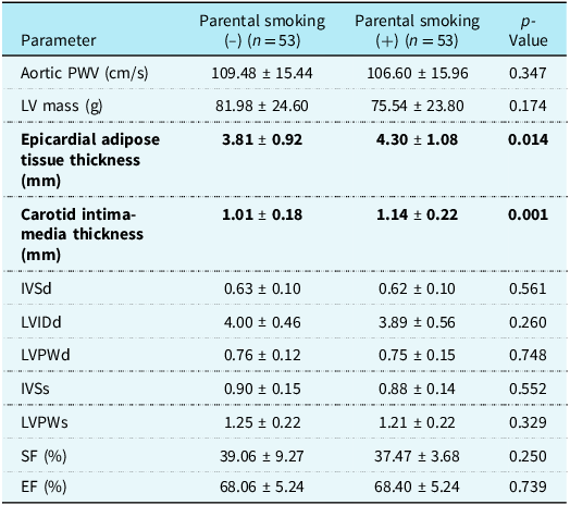
As shown in Table 1, there were no statistically significant differences between the smoke-exposed and non-exposed groups in aortic pulse wave velocity, left ventricular mass, interventricular septal thickness, posterior wall thickness (systolic or diastolic), shortening fraction, or ejection fraction (all p > 0.05). In contrast, children exposed to parental smoking demonstrated significantly greater epicardial adipose tissue thickness (p = 0.014) and carotid intima-media thickness (p = 0.001) compared with controls (Figure 1).
Comparison of echocardiographic parameters between the study and control groups

Independent t test. PW = pulse wave; LV = left ventricle; IVS = interventricular septum; LVPW = left ventricular posterior wall.
PWV = aortic pulse wave velocity; LV = left ventricle; IVSd = interventricular septum diastolic thickness; LVIDd = left ventricular end-diastolic internal diameter; LVPWd = left ventricular posterior wall diastolic thickness; IVSs = interventricular septum systolic thickness; LVPWs = left ventricular posterior wall systolic thickness; SF = shortening fraction; EF = ejection fraction.
Comparison of epicardial adipose tissue and carotid intima-media thickness. Both epicardial adipose tissue thickness and carotid intima-media thickness were significantly higher in the parental smoking (+) group compared with the parental smoking (–) group (p = 0.014 and p = 0.001, respectively).

Multivariable and covariate analyses
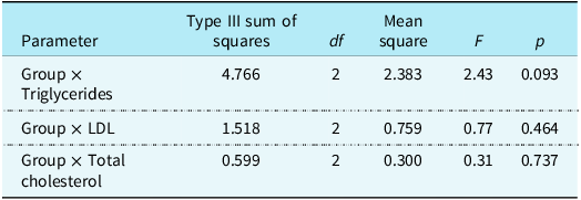
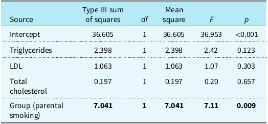
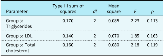
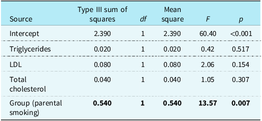
To evaluate whether lipid parameters influenced the association between parental smoking and subclinical atherosclerotic changes, triglycerides, low-density lipoprotein cholesterol, and total cholesterol were included as covariates in the analysis of covariance models. None of these lipid variables showed a significant independent association with epicardial adipose tissue thickness or carotid intima-media thickness (Tables 2 and 4).
Effect of triglycerides, low-density lipoprotein, and total cholesterol on epicardial adipose tissue thickness

Analysis of covariance (ANCOVA) was used with triglycerides, LDL, and total cholesterol as covariates to control for their effects on epicardial adipose tissue thickness (EAT). These covariates were found to be homogeneous.
LDL = low-density lipoprotein.
Regression analysis of the effect of triglycerides, low-density lipoprotein, and total cholesterol on epicardial adipose tissue thickness

Regression analysis was performed to evaluate the independent effects of triglycerides, LDL, and total cholesterol on epicardial adipose tissue thickness.
LDL = low-density lipoprotein.
Effect of triglycerides, low-density lipoprotein, and total cholesterol on carotid intima-media thickness

Analysis of covariance (ANCOVA) was used with triglycerides, LDL, and total cholesterol as covariates to assess their effects on carotid intima-media thickness (CIMT). These covariates were found to be homogeneous.
LDL = low-density lipoprotein.
In multivariable linear regression analyses adjusting for age, sex, lipid parameters, sleep duration, and maternal educational level, parental smoking remained a significant and independent predictor of both epicardial adipose tissue thickness (p = 0.009) and carotid intima-media thickness (p = 0.007) (Tables 3 and 5).
Regression analysis of the effect of triglycerides, low-density lipoprotein, and total cholesterol on carotid intima-media thickness

Regression analysis was performed to determine the independent effects of triglycerides, LDL, and total cholesterol on carotid intima-media thickness).
LDL = low-density lipoprotein.
When sleep duration and maternal educational level were included as covariates in repeated multivariable analyses, parental smoking remained independently associated with both epicardial adipose tissue thickness and carotid intima-media thickness.
Associations with sleep duration
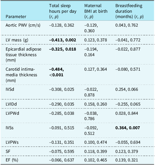
Correlations between cardiac measurements, maternal body mass index at birth, breastfeeding duration, and total daily sleep duration are presented in Table 6. No significant associations were observed between sleep duration and aortic pulse wave velocity, systolic wall thicknesses, shortening fraction, or ejection fraction (all p > 0.05). In contrast, daily sleep duration was negatively correlated with epicardial adipose tissue thickness (r = –0.325, p = 0.018) and carotid intima-media thickness (r = –0.484, p < 0.001) (Figure 2).
Correlation of cardiac function parameters with daily sleep duration, maternal body mass index at birth, and breastfeeding duration

Pearson’s correlation test was used for all analyses.
PWV = aortic pulse wave velocity; LV = left ventricle; IVSd = interventricular septum diastolic thickness; LVIDd = left ventricular end-diastolic internal diameter; LVPWd = left ventricular posterior wall diastolic thickness; IVSs = interventricular septum systolic thickness; LVPWs = left ventricular posterior wall systolic thickness; SF = shortening fraction; EF = ejection fraction.
Correlation between daily sleep duration and epicardial adipose tissue thickness and carotid intima-media thickness in children. Scatter plots show a significant negative correlation between total sleep hours per day and both epicardial adipose tissue thickness (blue line, r = –0.325, p = 0.018) and carotid intima-media thickness (red line, r = –0.484, p < 0.001).

Shorter sleep duration was also associated with lower left ventricular mass (r = –0.413, p = 0.002), reduced interventricular septal diastolic thickness (r = –0.308, p = 0.025), smaller left ventricular end-diastolic diameter (r = –0.290, p = 0.035), and reduced posterior wall diastolic thickness (r = –0.285, p = 0.038) (Figure 3). Breastfeeding duration showed a positive correlation with interventricular septal systolic thickness (r = 0.364, p = 0.007), whereas no significant correlations were observed with maternal body mass index at birth.
Correlation between total sleep hours and diastolic cardiac wall parameters. Scatter plots showing negative correlations between total sleep hours per day and interventricular septum diastolic thickness (IVSd, green line; r = –0.308, p = 0.025), left ventricular end-diastolic internal diameter (LVIDd, blue line; r = –0.290, p = 0.035), and left ventricular posterior wall diastolic thickness (LVPWd, red line; r = –0.285, p = 0.038). IVSd = interventricular septum diastolic thickness; LVIDd = left ventricular end-diastolic internal diameter; LVPWd = left ventricular posterior wall diastolic thickness.

Sociodemographic characteristics
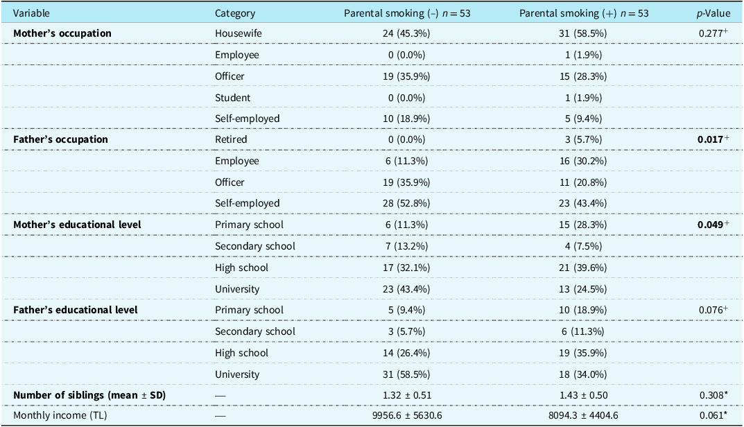
Sociodemographic and socio-economic characteristics of the families are summarised in Table 7. No statistically significant differences were observed between groups regarding maternal occupation, paternal educational level, number of siblings, or household income. However, paternal occupation (p = 0.017) and maternal educational level (p = 0.049) differed significantly between the groups. Fathers in the smoke-exposed group were more frequently retired or employed as manual labourers, while mothers more commonly had a primary school education. As illustrated in Figure 4, higher maternal educational attainment was more prevalent in the non-smoking group.
Socio-economic and demographic characteristics of families in the study and control groups

Independent t test () and chi-square test (+) were used for statistical analysis*.
Distribution of maternal educational level according to parental smoking status. Bar graph showing the distribution of mothers’ educational levels in the parental smoking (+) and parental smoking (–) groups. Mothers in the smoking group were more likely to have a primary school education, whereas those in the non-smoking group were more often university graduates.

Discussion
In this study, echocardiographic evaluation conducted in the absence of traditional atherosclerotic risk factors demonstrated significantly higher carotid intima-media thickness and epicardial adipose tissue thickness in children and adolescents with parental smoking. To our knowledge, this is the first clinical study to show early atherosclerotic changes in this population independent of conventional cardiovascular risk factors.
Epidemiological, observational, and experimental evidence has consistently shown that secondhand smoke exerts detrimental cardiovascular effects on children.Reference Raghuveer, White and Hayman4 Exposure to cigarette smoke impairs endothelial function, increases blood pressure and heart rate, and leads to heightened peripheral vascular resistance and catecholamine release. Furthermore, tobacco smoke reduces coronary flow-mediated dilation and promotes a prothrombotic state. Reactive oxygen species present in inhaled smoke oxidises the low-density lipoproteins, initiating an inflammatory cascade involving monocyte adhesion to the arterial intima, thereby promoting atherogenesis.Reference Somm, Schwitzgebel and Vauthay9,Reference Stocker and Keaney24,Reference Kallio, Jokinen and Raitakari25
The non-invasive identification of subclinical atherosclerosis is essential for preserving cardiovascular health during childhood. An increase in carotid intima-media thickness correlates with the severity of coronary atherosclerosis and is considered an indicator of elevated cardiovascular risk.Reference Lamotte, Iliescu, Libersa and Gottrand26 Although the predictive value of carotid intima-media thickness measured in childhood for future cardiovascular disease remains uncertain, the well-established association between childhood cardiovascular risk factors and adult vascular disease suggests that elevated carotid intima-media thickness in childhood carries important clinical significance.Reference Dratva, v. and Hodis27 In accordance with our findings, Geerts et al. Reference Geerts, Bots, Grobbee and Uiterwaal6 reported that parental smoking leads to persistent vascular injury in children, with both maternal smoking during pregnancy and postnatal passive exposure contributing to vascular damage. Similarly, West et al., Reference West, Juonala and Gall12 in the Young Finns Study, demonstrated that childhood exposure to environmental tobacco smoke is associated with carotid atherosclerotic plaque formation in adulthood. Lamotte et al. Reference Lamotte, Iliescu, Libersa and Gottrand26 reported significant associations between obesity, diabetes mellitus, dyslipidaemia, hypertension, and chronic kidney disease and increased carotid intima-media thickness. In contrast to our findings, their study identified positive correlations between carotid intima-media thickness and blood pressure, cholesterol levels, and body mass index. The Bogalusa Heart StudyReference Berenson, Srinivasan, Bao, Newman, Tracy and Wattigney28 further confirmed that body mass index, blood pressure, low-density lipoprotein, total cholesterol, triglycerides, and smoking are strongly associated with early atherosclerotic lesions in young individuals. Conversely, Kallio et al. Reference Kallio, Jokinen and Raitakari25 suggested that secondhand smoke may contribute to atherosclerosis partly through alterations in apolipoprotein B metabolism, although the precise mechanism remains unclear. In our analysis, the association between parental smoking and carotid intima-media thickness persisted independently of traditional cardiovascular risk factors, as demonstrated by multivariate analysis.
Epicardial adipose tissue thickness is increasingly recognised as an independent predictor of cardiovascular events, reflecting both cardiac and visceral adiposity. This tissue lies in direct contact with the myocardium and functions as a metabolically active endocrine organ, secreting a variety of cytokines and vasoactive peptides, including free fatty acids, interleukin-6, tumour necrosis factor-alpha, angiotensin II, and plasminogen activator inhibitor-1.Reference Iacobellis and Barbaro29–Reference Gastaldelli and Basta31
Under physiological conditions, epicardial adipose tissue functions as an energy reservoir and secretes anti-inflammatory mediators such as adiponectin and adrenomedullin. In contrast, under pathological conditions, epicardial adipose tissue releases proinflammatory adipokines, including resistin and leptin, thereby promoting lipotoxicity and increasing cardiovascular risk.Reference Gać, Czerwińska, Poręba, Macek, Mazur and Poręba32
Consistent with our findings, Yang Lu et al. Reference Lu, Wang and Zhan33 reported that both greater epicardial adipose tissue volume and higher tissue density are associated with structural and functional cardiac alterations, even in individuals without coronary artery disease. Similarly, Li et al. Reference Li, Wang and Wang3 reported in a cohort of Chinese children that exposure to parental smoking was associated with both hypertension and metabolic syndrome, independent of demographic and familial factors. In our study population, parental smoking was correlated with increased epicardial adipose tissue thickness but not with hypertension, suggesting a direct association between parental smoking and early vascular changes independent of metabolic syndrome.
Adipokine imbalance may play a key role in this association. In states of excessive adiposity, proinflammatory adipokines predominate, whereas anti-inflammatory adipokines such as adiponectin are downregulated, contributing to metabolic dysfunction. Previous studies have demonstrated that parental smoking increases circulating levels of leptin and retinol-binding protein-4 while reducing adiponectin and fibroblast growth factor-21, thereby predisposing children to metabolic syndrome.Reference Li, Wang and Wang3
Our findings are consistent with these results, demonstrating that parental smoking is associated with increased epicardial adipose tissue thickness, thereby contributing to early atherosclerosis independently of metabolic status. Similarly, Nagel et al.Reference Nagel, Arnold, Wilhelm, Link, Zoellner and Koenig34 reported that passive smoke exposure among German children was associated with an increased risk of obesity and adverse cardiometabolic biomarkers, both of which may heighten future cardiovascular risk.
In addition to passive smoke exposure, sleep duration appeared to influence early vascular changes. In our study, shorter total sleep time was negatively correlated with both carotid intima-media thickness and epicardial adipose tissue thickness, independent of parental smoking and other cardiovascular risk factors. Consistent with our findings, Kong et al. Reference Kong, Wing and Choi35 reported that reduced sleep duration among schoolchildren in Hong Kong was associated with atherogenic dyslipidemia.
Similarly, Navarro–Solera et al. Reference Navarro-Solera, Carrasco-Luna, Pin-Arboledas, González-Carrascosa, Soriano and Codoñer-Franch36 observed that obese children with shorter sleep durations exhibited greater adiposity and increased waist circumference, both of which contribute to higher atherosclerotic risk. Furthermore, Grandner et al. Reference Grandner, Alfonso-Miller, Fernandez-Mendoza, Shetty, Shenoy and Combs37 demonstrated that insufficient sleep promotes obesity, inflammation, cardiovascular disease, diabetes, and an increased risk of mortality.
Collectively, these findings, together with our results, underscore the critical role of adequate sleep in maintaining cardiovascular health during childhood and adolescence.
Our study found no significant differences in either systolic or diastolic blood pressure between the groups, consistent with the findings of Housberg et al., Reference Hausberg, Mark, Winniford, Brown and Somers38 who reported that passive smoke exposure increases sympathetic nerve activity without altering blood pressure. Similarly, Weitzman et al. Reference Weitzman, Cook and Auinger39 observed that although passive smoking was associated with metabolic syndrome among children in the United States, it was not linked to hypertension.
Socio-economic factors also appear to influence smoking exposure. Cigarette use has been reported to be more prevalent among individuals with lower income levels.Reference Özcan40 In our study, fathers in the parental smoking group were more frequently retired or employed as manual labourers, while mothers more often had only primary school education. These findings are consistent with previous reports indicating that lower socio-economic and educational status are associated with higher rates of parental smoking.Reference Kaneita, Yokoyama and Miyake41,Reference Kelishadi, Noori and Qorbani42
Conclusion
Parental smoking was found to significantly increase mean carotid intima-media thickness and epicardial adipose tissue thickness in children. Although previous studies have investigated early atherosclerotic changes within specific disease populations, the present study is the first to demonstrate that parental smoking alone is associated with increased carotid intima-media thickness and epicardial adipose tissue thickness in children and adolescents, independent of traditional atherosclerotic risk factors. These findings suggest that parental smoking contributes to early subclinical atherosclerosis, even in the absence of other metabolic or cardiovascular risk factors. Furthermore, we found that shorter sleep duration, independent of parental smoking and conventional cardiovascular risk factors, was significantly associated with early atherosclerotic changes. This underscores that adequate and regular sleep during childhood and adolescence plays a critical role in maintaining cardiovascular health, aligning with its established importance in growth and development. Lower parental educational and socio-economic status were associated with greater exposure to smoking, highlighting the need for targeted public health interventions. Early identification and management of atherosclerosis and its related risk factors in childhood are essential to prevent long-term cardiovascular morbidity and mortality.
Acknowledgements
None.
Financial support
This research received no specific grant from any funding agency, commercial, or not-for profit sectors.
Competing interests
None.
Ethical standard
The study was conducted in accordance with the Helsinki Declaration, and written informed consent was obtained from the parents in the study and control groups.