Aïstopoda is a clade of small, elongate (up to 230 vertebrae), fully limbless Palaeozoic tetrapods. They are characterised by skulls with highly reduced ossification and broad openings in the cheek, holospondylous vertebrae with intravertebral foramina for spinal nerves, and ribs with an accessory process (although this can be reduced in size in some), giving them a k-shape (Baird Reference Baird1964; Carroll Reference Carroll and Wellnhofer1998a). Traditionally, they have been classified with other small, derived tetrapods with holospondylous vertebrae (although some retaining thin, strap-like intercentra) in the Lepospondyli (Romer Reference Romer1945; Baird Reference Baird1965; Carroll Reference Carroll, Heatwole and Carroll2000). This classification has received some cladistic support as well, with lepospondyls placed as either stem amniotes (e.g., Ruta et al. Reference Ruta, Coates and Quicke2003; Ruta & Coates Reference Ruta and Coates2007), stem-amphibians (e.g., Vallin & Laurin Reference Vallin and Laurin2004), or stem-caecilians (Anderson Reference Anderson2001, Reference Anderson, Anderson and Sues2007a; Anderson et al. Reference Anderson, Reisz, Scott, Froebisch and Sumida2008) within crown-group Tetrapoda. However, recent detailed anatomical work on the braincases of lepospondyls has shown strong evidence that a stem tetrapod placement is correct and that Lepospondyli is likely not a monophyletic group (Pardo et al. Reference Pardo, Szostakiwskyj, Ahlberg and Anderson2017).
Aïstopods are composed of five families: the Lethiscidae, Ophiderpetontidae, Oestocephalidae, Pseudophlegethontiidae, and Phlegethontiidae. Lethiscus, the only known representative of Lethiscidae, is the oldest, phylogenetically earliest diverging, and possesses a general cranial architecture from which the remaining groups can be derived (Wellstead Reference Wellstead1982; Anderson et al. Reference Anderson, Carroll and Rowe2003; Pardo et al. Reference Pardo, Szostakiwskyj, Ahlberg and Anderson2017). Phlegethontiids have lightly built skulls, have the greatest number of vertebrae, and lack a lot of the scalation present in the other groups (McGinnis Reference McGinnis1967; Anderson Reference Anderson2002, Reference Anderson2007b). They are rare elements of many late Carboniferous and early Permian faunas. Pseudophlegethontia, known from a single specimen from Mazon Creek (Anderson Reference Anderson2003a), appears to be intermediate between Lethiscus and Phlegethontia; additional material from both taxa would be welcome. Oestocephalidae are characterised by thick carpets of densely ossified rhomboidal dorsal scales and wheat-shaped ventral ‘gastralia' (tightly packed individual spindle-shaped grains arranged en chevron), particularly prominently k-shaped ribs, and highly derived skulls with extremely elongate postorbital regions unique among tetrapods (Carroll Reference Carroll1998b; Anderson Reference Anderson2003b). Ophiderpetontids appear to have less derived versions of this cranial morphology and are probably more closely related to oestocephalids than other aïstopods (Huxley & Wright Reference Huxley and Wright1867; Boyd Reference Boyd1982; Milner Reference Milner1993).
Whereas aïstopod vertebrae have been found in many assemblages of the late Carboniferous to early Permian, body fossils are restricted to particular localities, often associated with coal mining. The oldest aïstopod Lethiscus is nearly completely preserved in an elongate nodule (the only known specimen) from the mid-Viséan-aged Wardie Shale – the only tetrapod from this otherwise well-known fish locality (Wellstead Reference Wellstead1982; Anderson et al. Reference Anderson, Carroll and Rowe2003; Pardo et al. Reference Pardo, Szostakiwskyj, Ahlberg and Anderson2017). Ophiderpeton is found at the Jarrow Colliery, Ireland (Huxley & Wright Reference Huxley and Wright1867), Newsham, Northumbria, England (Boyd Reference Boyd1982), and the late-Viséan-aged lagerstätten at East Kirkton, Scotland (Milner Reference Milner1993). Oestocephalus is best known from the cannel coals of Linton, Ohio (Steen Reference Steen1930; Baird Reference Baird1964; Hook & Baird Reference Hook and Baird1988), but also occurs at the slightly older nodule locality from coal mine spoil-piles along the Mazon Creek (Westphalian C) SE of Chicago (Baird et al. Reference Baird, Shabica, Anderson and Richardson1985; Carroll Reference Carroll1998b), and the Linton-like locality of Nyrany (Westphalian D), in the Bohemia coal mining region of the Czech Republic (Fritsch Reference Fritsch1875; Steen Reference Steen1938; Milner Reference Milner and Panchen1980). Coloraderpeton, another oestocephalid, is a singleton taxon from limestones from the Sangre de Cristo Formation of Colorado (Vaughn Reference Vaughn1969; Gallup Reference Gallup1983; Anderson Reference Anderson2003b; Pardo et al. Reference Pardo, Szostakiwskyj, Ahlberg and Anderson2017). Phlegethontiids are the most dispersed of aïstopods, also known from Linton, Mazon Creek, Nyrany, and isolated braincases and associated material attributed to the genus Sillerpeton is known from the early Permian-aged fissure-fill deposits at Richard's Spur (Ft. Sill; McGinnis Reference McGinnis1967; Lund Reference Lund1978; Anderson Reference Anderson2002).
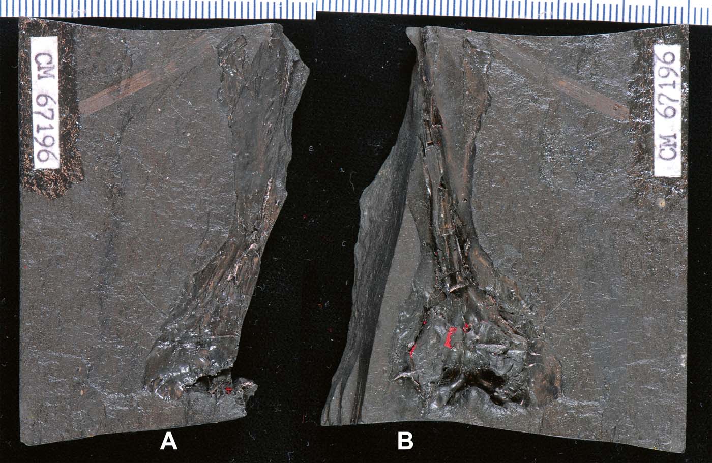
Material from spoil-piles from another coal mine in Ohio at Five Points was collected in the 1980s–1990s (Hook & Baird Reference Hook and Baird1986). As at Linton (Hook & Baird Reference Hook and Baird1988), the fossiliferous layers at Five Points comprise cannel coals (an extremely sediment-rich lower level of a productive coal seam). This material was first worked in the field by teams lead by the late Don Baird and Robert Hook at the spoil heaps from the mine, but with a National Geographic Grant to David S Berman and the Carnegie Museum much of this material was shipped to Pittsburgh and systematically worked by hand. Fossils found were prepared following the technique of Baird (Reference Baird1955), where infilling kaolinite and any remaining bone material was removed, leaving a high-resolution natural mould from which red latex casts were taken (Fig. 1). Following this approach, a large collection of vertebrates was made, and has recently become available for study at the Carnegie Museum.
Part and counterpart of CM 67196. (A) Skull roof. (B) Braincase in ventral view. Bone and infilling clay minerals were removed to produce a natural mould, from which a red latex peel was taken. Remnants of the peel remain, underscoring how latex peel production is not specimen neutral. Scale shows millimetres.

One specimen, which is the subject of the present study, was immediately recognised as important by Baird. It preserves much of an elongate braincase and partial skull roof of an enigmatic tetrapod. Initially, the generalised, superficially basal morphology of the braincase led Baird to assume that it belonged to an embolomere and approached one of us (RH) to study the specimen. Specimen drawings were made from the peel, but it was apparent, based largely on the morphology of the dermal skull, that the specimen was not an embolomere but an aïstopod. When informed of this, Baird set the specimen aside for future study. Given recent work on aïstopod braincases, we now have greater context to interpret the morphology that had caused such taxonomic confusion at the time. Considering the importance of the formative work by Clack on embolomere braincases (Clack Reference Clack1987; Clack & Holmes Reference Clack and Holmes1988) upon which we draw for our interpretation, it is fitting that this specimen be formally described and discussed in this volume.
1. Materials and methods
Abbreviations: CM=Carnegie Museum of Natural History, Pittsburgh.
The specimen, CM 67196, part and counterpart of a partial skull, was collected in 1994 and had previously been prepared following Baird (Reference Baird1955), and it was from these original peels that the specimen drawings were made. However, each time a red latex peel is taken from a specimen from Linton and Five Points, some of the matrix is broken, slowly degrading the specimen (Fig. 1). We undertook the creation of a virtual cast using micro-computed tomography (Figs 2b, 3b). Both parts of the specimen were scanned on a SkyScan 1173 at the Anderson Lab in the McCaig Institute for Bone and Joint Health at the University of Calgary. The specimen was scanned at 71 kV and 104μA, with no filter, and at a voxel size of 69.58 μm, producing 436 slices. After the scan data were reconstructed using NRecon 1.7.1.6 into stacked jpgs, they were cropped and downsampled using ImageJ. The image stacks were opened in Amira 5.3.3, and virtual casts were made using threshold values for air along the mould surface using the LabelFields module. A 2×model of the braincase was made using a three-dimensional (3D) printer in the Ungrin Lab in the Department of Comparative Biology and Experimental Medicine at the University of Calgary, which facilitated the description.
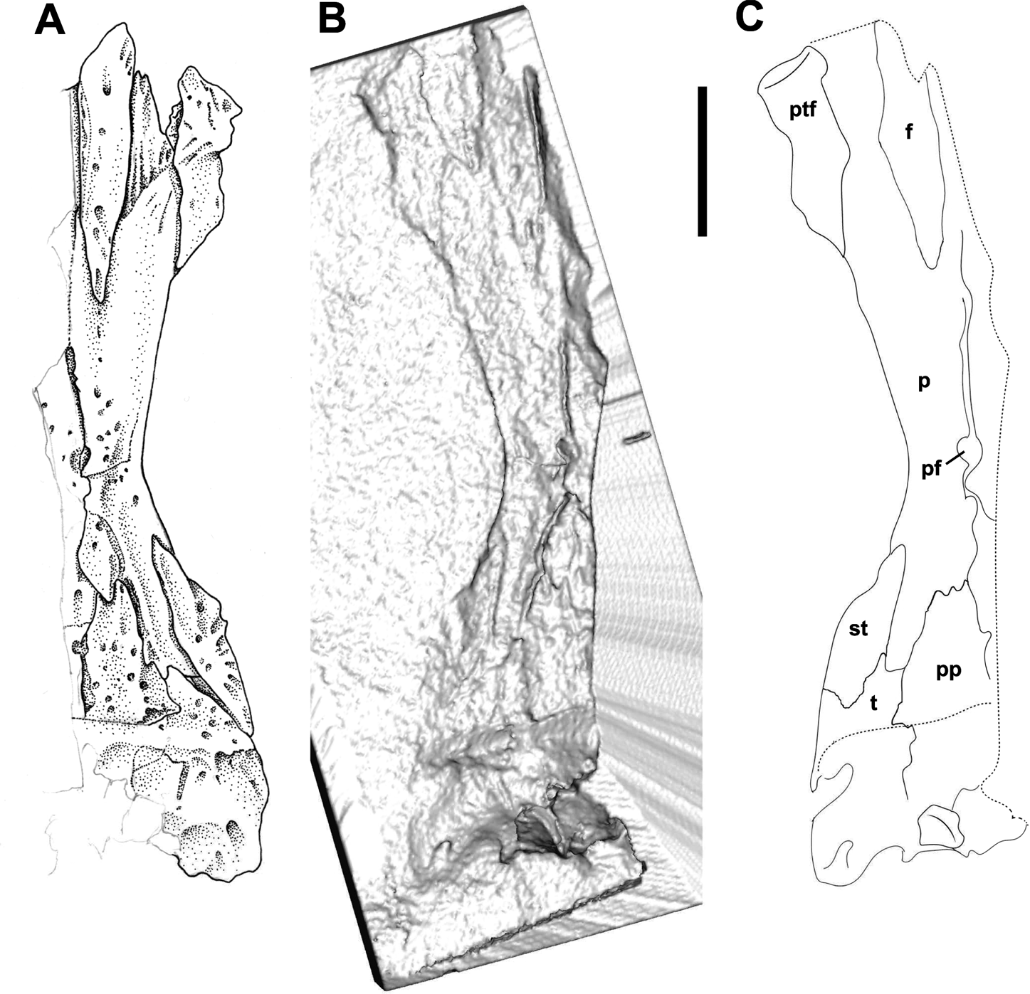
Skull roof of CM 67196. (A) Interpretive drawing made from red latex peel. (B) Virtual cast produced from volumetric rendering of empty space in CT scans. (C) Interpretive drawing from virtual cast, showing broad agreement between the two approaches. Note, CT data are stacked radiotomographs and so anatomy in (B) and (C) is shown flipped from natural position. Abbreviations: f=frontal; p=parietal; pf=parietal foramen; pp=postparietal; ptf=postfrontal; st=supratemporal; t=tabular. Scale bar=1cm.

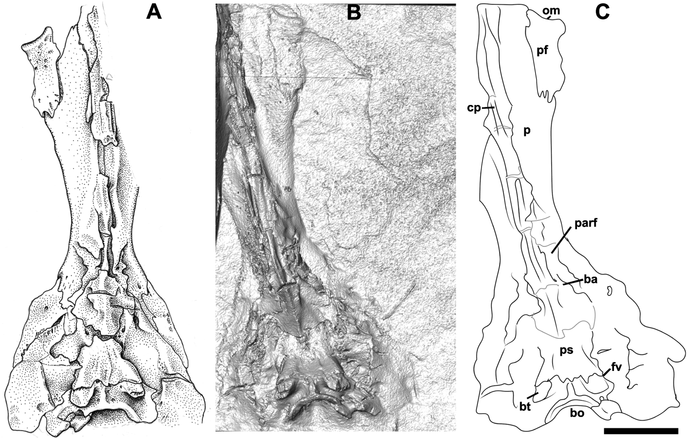
Ventral view of CM 67196. (A) Interpretive drawing made from red latex peel. (B) Virtual cast produced from volumetric rendering of empty space in CT scans. (C) Interpretive drawing from virtual cast, showing broad agreement between the two approaches. Note, CT data are stacked radiographs and so anatomy in (B) and (C) is shown flipped from natural position. Abbreviations: ba=basicranial articulation; bo=basioccipital; bt=‘basal tuberosity'; cp=cultriform process of parasphenoid; fv=fenestra vestibularis; om=orbital margin; p=parietal; parf=ventral flange of the parietal; pf=postfrontal; ps=parasphenoid (basal plate). Scale bar=1cm.

2. Systematic palaeontology
Tetrapoda Goodrich, Reference Goodrich1930
Aïstopoda Miall, Reference Miall1875
Oestocephalidae Anderson, Reference Anderson2003b
Oestocephalus amphiuminus (Cope, Reference Cope1868)
2.1. Referred specimen
CM 67196, part and counterpart of a partial skull, in cannel coal. Bone and kaolinite were removed previously to produce latex peels.
2.2. Locality
Spoil heaps at now closed coal mine at Five Points, Ohio, USA.
2.3. Description
The specimen preserves the posterior skull. Of the skull roof, the right side is preserved from just posterior to the orbit to the occiput, and to just left of the midline (Fig. 2). All bones possess light pitted ornament around the presumed centre of ossification; the postfrontal has a light vermiform ornament. On the ventral surface (Fig. 3) nearly the entire posterior braincase is preserved but ossifications of the ethmoid region are not present, either because they are obscured by other elements or because that portion of the skull has been lost. The braincase is somewhat cracked from compression, creating a series of laterally directed fractures and some collapse of the brain cavity. The skull roof is relatively undamaged, although there is some displacement of elements anteriorly. The specimen is 57.5 mm long and, estimating from complete skulls, we reconstruct a total skull length of approximately 97 mm, making this the largest described aïstopod skull.
2.3.1. Skull roof
The right parietal is preserved in its entirety (Fig. 2). It possesses the characteristic oestocephalid posterolateral flange that articulates with the tabular and supratemporal. Identification of the specimen as Oestocephalus is confirmed by the tabular occupying the posterolateral corner of the skull roof, as opposed to Coloraderpeton, where this is taken up by a posteromedial extension of the supratemporal. On the rostral margin the parietal is in articulation with the frontal, which is at a sharp point that would have created the distinctive W-shaped rostral margin of the parietal had the frontal not been slightly medially displaced from its articular facet. It is possible that what is interpreted as the facet is a portion of the frontal, where it would underlap the postfrontal. To the right of this frontal articular facet the posterior portion of the postfrontal can be seen. It is distally fractured so the nature of its articulation with the jugal remains unknown, but it forms a portion of the temporal fenestra, again consistent with Oestocephalus and not Coloraderpeton. In ventral view, a portion of the orbital margin can be seen on the rostral surface of the postfrontal. Along the interparietal suture a parietal foramen is present just posterior to the midpoint of the suture's length, as is typical for oestocephalids.
The posterior skull roof is characteristic of Oestocephalus. The large tabular occupies the posterolateral margin of the skull roof, excluding the spindle-shaped supratemporal. The postparietal is broadly triangular, with the base at the occiput and apex extending rostrally, deeply incising the posterior margin of the parietal, as is typical for oestocephalids. There is a large occipital exposure of both postparietal and tabular, but this area is heavily co-ossified and lacks clearly defined sutures between elements.
2.3.2. Braincase
The nearly complete posterior braincase is seen in ventral view (Fig. 3). The plane of preservation begins from the underside of the dermal skull roof rostrally to the basioccipital dorsally to the midpoint of the occipital cotyle and ventral half of the otic capsules. The skull is collapsed into the braincase posteriorly, so that in lateral view the posterior braincase flexure is overemphasised, and many laterally extending fractures are found throughout the length of the specimen. More could be preserved dorsally, but it is fractured to the point it becomes undistinguishable.
The basioccipital is fused to the exoccipitals and opisthotic, as is the case for aïstopods generally. The occipital articulation is concave and notochordal, but the notochord presumably was pinched off by basioccipital ossification. The articulation is broad. Just rostral to the cotylar margin, a deep sulcus runs around the perimeter of the cotyle, creating a prominent cotylar rim. At the ventral midline, the raised rim extends rostrally as a ridge (seen in many early tetrapods with an exposed basioccipital), spanning rostrally to its ending at the suture with the parasphenoid. The ‘basal tuberosities' are robust, rounded, solid structures extending ventrally and posterolaterally from the floor of the cranium formed from the endochondral basioccipital. No lateral depressions, or ‘pockets' like described by Clack & Holmes (Reference Clack and Holmes1988) in Archeria, are present. The tuberosities are distally capped by smooth, crescentic, flat surfaces. The concave side of the crescentic surfaces, which face rostrally, appear to be articular facets (see Section 3). Each tuberosity is located posteroventral to the laterally facing fenestra vestibularis, which is walled posteriorly by the basioccipital–opisthotic complex, and ventrally by the parasphenoid. No stapes is preserved.
The parasphenoid is an elongate midline structure, spanning from the basioccipital to the anterior margin of the specimen. No teeth or denticles are present anywhere on the parasphenoid. The rectangular basal plate, like in all aïstopods (except Phlegethontia, where it is coossified with the endochondral braincase; Anderson Reference Anderson2002), terminates posteriorly with two flanges that overlie the anterior surface of the ‘basal tuberosities', exposing the majority of the basioccipital in ventral view. The plate extends forward and tapers to form the cultriform process, at which point the braincase bears a pair of ill-defined processes that form weakly developed basicranial articulations for the massive oestocephalid palatoquadrate ossification (not preserved in this specimen). Given the extent of the basal plate and lack of access to information from the lateral braincase wall, it cannot be determined whether the basisphenoid was a separate ossification or coossified to the basioccipital, thus closing the ventral cranial fissure (as in Coloraderpeton).
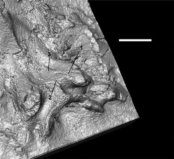
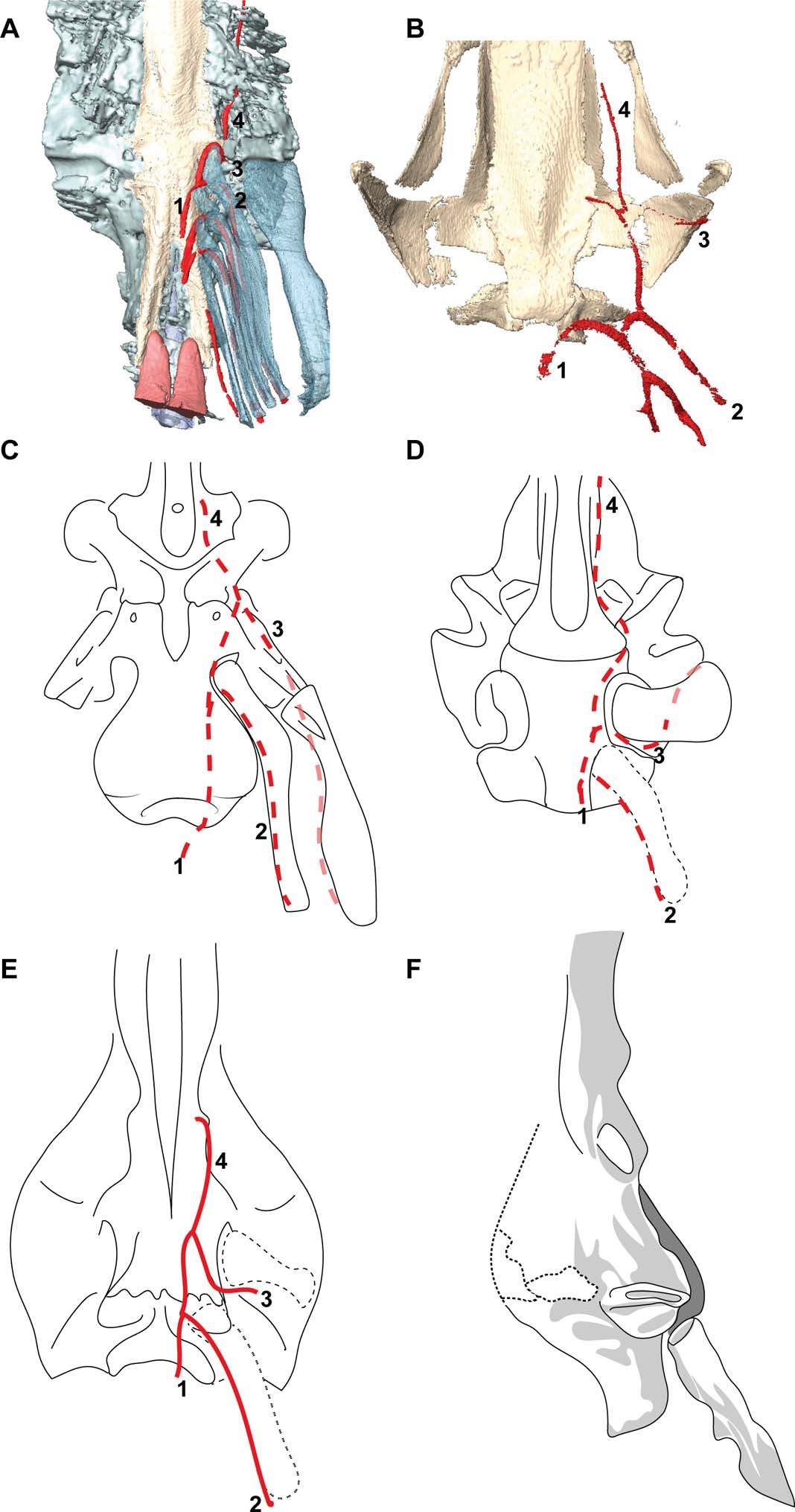
On the superficial surface of the basal plate run channels for vasculature (Fig. 4). Two channels begin at the posterior margin in the space between the ‘basal tuberosities' to either side of the median keel of the basioccipital and pass rostrally, another channel passes down the tuberosities and meets the first to form a common channel that passes rostrally until becoming indistinct along the ventral surface of the basal plate. Presumably these channels are associated with the dorsal aorta and associated basicranial circulation Fig. 5a, b), but no obvious foramina are visible except two notches along the margin of the basal plate approaching the basicranial articulations.
Ventral view of parasphenoid and basioccipital complex rendered from μCT, showing light grooves for possible vessels. 1. Groove passing along the ‘basal tuberosity' interpreted for the efferent artery of the branchial arch. 2. Groove passing towards the basioccipital sulcus interpreted for the dorsal aorta. 3. Groove passing rostrally interpreted as for internal carotid artery. Scale bar=5mm.

Pharyngeal and basicranial arterial circulatory reorganisation on the tetrapod stem, in ventral view. (A) Cranial and dorsal pharyngeal circulation in the plesiomorphic actinopterygian Polyodon spathula, rendered from contrast μCT. (B) Cranial and dorsal pharyngeal circulation in the salamander Ambystoma mexicanum, rendered from contrast μCT. (C) Interpretation of the elpistostegalid Tiktaalik roseae (modified from Downs et al. Reference Downs, Daeschler, Jenkins and Shubin2008), precise position of the branchial skeleton and vasculature is hypothetical. (D) Interpretation of stem tetrapod Acanthostega gunnari (modified from Clack Reference Clack and Holmes1998 and Coates & Clack Reference Coates and Clack1991), known to have internal gills, showing transition to stapes from hyomandibula, precise position of vasculature is hypothetical. (E) Aïstopod Oestocephalus, reconstruction from the present study. (F) Ventral braincase of the embolomere Neopteroplax conemaughensis, after Romer (Reference Romer1963), showing an anomalous bone, possibly an infrapharyngobranchial or epibranchial, in close association with the ‘basal tuberosity'. Vessels: 1. dorsal aorta; 2. first efferent branchial artery, putatively supplying oxygenated blood to dorsal aorta from gills; 3. hyomandibular/stapedial artery; 4. common carotid artery. With the loss of the function of the hyomandibula, there is an expansion of the base of the stapes that is associated with a posterior displacement of the articulation of the first branchial arch. Note the absence of a dorsal branchial skeleton in association with the efferent branchial arteries in the crown tetrapod Ambystoma.

Where the parasphenoid begins to taper, a keel develops on the midline; this grows in prominence rostrally, becoming quite sharp at the level of the basicranial articulations. Onto the cultriform process the keel becomes more robust, and continues forward towards the optic region, slightly flattening at the level where the dermal skull roof flares laterally towards the orbits. The metopic region of the braincase is laterally invested, or perhaps invaded by, substantial longitudinal descending flanges of the parietals, that extend ventrally to meet the parasphenoid on each lateral surface through the narrowest portion of the skull roof. Similar flanges are present in Coloraderpeton brilli and, to a lesser extent, Lethiscus stocki, where they roughly flank the cerebral hemispheres (Pardo et al. Reference Pardo, Szostakiwskyj, Ahlberg and Anderson2017). These flanges begin rostrally where the parietals begin to narrow and extend posteriorly to near the basicranial articulations. There is not an obvious opening for the bucco-hypophyseal canal in the parasphenoid, but the hypophyseal region of the parasphenoid is heavily fractured.
3. Discussion
Several morphological features justify the attribution of the specimen to Oestocephalus amphiuminum. These include: the tabular restricting the supratemporal from the posterolateral margin of the skull table (occupied by a posterior expansion of the supratemporal in Coloraderpeton), the postparietals forming together a single rostral projection into the margin of the parietal (Coloraderpeton has each postparietal separately projecting into the parietal), and posteriorly broader postparietals. Given overall similarities in faunal composition between the Five Points fauna and the better-known Linton fauna (pers. obs.), we find it unsurprising that Oestocephalus is present in the former locality given its prominence in the Linton fauna. In the future, should more complete specimens be found, close comparisons of Linton and Five Points aïstopods could test the specific assignment.
CM 67196 demonstrates a variation of the morphology described in other aïstopods. The parasphenoid is dorsoventrally tall in all aïstopods, but it is particularly striking in CM 67196. One possibility is that, given the extreme elongation of the postorbital region of the skull, these high dorsal walls of the parasphenoid may have structurally reinforced the braincase between the more robust otoccipital and sphenethmoid regions. Future work could test this using 3D models and engineering analytics such as Finite Elements Analysis. Alternately, the vaulted parasphenoid may have served to allow more dynamic movement of the palatoquadrate as suggested by the weakly developed basipterygoid processes. However, more moderate vaulting is more widely distributed among embolomeres (Clack Reference Clack1987; Clack & Holmes Reference Clack and Holmes1988), so the presence of this morphology in aïstopods may be due to phylogenetic, rather than functional, causes.
The absence of teeth on the parasphenoid is striking and suggests some variability in this feature across aïstopods. A denticle field is present on the cultriform process of Coloraderpeton brilli (Pardo et al. Reference Pardo, Szostakiwskyj, Ahlberg and Anderson2017) and a possible row of midline teeth is present in a specimen of Oestocephalus from Mazon Creek (Carroll Reference Carroll1998b), but teeth are entirely absent from the parasphenoid in Lethiscus stocki (Anderson et al. Reference Anderson, Carroll and Rowe2003; Pardo et al. Reference Pardo, Szostakiwskyj, Ahlberg and Anderson2017) as well as Phlegethontia (Anderson Reference Anderson2002), and appears to be absent here as well. It is unclear whether the presence or absence of parasphenoid dentition is taxonomic (in which case CM 67196 may represent a previously unrecognised species of oestocephalid) or ontogenetic (in which case the large size of CM 67196 may indicate ontogenetic resorption of the dentition of the parasphenoid dentition). Description of ophiderpetontid and oestocephalid ontogenetic trajectories may provide insight into this problem.
3.1. ‘Basal tuberosities'
Aïstopods have historically been described as having a ventrally facing stapes with a low flat ridge rather than a columella. Although this anatomy is clearly visible in phlegethontiids (McGinnis Reference McGinnis1967; Anderson Reference Anderson2002; Clack & Anderson Reference Clack, Anderson, Clack, Fay and Popper2016), recently described anatomy of Lethiscus stocki shows that more generalised aïstopods have a more traditional stapedial morphology, with a laterally facing fenestra vestibularis and a columella that extends dorsolaterally towards the spiracular notch (Pardo et al. Reference Pardo, Szostakiwskyj, Ahlberg and Anderson2017). Our observations here of Oestocephalus, and previously figured morphology of Coloraderpeton brilli (Pardo et al. Reference Pardo, Szostakiwskyj, Ahlberg and Anderson2017), show that this is a more widely distributed organisation of the aïstopod ear. However, this conflicts with previous descriptions of the braincase of ophiderpetontids, where prominences in the ventral otic region have been interpreted as ventrally facing stapedes (Carroll Reference Carroll1998b). Our study of CM 67196 shows that these structures are not the stapedes, but rather are ventral projections of the neurocranium.
Ventral projections in the otoccipital region of early tetrapods have generally been named basal tuberosities, or basal tubera, and have traditionally been considered attachment sites for the hypaxial musculature, especially needed when the occipital articulation is expanded, as is the case for aïstopods and embolomeres (see the discussion in Clack & Holmes Reference Clack and Holmes1988). Their occurrence is relatively sporadic among early tetrapods, but they have been described in embolomeres (Clack Reference Clack1987; Clack & Holmes Reference Clack and Holmes1988), seymouriamorphs (White Reference White1939), temnospondyls (e.g., Maddin et al. Reference Maddin, Reisz and Anderson2010), and early amniotes (e.g., Heaton Reference Heaton1979). However, the specific anatomy of Oestocephalus (and of Coloraderpeton) suggests that these structures are not homologous. Where basal tuberosities are known to participate in hypaxial muscle insertion, the insertion site is deeply pocketed with extensive rugose participation of the parasphenoid. This is not the case in aïstopods, where the entire tuberosity comprises smooth endochondral bone, with the exception of a joint-like facet on the posterioventral surface of the tuberosity. The tuberosities are further rounded on their surface facing posterior, which would make interpreting these structures as muscle insertion points less likely. Furthermore, faint grooves are present along the anterior surface of the tuberosity and on the basal plate of the parasphenoid (Fig. 4), suggesting paths for vasculature.
Articulation surfaces on the ventral surface of the otoccipital region in the generalised osteichthyan condition exemplified by the modern actinopterygian Polyodon spathula (Fig. 5a) and tetrapodomorphs spanning the fin-to-limb transition, such as Tiktaalik (Downs et al. Reference Downs, Daeschler, Jenkins and Shubin2008; Fig. 5c) and Acanthostega (Fig. 5d), generally represent the articulation for the first infrapharyngobranchial, with an associated groove or canal marking the path of the first efferent branchial artery, and we suggest here that this is the most reasonable interpretation of this structure in aïstopods as well (Fig. 5). Furthermore, their position ventral, slightly posterior, and medial to the fenestra vestibularis is consistent with the relative position of the hyomandibular articulation and articulation for the infrapharyngobranchial in these taxa, offering further support for this interpretation of the posterior braincase in aïstopods. This is consistent with other sarcopterygian plesiomorphies recently identified in the aïstopods Lethiscus and Coloraderpeton, including an open bucco-hypophyseal canal, a persistent cranial notochord, and open ventral cranial fissure (Pardo et al. Reference Pardo, Szostakiwskyj, Ahlberg and Anderson2017), as well as the suggestion that an epibranchial and likely infrapharyngobranchial skeleton persisted in the Devonian tetrapods of the fin-to-limb transition (Coates & Clack Reference Coates and Clack1991).
Basal tuberosities in temnospondyls, seymouriamorphs, and amniotes lack the anatomy and relative position of the first infrapharyngobranchial articulation in aïstopods and, therefore, likely do represent muscle attachments as traditionally interpreted. This is consistent with the absence of dorsal branchial elements in modern crown-tetrapods as exemplified by the salamander Ambystoma mexicanum (Fig. 5b), although substantial reorganisation of the pharynx occurs in amniotes. We do suggest, however, that the anatomy of basal tuberosities in embolomeres and ‘whatcheeriids' be revisited with specific attention to the criteria we have identified here. Although embolomeres have traditionally been interpreted as stem-amniotes, recent work has identified the presence of supraneural spines (unknown outside Devonian stem tetrapods) in at least one embolomere (Clack Reference Clack2011), suggesting that plesiomorphic anatomy in embolomeres may have been overlooked or misinterpreted. Notably, Clack & Holmes (Reference Clack and Holmes1988) stated that normally rugose terminations of the basal tuberosities were ‘ossified' in the embolomere Archeria crassidisca. A similar morphology is figured in specimen drawings of Neopteroplax conemaughensis (Romer Reference Romer1963) and Eogyrinus attheyi (Panchen Reference Panchen1972). Of particular interest, N. conemaughensis in particular preserves an anomalous slender element behind the quadrate ramus of the pterygoid and in articulation with the basal tuberosity in a position essentially equivalent to how we predict the first infrapharyngobrachial might be oriented in Oestocephalus (Fig. 5f). Although this element was interpreted by Romer (Reference Romer1963) to be a fragment of the pterygoid ramus, he illustrates a conspicuous proximal facet as well as a posteromedial flange which, if this element is indeed an infrapharyngobranchial or epibranchial, might serve as the origin for branchial constrictor musculature.
If our interpretation here is correct, and if this morphology is more broadly distributed among embolomeres, this would add to growing morphological evidence for an early divergence for embolomeres within the tetrapod stem-lineage. Furthermore, this may also help reconcile certain discrepancies in the braincase morphology of Lethiscus and other aïstopods. The braincase of Lethiscus shows a number of features (open bucco-hypophyseal canal, rostral notochord reaching the bucco-hypophyseal canal, open basicranial fissure) that are consistent with traditionally ‘plesiomorphic' tetrapod braincases (Pardo et al. Reference Pardo, Szostakiwskyj, Ahlberg and Anderson2017), whereas Oestocephalus and, to a lesser extent, Coloraderpeton show a more general suite of characters more consistent with those seen in a range of ‘advanced' tetrapods, particularly embolomeres. This presents a series of challenges in conceptualising both early tetrapod phylogeny and evolution of traditionally conceived ‘advanced' tetrapod features. One possibility is that embolomeres diverged earlier in tetrapod evolution than previously appreciated, and therefore represent part of the original Devonian diversification. This may itself find circumstantial support in embolomere-like morphology in the Fammenian tetrapod Tulerpeton (Lebedev & Coates Reference Lebedev and Coates1995) and in some disarticulated tetrapod elements from the Tournaisian of the Horton Bluff Formation, Nova Scotia (Anderson et al. Reference Anderson, Smithson, Mansky, Meyer and Clack2015). If so, the supposedly advanced morphology described in some embolomeres might represent early experimentation or mosaicism rather than emergence of advanced tetrapod anatomy. However, considering that phlegethontiids have a fully coossified braincase, and considering some unpublished data on possible soft tissue preservation in Lethiscus, it may be that what we consider ‘advanced' braincase morphology might represent degrees in the extent of ossification of the braincase at maturity, which becomes more extensive within multiple lineages, perhaps as a result of increasingly terrestrial lifestyles. These hypotheses can be tested through the study of braincase morphology across more taxa on the tetrapod stem.
Another possibility is that aïstopods are themselves polyphyletic, with Lethiscus representing a separate origin of limblessness from oestocephalids and ophiderpetontids, with phlegethontiids and pseudophlegethontiids allied with one, both, or neither limbless clade. This is likely not the case given the presence of a number of characters uniting all aïstopods, including a temporal fenestra (lost in Coloraderpeton), tabular process of the parietal (shared with embolomeres, lost in phlegethontiids, which apparently lack distinct parietals and tabulars), and a number of other features. There are, however, a few characteristics that are not shared between all aïstopods, which deserve additional consideration. Lethiscus preserves a dorsomedially facing adductor fossa and a relatively large Meckelian fenestra (Pardo et al. Reference Pardo, Szostakiwskyj, Ahlberg and Anderson2017), similar to the condition seen in many embolomeres and colosteids, whereas Coloraderpeton, oestocephalids, and ophiderpetontids have a dorsally facing adductor fossa and lack Meckelian fenestrae. Furthermore, the ventral cranial fissure is mostly closed in both Coloraderpeton (Pardo et al. Reference Pardo, Szostakiwskyj, Ahlberg and Anderson2017) and Oestocephalus (this paper), although both braincases still retain a range of other more plesiomorphic tetrapod characteristics. Although the general pattern of commonalities between major aïstopod morphotypes does seem to strongly support aïstopod monophyly, recent findings of lepospondyl polyphyly (Pardo et al. Reference Pardo, Szostakiwskyj, Ahlberg and Anderson2017) show that polyphyly within individual lepospondyl orders, including aïstopods, should not be completely excluded a priori at this point in time and should continue to be tested as more data on both aïstopods, and early tetrapods in general, become available.
4. Conclusion
Traditionally, it has been considered curious that despite their highly derived rib and vertebral structures, as well as their greatly elongate, legless bodies, aïstopods, with the exception of the Devonian taxa, are among the first tetrapod groups to occur in the fossil record (e.g., Romer Reference Romer1945). It has also been noted that ‘the skulls of these forms are highly specialized' (Carroll Reference Carroll1988, p. 176). This statement no doubt alludes to the skull of Phlegethontiidae, in which the highly ossified braincase superficially resembles more closely that of a snake than any known Palaeozoic tetrapod. In contrast, the braincase and skull roof of CM 67196 is substantially more conservative, permitting comparison with other early diverging Palaeozoic tetrapods such as embolomeres, colosteids, whatcheeriids, and baphetids.
Description of the anatomy of the aïstopod braincase has proven critical in resolving the difficult question of aïstopod relationships, but some challenges remain. The anatomy of Lethiscus is extremely primitive, seen otherwise among early diverging members of the tetrapods stem. For example, the open bucco-hypophyseal canal seen in Lethiscus and Coloraderpeton is widely distributed among early gnathostomes but is lost early in the fin-to-limb transition, but this appears to be absent in Oestocephalus and may also be absent in Phlegethontia. Conversely, Lethiscus retains an open ventral cranial fissure, whereas this appears to have closed to sphenotic suture in Coloraderpeton. Oestocephalus appears more similar to Coloraderpeton in braincase morphology, which makes sense given their apparent sister-group relationship found in previous studies (Anderson Reference Anderson2003b) and similar cranial construction, but it remains to be seen whether Oestocephalus shares a closed ventral cranial fissure. Integration of phlegethontiids into this framework of braincase evolution may be more successful than previous attempts focusing on the dermal skull, which is highly reduced in Phlegethontia and unknown in Sillerpeton. The phlegethontiid braincase exhibits more or less complete coosification of the entire neurocranium and associated parasphenoid, but is otherwise not dissimilar to that of more generalised aïstopods. However, the ventral rotation of the phlegethontiid fenestra vestibularis demands further explanation.
5. Acknowledgements
Our deepest thanks for the collegiality, inspiration, and friendship of the subject of honour of this volume, Jennifer Clack. In her work, she was never afraid to ask big questions and come to, perhaps at first, blush startling conclusions, as the data took her; may we continue as fearlessly in our own work. Many thanks to the editors, P. Ahlberg, M. Ruta, and T. Smithson, for the invitation to participate in this volume and for their patience during the production process. Thanks to D. Berman and A. Henrici for the loan of the specimen, and M. Ungrin for making the 3D printout of the braincase. The study was improved by review from T. Smithson and an anonymous reviewer. This study was supported by a Natural Sciences and Engineering Research Council of Canada Discovery Grant to JSA.