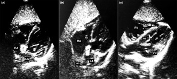
A 9-month-old male diagnosed with Down syndrome and secondary West syndrome required progressive up-titration of adrenocorticotropin hormone dosage (maximum 0.5 mg/day) for the complete control of infantile spasms. The patient was asymptomatic from a cardiological viewpoint, but Cushing syndrome cursing with obesity (body mass index 30.77), buffalo hump, moon face, and high blood pressure (117/89 mmHg) were noticed on physical exam with this dosage. On the hypertension work-up, the electrocardiogram was uneventful, but the echocardiography revealed a large hyper-echogenic non-obstructive mass at the inter-atrial septum and left ventricular non-obstructive hypertrophic cardiomyopathy (Fig 1). Both findings were not observed in previous echocardiographic studies carried out due to the Down syndrome at 1 and 6 months old. The differential diagnosis of the mass was made with infective endocarditis, cardiac tumours, and cardiac thrombus, but these entities were initially ruled out based on the absence of suggestive clinical-laboratory picture and risk-factors such as prolonged fever or previous central catheter lines. Confirmatory cardiac computed tomography scan or magnetic resonance imaging were not performed due to the risk of radiation, the need for sedation and overall, the presence of a “dumbbell-shaped” morphology was very suggestive of lipomatous atrial septum hypertrophy. Reference Laura, Donnino, Kim, Benenstein, Freedberg and Saric1,Reference Heyer, Kagel, Lemburg, Bauer and Nicolas2 Both echocardiographic findings were considered as side effects of adrenocorticotropin hormone. Clinical follow-up with serial Holter monitoring and echocardiographic studies was decided to monitor the cardiological evolution along with the progressive withdrawal of adrenocorticotropin hormone. The patient did not present any cardiac complication, and the discontinuation of adrenocorticotropin hormone at 12 months old was followed by a progressive regression of Cushing’s syndrome and echocardiographic findings, which entirely disappeared at the age of 24 months.
Echocardiography (longitudinal subcostal axis view) performed under treatment with adrenocorticotropin hormone (0.75 mg/day). Panel A and B show a hyperrefringent mass (8 × 10 mm) located at the centre of the inter-atrial septum extended also to the lateral tricuspid ring (black stars). Panel B shows the pathognomonic “dumbbell-shaped” morphology of the mass (black stars) with sparing of the fossa ovalis (intermittent white arrow). Characteristically, the cephalic portion of the mass is usually larger that the caudal portion. Panel C shows a longitudinal subcostal axis view revealing a left ventricular concentric hypertrophy (IVS diameter 11 mm (z score 4.38); PW diameter 8 mm (z score 4.14)). LA (left atria); RA (right atria); IAS (inter-atrial septum); IVS (inter-ventricular septum); LV (Left ventricle); RV (right ventricle); IVS (inter-ventricular septum); PW (posterior wall). FO (fossa ovalis).

The occurrence of hypertrophic cardiomyopathy related to steroids administration has been widely described in infants with bronchopulmonary dysplasia and infantile haemangioma with a dose-dependent and reversible nature in 3–6 weeks. Reference Rodriguez-Gonzalez, Castellano-Martinez, Grujic and Prieto-Heredia3–Reference Thédenat, Léauté-Labrèze and Boralevi5 The hyperinsulinism and arterial hyper-tension secondary to steroids are the major hypothesised pathogenic mechanisms for the development of hypertrophic cardiomyopathy in these patients.
The detection of cardiac masses in echocardiographic studies in children is a rare but alarming and usually challenging finding. Differential diagnosis includes a wide range of diseases (malignancies, benign histological tumours, thrombi, endocarditis, etc.) usually requiring aggressive diagnostic and therapeutic interventions. Lipomatous atrial septum hypertrophy was first described in 1964 as a necropsy finding. Reference Prior6 The increasing availability of cardiac imaging studies is leading to an increasing recognition of this entity, with an estimated incidence of 2–8% of adults in whom cardiac imaging studies are carried out. Reference Laura, Donnino, Kim, Benenstein, Freedberg and Saric1,Reference Heyer, Kagel, Lemburg, Bauer and Nicolas2 Lipomatous atrial septum hypertrophy consists of the thickening of the upper (usually the larger part of the mass) and lower part of the atrial septum with a tendency to bulge into the right atrium, typically sparing the fossa ovalis, leading to a pathognomonic dumbbell-shaped morphology. It is a non-neoplastic mass secondary to a non-capsulated accumulation of adipose tissue. Reference Nadra7,Reference O’Connor, Recavarren, Nichols and Parwani8 The development of lipomatous atrial septum hypertrophy is attributed to the ability of the atrial septum cells to differentiate into adipocytes with appropriate stimuli. Associated accumulation of adipose tissue can also be observed in different localisations such as the crista terminalis, endocardium, and mediastinum. Reference Nadra7,Reference O’Connor, Recavarren, Nichols and Parwani8
Older age, obesity, female sex, prolonged parenteral nutrition, and chronic steroid administration are known risk-factors. Reference Laura, Donnino, Kim, Benenstein, Freedberg and Saric1,Reference Heyer, Kagel, Lemburg, Bauer and Nicolas2,Reference Illade, Plaza and Molina9,Reference Tutar, Çiftçi and Fitoz10 The vast majority of cases are incidentally detected in routine cardiac imaging in asymptomatic patients. However, heart failure and superior caval vein syndrome secondary to the right atrial invasion, and cardiac arrhythmia and sudden cardiac death episodes have been also described. Reference Laura, Donnino, Kim, Benenstein, Freedberg and Saric1,Reference Heyer, Kagel, Lemburg, Bauer and Nicolas2,Reference Illade, Plaza and Molina9,Reference Tutar, Çiftçi and Fitoz10 The diagnosis is based on the combination of risk-factors, cardiological symptoms, and suggestive findings on cardiac imaging (echocardiography, computed tomography scan or cardiac magnetic resonance imaging). Reference Laura, Donnino, Kim, Benenstein, Freedberg and Saric1,Reference Heyer, Kagel, Lemburg, Bauer and Nicolas2 The recognition of lipomatous atrial septum hypertrophy seems to be relevant before carrying out invasive cardiological interventions involving trans-septal catheterisation access, when the presence of the mass can make the procedure particularly challenging. Reference Laura, Donnino, Kim, Benenstein, Freedberg and Saric1,Reference Heyer, Kagel, Lemburg, Bauer and Nicolas2
Treatment depends on the rare occurrence of cardiological complications. To date, there are only two previous paediatric cases of lipomatous atrial septum hypertrophy reported. Reference Illade, Plaza and Molina9,Reference Tutar, Çiftçi and Fitoz10 Both patients debuted with supra-ventricular tachycardia episodes. One case was associated with chronic steroid therapy and the other with the prolonged administration of parenteral nutrition. Of note, one patient presented clinical signs of Cushing syndrome as observed in our patient. In both cases, the mass persisted after 9 and 12 months without associated complications. The death of one of the patients was not related to lipomatous atrial septum hypertrophy complications.
Our case documents the occurrence of previously not reported cardiological complications associated with one of the essential treatments of West syndrome. Remarkably, lipomatous atrial septum hypertrophy can be misdiagnosed as another type of cardiac masses leading to aggressive procedures such as myocardial biopsy, computed tomography scan (radiation), cardiac magnetic resonance imaging (sedation), or prolonged antibiotic regimens. Therefore, to know the existence of this usually benign complication of adrenocorticotropin hormone would allow for conservative approach in this setting. It would be sufficient to monitor the otherwise rare appearance of arrhythmias and heart failure, as well as the echocardiographic stabilisation or regression of the mass after the discontinuation of the treatment. Transthoracic echocardiography could not be accurate for the precise diagnosis of cardiac masses, and ideally advanced cardiac imaging techniques such as computed tomography scan and magnetic resonance imaging are better for this purpose. However, in cases like our patient, a small infant with clear predisposing factor for the development of lipomatous atrial septum hypertrophy and with good acoustic window allowing to obtain high-quality images, echocardiography could be enough to establish the diagnosis. In the case of significant arrhythmia or mechanical obstruction with heart failure, antiarrhythmic drugs, electrophysiological study, or surgical resection would be indicated. Reference Laura, Donnino, Kim, Benenstein, Freedberg and Saric1,Reference Heyer, Kagel, Lemburg, Bauer and Nicolas2,Reference Illade, Plaza and Molina9,Reference Tutar, Çiftçi and Fitoz10
Lipomatous atrial septum hypertrophy is not currently listed as a side effect of adrenocorticotropin hormone. Furthermore, the prevalence and the impact of lipomatous atrial septum hypertrophy and hypertrophic cardiomyopathy are unknown in West syndrome under adrenocorticotropin hormone therapy, since echocardiography is not currently included in most West syndrome work-up protocols unless there is previous underlying disease, such as Down syndrome in our patient. Therefore, we propose that echocardiographic follow-up would be advisable in children with West syndrome before and during treatment with adrenocorticotropin hormone, especially if associated Cushing syndrome is noticed.
Financial support
This research did not receive any specific grant from funding agencies in the public, commercial, or not-for-profit sectors.
Conflicts of interest
None.
Ethical standards
The authors assert that all procedures contributing to this work comply with the Helsinki Declaration of 1975, as revised in 2008, and has been approved by the regional ethics board in Gothenburg, Sweden.