INTRODUCTION
Intensity modulated radiation therapy (IMRT) along with multileaf collimators (MLC) represents one of the most significant technical tools in the management of carcinoma prostateReference Ezzell, Galvin and Low1. IMRT utilizes inverse treatment planning through computer-based optimization processes to deliver user-specified absorbed dose and dose-volume constraints in specified target volumes as well as in normal tissuesReference Jeraj and Robar2,Reference Pugachev, Boyer and Xing3. MLCs have simplified the effort required to generate beam shapes that conform to the target shapeReference Boyer, Biggs and Galvin4. Conventional IMRT has physical limitations which include tongue-and-groove effects and larger number of monitor units required for a treatment can result in increased peripheral dose (PD), or the body dose defined outside the geometrical boundaries of the radiation field. To improve upon existing MLC-based IMRT techniques, collimator rotation can be incorporated into IMRT delivery. The potential of rotational collimator IMRT provides an additional degree of freedom and flexibility when delivering a desired fluence mapReference Milette and Otto5,Reference Otto and Clark6.
Brahme et al.Reference Brahme7 showed that the best orientation of the collimator is when the direction of the leaves is parallel with the direction in which the target volume has the smallest cross section. Otto et al.Reference Otto and Clark6 and Milette et al.Reference Milette and Otto5 reported new methods to deliver an MLC based IMRT plan by incorporating collimator rotation between different segments in each field. It was shown that a number of improvements could be attained through the use of collimator rotation over conventional techniques; improved target conformity and healthy tissue sparing, reduced total number of monitor units (MU) and apertures, improvements in spatial resolution, reduced interleaf effects and maximum deliverable field size over conventional techniques. This approach resulted in increased total treatment time due to mechanical limitations of the collimator rotation speed between segmentsReference Milette and Otto5,Reference Otto and Clark6 which may have restricted implementation. In a separate case studyReference Fung, Enke and Ayyangar8, an algorithm to automatically determine the optimal collimator angle for each beam was utilized to include the nodes in the beams eye view (BEV) for one patient. It showed that plans with optimized collimator angles were worse than the original plan with collimator 0° since all the organs at risk received higher dose in the collimator rotated plan.
Volumetric modulated arc therapy (VMAT) plans exploited a 45° collimator angle for nasopharynx carcinoma patients as it was shown to be more effective in blocking organs at risk while allowing the targets to receive dose from an open beamReference Otto9. As discussed, investigators corroborate that collimator rotation utilized in various sites could provide an additional degree of freedom in inverse treatment planning to improve target dose conformality, reduce total MU while maintaining acceptable dose to OARs and healthy tissuesReference Milette and Otto5–Reference Brahme7.
The aim of this work is to exploit the collimator angle rotation in IMRT plans in the management of carcinoma prostate. Inverse plans, created using conventional collimator angle 0° (CA0) for eight prostate patients, were compared to plans using collimator angle 70° (CA70) for all fields and also with plans utilizing an automatic collimator angle optimization tool (CAopt) for each field. A methodology was developed to ensure that no additional time was added to the overall treatment time due to the change in collimator angle between fields. The planning efficiency and quality was measured in terms of total number of MU, conformity index, dose to organs at risk (OARs) and peripheral dose (PD). Dose to targets and OARs were evaluated by parameters defined in the Conventional or Hypofractionated high-dose intensity-modulated radiotherapy for prostate cancer (CHHiP) trialReference Khoo and Dearnaley10.
METHODS AND MATERIALS
In this retrospective study, seven intermediate risk patients and one high risk patient with prostate cancer were included. The mean target volume was 61.9 ± 9.7 cc. Each patient was planned according to CHHiP trial guidelinesReference Khoo and Dearnaley10. Optimal inverse IMRT plans for all patients were initially produced with collimator angle 0° (CA0). In order to ensure consistency in the comparison, plans were then created using collimator angle 70° (CA70) and optimized collimator angles for each field (CAopt). Hence for each patient, three plans were generated at collimator positions 0°, 70° and optimized angles resulting in a total number of 24 optimized plans.
Treatment planning
A five-field 6 MV photon beam arrangement was used for each plan in the proposed study. The beams were placed at the specified gantry angles (180°, 100°, 35°, 325°, 260°) for all plans with collimator angle for the ‘standard’ plan set to 0°. For a single non-zero collimator angle approach, a beam’s eye view (BEV) encompassing the target and OARs was considered on deciding the most appropriate collimator angle for all beams with pre-specified gantry angles. At collimator angle 70°, the critical structures were effectively blocked by the secondary jaws rather than the multileaf collimator (MLC) in the beam’s eye view (BEV). Similar angles have been used in volumetric modulated arc therapy (VMAT) in order to avoid tongue and groove effectsReference Otto9,Reference Pengpeng, Laura, Yingli, Yoshiya, Gig and Margie11.
All patients were CT scanned (GE Medical Systems, Milwaukee, WI) with 2.5 mm slice thickness. Outlining of target volumes and organs at risk (OARs) was undertaken on each CT image according to the CHHiP guidelinesReference Khoo and Dearnaley10. The inverse treatment planning system (TPS) used was Oncentra® v3.3 sp1 (Nucletron BV, Veenendal, The Netherlands).
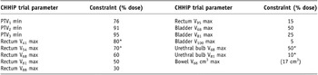
As per the CHHiP protocol, PTV1 was constructed by growing a 1 cm isotropic margin around the outlined prostate with all or part of the seminal vesicles. For PTV2, a uniform margin of 1 cm was added to the prostate alone except towards the rectum where a 5 mm margin was used. PTV3 was grown from the prostate by using a 5 mm margin except posteriorly where it was 0 mm. Normal tissues delineated for the study included bladder, rectum, bowel, femoral heads and penile bulb. The aim of planning was to deliver a prescribed dose of 60 Gy in 20 fractions to PTV3 for each patient. Three dose levels were prescribed to the three different planning target volumes as detailed in Table 1.
Dosimetric quality parameters outlined in the CHHIP trial protocol (e.g. V41 – 41% of rectum can receive a maximum of 80% of the prescribed dose)

*Advisory constraint.
Collimator optimization
Oncentra® v3.3 sp1 (Nucletron BV, Veenendal, The Netherlands) treatment planning system is equipped with an algorithm to determine an optimal collimator angle other than 0° for each field around the selected structure (PTV1 in this study). The algorithm aims to minimize the field size in order to cover the target structure with the intended margin. The orientation of the collimator angles are calculated automatically such that the directions of MLC leaves are parallel to the direction in which the target volume has the smallest cross section. Rotating the collimator for different gantry angles can have the limitation of extending delivery times. As the beams were defined with specified gantry angles for all patients, the maximum time taken for the gantry rotation (average speed of 6°/sec) between any two fields was found to be 10 seconds. Hence to ensure that the time taken for collimator rotation (average speed of 4°/sec) between two fields did not exceed the gantry rotation time, the maximum difference between collimator angles between any two fields was fixed at 40°. However, on some occasions, if the collimator angles produced with CAopt for any two fields exceeded 40°, then a shift of 90° or 180° was applied. Hence, it was not possible for the overall treatment times to be extended.
Plan evaluation
All plans were optimized and evaluated as per objectives and constraints of the CHHiP protocolReference Boylan, Golby and Rowbottom12. The minimum dose to be achieved by different PTVs (100%) and maximum dose levels for volumes of OARs are detailed in Table 1.
The dose conformity of PTV3 was articulated by the conformity index (CI) defined as the volume of body receiving more than 95% of the prescribed dose, divided by the volume of the PTV3. CI will be larger than one, and will increase with decreasing plan conformityReference Verbakel, Cuijpers and Hoffmans13. CI is defined as follows:

Similarly, for PTV2 and PTV1, the volume of body receiving higher than 91% and 76% were measured, respectively. For each patient the volume of patient receiving 20% of the highest prescribed dose (V20), excluding the planning target volume but including critical structuresReference Bedford14 was considered to be the peripheral dose (PD).
Efficiency in plan delivery and physical properties such as total number of MU, total field area and total MLC defined area were analyzed. The MU generated for each beam was summed to obtain the total number of MU for each patient plan. The total field area was defined as the product of the x and y jaw sizes of the linear accelerator for each beam. The MLC area was generated by the treatment planning system for each segment. This MLC area for each segment was then summed for every beam and the average taken for each patient.
Planning time was recorded for all patients and was defined as the time from starting a plan, including beam selection, optimization (single iteration) and final dose calculationReference Oliver, Ansbacher and Beckham15. More iterations are typically required to achieve an optimal plan although it is assumed that a single iteration will result in the minimum planning time. This was recorded for standard and optimized collimator angle techniques.
Statistical analysis was performed using two-tailed student t-tests to obtain the significant difference between all measured parameters of the three techniques.
RESULTS
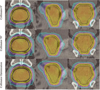
Eight prostate patients were retrospectively planned according to the CHHiP trial protocol. The plans created with the rotational collimator approach, CA70 and CAopt, were compared to CA0 for segmental step-and-shoot IMRT. The mean planning time for 0°/70° collimator rotation and optimized collimator rotation was 12.6 ± 1.10 minutes and 15.1 ± 1.18 minutes, respectively. All three techniques met the desired tolerances as per the CHHiP protocol for each patient. Figure 1 shows the dose distribution generated by CA0, CA70 and CAopt to PTVs for a patient and Figure 2 illustrates DVH analysis with dose to organs at risk and PTVs for the same patient.
Isodose distribution of planning target volumes (PTVs) in all views produced by three techniques for one of the patients. Dashed lines in sagittal and coronal view are contours, whereas continuous lines are isodose lines (i) PTV3 – dark blue and 95% isodose – yellow (ii) PTV2 green and 91% isodose – pink (iii) PTV1 – blue and 76% isodose - sky blue.

Dose-volume analysis of patient no. 5 created with all three techniques: 0° collimator angle CA0 (solid lines), CA70 (dotted lines) and CAopt (dashed lines) with organs at risk

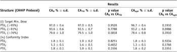
A summary of dose-volume indices is shown in Tables 2 and 3. As shown in Table 2, no significant difference was found between the techniques for target dose conformity.
Target dose and conformity achieved with collimator angle 0°, 70° and optimized collimator angles CAopt for six patients

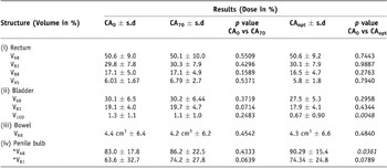
Volumes receiving set doses of organs at risk with each technique. e.g. V95% = Volume receiving 95% of prescribed dose; * - advisory constraint

Table 3 shows the average dose to relevant organs at risk including rectum, bladder, bowel, and penile bulb. The constraints to all organs at risk except penile bulb were fulfilled by each technique. The average penile bulb volume was 3 cmReference Pugachev, Boyer and Xing3. The V68 and V81 dose constraint for penile bulb were advisory as per the CHHiP protocol (Table 1) and couldn’t be achieved with any of the techniques. Our study shows that the dose to penile bulb (V68) with CAopt was significantly higher (p value = 0.036) than the other two techniques. The dose to bladder volume (V100) was significantly reduced with CAopt compared to CA0 (p value = 0.0048).
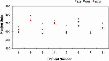
The proposed approaches, CA70 and CAopt, produced similar dose distributions to CA0 and at the same time significantly reduced the total number of MU as shown in Figure 3. The result shows that MU could be reduced by up to 20% with collimator rotational techniques without compromising the plan quality. However some plans were challenging for all collimator angles as large volumes of bladder, bowel and rectum overlapped with PTV structures.
Comparison of total number of MU in plans created with CA0, CA70 and CAopt.

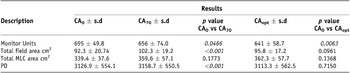
Although there is a reduction in the total number of MU, PD increased significantly for CA70 by 1%, whereas it decreased with CAopt by 0.4% compared to CA0. Also, it was found in general that the total number of MU increased with a decrease in average MLC area for all three techniques. The total number of MU and the average MLC area are given in Table 4.
Comparison of the physical properties between three techniques. Results are presented as mean (s.d - standard deviation; PD – Peripheral dose).

DISCUSSION
The feasibility of exploiting a practical collimator optimization technique for prostate IMRT was analyzed. The results presented indicate that plans generated with collimator angle 70° (CA70) and optimized collimator angle (CAopt) produced plans with similar quality compared to collimator angle 0° (CA0) whilst at the same time significantly reducing total MU by 6% and 9%, respectively. Other investigators have shown that total MU could be reduced significantly by rotating the collimator between segmentsReference Milette and Otto5,Reference Otto and Clark6. However, the disadvantage of the technique was increased total treatment time due to the mechanical limitation of collimator rotation between each segment of the field. Our results show that the collimator rotation between fields reduced total MU without increasing the time to rotate the collimator which would result in reduced delivery time. This would be of benefit because studies have estimated that the increase in MU associated with IMRT may raise the probability of long-term complications including secondary malignanciesReference Kry, Salehpour and Followill16. Furthermore, decrease in MU may reduce the total treatment time and thereby decrease the possibility of spatial inaccuracy from patient movementReference Kry, Salehpour and Followill16,Reference Hall and Wuu17.
The collimator angle produced with the algorithm could be practically delivered without the need of additional time between any two fields. Results show that CAopt created optimal plans and also reduced the total number of MU consistently with increased target conformity compared to CA0. Apart from the penile bulb, an advisory constraint, all other parameters were comparable between CA0 and CAopt. The work presented in this paper shows that the current limitation of collimator optimization is that it needs additional time (∼2.5 minutes) for treatment planning as compared to conventional technique with no collimator rotation. However, this can be improved in future with software upgrade in TPS and is not a long time considering that treatment planning and checking times can typically range from 339 to 908 minutesReference Thomas, Vinall, Poynter and Routsis18. Studies have shown that the axis of rotation of the collimation system and collimator speed is highly reproducible and is therefore not a significant source of errorReference Milette and Otto5,Reference Otto and Clark6.
Peripheral dose is introduced through internal scatter, head scatter, transmission through collimation, head leakage, and room scatter. A strategic orientation of the collimator with a tertiary MLC may reduce PD distributions by more than a factor of twoReference Stern19. Our results found an increase in PD for CA70 compared to CA0 although there was a slight reduction in PD with CAopt with no penalty on treatment time. This may be due to the increase in total area created by the jaws and higher spatial resolution in fluence map generation with CA70. PD distribution with collimator rotation was found to be higher in the entrance region of anterior oblique beams and reduced in the lateral beams. Reduction in PD may result in reduced complication rates to normal tissues outside the treatment fieldsReference Stern19. The work presented in this paper has shown it is important that MU and PD should both be investigated.
CONCLUSION
It has been shown that the rotational collimator technique has a potential advantage over CA0 in the inverse IMRT treatment planning of carcinoma prostate. The proposed technique produced comparable plan to CA0 while minimizing the total number of MU. It has been shown that reduced MU per plan does not always lead to reduced scattered dose to patients.
Acknowledgements
The authors would like to thank all radiotherapy physics staff of Belfast Health and Social Care Trust for their support on this project and Dr. Alan R. Hounsell and Dr. Christina Agnew for reading the manuscript. C.K. McGarry is supported by a Health & Social Care Research & Development Office of the Public Health Agency Training Fellowship Award. We would like to thank Ms. Sharon Hynds for her help.