Introduction
Human lymphatic filariasis is a mosquito-borne tropical disease. It has been estimated that 129 million people are infected and 1.2 billion people are at risk of this infection (Michael & Bundy, Reference Michael and Bundy1997). Due to the possibility of the emergence of drug-resistant parasites and insecticide-resistant arthropod vectors, other forms of intervention need to be considered.
Vaccination is one such tool that may control and even eradicate filarial infection from the world (Shi et al., Reference Shi, Hasnanin, Sacci, Holloway, Fujioka, Kumar, Wohlhueter, Hoffman, Collins and Lal1999). Due to the complex life cycle and antigenic diversity of the filarial parasite, polyvalent filarial vaccines containing multiple protective domains or epitopes from different antigens may be necessary and these may be constructed as chimeric proteins (Rainczuk et al., Reference Rainczuk, Scorza and Spithill2004). An alternative strategy would be to identify the gene products critical to parasite establishment in the host, based on highly expressed genes within the infective stage of the parasite, L3 or infective larvae (Gregory et al., Reference Gregory, Blaxter and Maizels1997).
Several decades ago a number of possible Brugia malayi vaccine candidate antigens were identified. Although studies of immunogenicity and the results of in vitro protection experiments have authorized many of the single specific vaccine candidate antigens, in vivo protection has not always been as satisfactory. There is consensus, however, that a highly effective filarial vaccine would require a combination of key antigens and induction of both humoral and cellular immune processes for optimal efficacy. Multiple component vaccines would also circumvent the problems associated with host genetic restriction and antigenic variability. The use of a multivalent DNA vaccine expression system (bicistronic vectors) may enable more efficient delivery of the antigen and promote a synergistic response among filarial antigens. The testing of viral bicistronic and polycistronic vectors in cancer therapy has been widely used (de Felipe, Reference de Felipe2002). The bicistronic delivery of filarial DNA vaccines may have the potential to enhance an immune response prior to filarial infection.
In earlier studies 24 kDa abundant larval transcript-2 (ALT-2) and 27 kDa vespid allergen homologue (VAH) of B. malayi have been suggested as promising vaccine candidate antigens (Gregory et al., Reference Gregory, Blaxter and Maizels1997; Murray et al., Reference Murray, Gregory, Gomez-Escobar, Atmadja and Maizels2001; Munirathinam et al., Reference Munirathinam, Kakaturu, Yi-Xun, Pankaj, Nutman, Kaliraj and Ramaswamy2004; Ramachandran et al., Reference Ramachandran, Mishra, Reddy, Harinath, Nutman, Kaliraj and McCarthy2004). ALT-2 has been shown to be an L3-stage-specific 24 kDa protein and has no mammalian homologue. Immunization of BALB/c mice with DNA as well as recombinant ALT-2 was able to provide partial (DNA) to 70% (protein) protection against challenge with B. malayi (Ramachandran et al., Reference Ramachandran, Mishra, Reddy, Harinath, Nutman, Kaliraj and McCarthy2004). Similar immunization experiments with ALT-2 in jirds also generated the same level of protection (Munirathinam et al., Reference Munirathinam, Kakaturu, Yi-Xun, Pankaj, Nutman, Kaliraj and Ramaswamy2004); however, DNA priming followed by boosting with rALT-2-failed to protect them.
Similarly, VAH has also been shown to be an L3 expressed protein in its recombinant form, and has been shown to be immunogenic in mice when given with complete Freund's adjuvant and able to induce a substantial (64%) reduction in the development of adult worms (Murray et al., Reference Murray, Gregory, Gomez-Escobar, Atmadja and Maizels2001).
In the present study, an attempt was made to enhance the efficacy of protein and DNA vaccine constructs with BmALT-2 and BmVAH by the cocktail approach against B. malayi infections using jirds as an animal model. Further, their immunological and protective responses were assessed in comparison with single vaccine constructs by the in vitro antibody-dependent cellular cytotoxicity (ADCC) assay and the in situ micropore chamber method.
Materials and methods
Construction and preparation of DNA vaccines
Recombinant plasmids BmALT-2 (accession number U84723) and BmVAH (accession number AF042088) were a kind gift from Dr Thomas B. Nutman, National Institutes of Health, Bethesda, Maryland, USA, and were cloned in the pRSET B vector as described previously (Ramachandran et al., Reference Ramachandran, Mishra, Reddy, Harinath, Nutman, Kaliraj and McCarthy2004). In the present study pBUDCE4.1, a dual promoter vector (Invitrogen, Carlsbad, California, USA), was used to make the vaccine construct by inserting the BmALT-2 and BmVAH genes between the Kpn1, Xho1 and Pst1, BamHI sites, respectively. The vaccine construct was maintained and amplified in Escherichia coli TOP10 cells grown in Luria–Bertani (LB) medium, containing 35 μg/ml Zeocin™. Endotoxin-free plasmid DNA was purified from E. coli TOP10 cells using an Endo-free plasmid Giga kit (Qiagen, Chatsworth, California, USA). Spectrophotometric analysis revealed 260/280 nm ratios to be ≥ 1.80. The purity of DNA preparations was checked on a 1% agarose gel and the presence of antigen genes in proper orientation was confirmed by DNA sequencing.
Expression in Chinese hamster ovary cell lines
Plasmids pBUDALT-2 and pBUDVAH were transfected into Chinese hamster ovary (CHO) cells obtained from the National Centre for Cell Science, Pune, India to confirm expression, along with appropriate positive and negative controls of pEGFPN3 (Clontech, California, USA) and pBUD plasmids, respectively. In brief, CHO cells were seeded at 106 cells/dish in six-well dishes before transfection (Rout et al., Reference Rout, Kumar, Jaganmohan and Murugan2007). A transfection of 10 μg of each plasmid/well was performed using lipofectamine (Invitrogen). At 48 h after transfection, RNA was extracted from cells using TriZOL and converted into cDNA using a cDNA synthesis kit (New England Biolabs, Ipswich, Massachusetts, USA) as per the manufacturer's protocol. The cDNA was verified using ALT-2 and VAH gene-specific forward and reverse primers.
Expression and purification of recombinant BmALT-2 and BmVAH in Escherichia coli
BmVAH expression and purification from salt-inducible E. coli GJ1158 cells (Bangalore Genie, India) was as described earlier (Anand et al., Reference Anand, Munirathinam, Mani, Prabhu, Kaliraj and Ramaswamy2007). BmALT-2 recombinant plasmid was expressed and purified as described previously (Ramachandran et al., Reference Ramachandran, Mishra, Reddy, Harinath, Nutman, Kaliraj and McCarthy2004). Prior to vaccination, the purified recombinant proteins were shown to be negative for bacterial endotoxin using a Limulus Amoebocyte Lysate assay (E-toxate kit, Sigma, St. Louis, Missouri, USA) according to the manufacturer's instructions.
Maintenance of the Brugia malayi life cycle
In the present study, jirds were not used to maintain the complete life cycle of B. malayi. The control and immunized groups of jirds were injected (intraperitoneally) with B. malayi L3 stage larvae only, to test the effect of vaccination in preventing the establishment of B. malayi infection. However, to obtain B. malayi L3 stage larvae for different experiments in the present study, we regularly maintained the life cycle of diurnally sub-periodic B. malayi infection in Mastomys coucha using Aedes aegypti (black-eyed Liverpool susceptible strain) as the mosquito vector, as described by Murthy et al. (Reference Murthy, Tyagi, Choudhary and Sen1983) with a few modifications. Mosquitoes aged 4 days were fed on cotton pads soaked in 6% sucrose solution containing 0.05% para-amino benzoic acid (PABA). Sugar pads were removed from the mosquito cages 1 day before allowing the mosquitoes to feed on infected Mastomys having 80–100 microfilariae per 20 μl of blood. The feeding of mosquitoes on infected Mastomys was carried out (at around 12.00 h) for 1 h, when the microfilariae (mf) appear at peak levels in the peripheral circulation. The mosquitoes were then fed on cotton pads soaked with sucrose and PABA solution. After 9–10 days, infected mosquitoes were mass-dissected and an L3 stage larva of B. malayi was isolated using Barman's apparatus (Suzuki & Seregeg, 1979). Fresh male Mastomys, aged 6–8 weeks, were injected subcutaneously with 100 L3 larvae per animal and monitored for patent infection by regularly checking the tail blood. The infected Mastomys with sufficient number of mf (80–100 per 20 μl) in tail blood were further used to infect fresh mosquitoes.
Immunization
Male outbred Mongolian gerbils (jirds) weighing 35–40 g were maintained at the Mahatma Gandhi Institute of Medical Sciences, Sevagram, India, and five jirds were used per group. For protein vaccine immunization, four groups of jirds (each group containing five jirds) were injected intraperitoneally with 25 μg of endotoxin-free recombinant antigens: group 1, rALT-2; group 2, rVAH; group 3, rALT-2+ rVAH with alum adjuvant (Sigma); group 4, control group, adjuvant only (alum). Another four groups of jirds were immunized (each group containing five jirds) separately (intramuscularly) with 150 μg of endotoxin-free DNA vaccine constructs: group 1, pBUDALT-2; group 2, pBUDVAH; group 3, pBUDALT-2+VAH; group 4, pBUD vector (control), respectively, followed by three booster doses at 2-week intervals. The serum samples were collected from the tail vein every 2 weeks to determine the titre of anti-ALT-2 and anti-VAH antibodies using ELISA.
Antigen-specific antibodies in animal models
The serum samples were collected from immunized animals and control animals after final immunization in order to analyse antibody response by ELISA as described (Reddy et al., Reference Reddy, Alli and Devi1996) with minor modifications. For isotype-specific ELISA, after 1 h incubation with optimum diluted (1:1000) mouse sera, plates were incubated with goat anti-mouse IgG1, IgG2a, IgG2b or Ig3 for 1 h at 37°C. After washing, plates were incubated with rabbit anti-goat IgG labelled with alkaline phosphatase for 1 h at 37°C. Plates were washed and developed with substrate (pNPP) for 30 min at room temperature. Optical density was measured against a phosphate-buffered serum (PBS) blank at 405 nm. Antibody titres were defined as the highest serum dilution that produced an optical density greater than the mean ± 3 SD above the value obtained with the control sera.
In vitro antibody-dependent cellular cytotoxicity assay
The cytotoxicity assay was carried out as described previously (Chandrasekhar et al., Reference Chandrasekhar, Rao, Parab and Subramanyam1985a, Reference Chandrasekhar, Rao and Subramanyamb). Briefly, ten L3 in 5 μl of RPMI 1640 medium (Sigma) were incubated with 50 μl of normal jird peritoneal exudate cells (5 × 10⁵ cells/ten L3) and 5 μl of normal or immunized jird serum in a 96-well culture plate (Nunc, Denmark). Plates were incubated in an atmosphere of 5% CO2 at 37°C. At different periods of incubation (24 and 48 h) the samples were examined microscopically for cellular adherence and cytotoxicity to infective larvae. The percentage of cytotoxicity was expressed as the ratio of number of immobile or dead parasites by the adherence of effector cells to the number of parasites that were recovered within the experimental period.
In situ cytotoxicity studies (micropore chamber technique)
The cytotoxic immune responses against filarial parasites in immunized and control groups of jirds were analysed by the micropore chamber technique as described previously (Anand et al., Reference Anand, Murugan, Prabhu, Anandharaman, Reddy and Kaliraj2008). Micropore chambers were implanted intraperitoneally into the immunized and control jirds under anaesthesia (sodium metapentathiol at 50 mg/kg body weight) through an incision of 2–3 cm and the skin was sutured. After 48 h, animals were anaesthetized and the chambers were taken out, and washed in normal saline. The contents were removed to a glass slide and examined microscopically for cell adherence and cytotoxicity. The percentage of cytotoxicity was expressed as the number of dead parasites/number of larvae placed into the micropore chamber × 100.
Parasite challenge studies
Four groups of jirds (each group containing five jirds) were used: (1) those immunized with rBmALT-2+VAH protein; (2) those immunized with pBUDALT-2+VAH; (3) those immunized with alum control; and (4) those immunized with pBUD vector alone (control). In each group, B. malayi larvae were injected into jirds by intraperitoneal injection of approximately 100 larvae per animal, 10 days after the administration of the last immunization dose. Jirds were checked for the development of patent infection by examining the blood samples collected from cut ends of tails for mf (Chenthamarakshan et al., Reference Chenthamarakshan, Cheirmaraj, Reddy and Harinath1997). After 120 days jirds were killed and adult worms were collected from peritoneal lavage and washings of abdominal organs. The arithmetic mean was calculated for the total worm burden and percentage protection was calculated as [(u − v)/u] × 100, where u is the mean value for the control group and v is the mean value for the experimental group.
Antigen-specific cellular immune response
Jirds were vaccinated and killed, and their spleens were removed under aseptic conditions, Single-cell suspensions were prepared from the spleens according to standard procedures (Onate et al., Reference Onate, Cespedes, Cabrera, Rivers, Gonzalez, Munoz, Folch and Andrews2003). Splenocytes were cultured at 37°C with 5% CO2 in a 96-well flat-bottomed tissue culture plate (Nunc) at a concentration 2 × 10⁵ viable cells/well in the presence of 10 μg/well of ConA (mitogen), 10 μg/well of rALT-2, 10 μg/well rVAH, 10 μg/well rALT-2+VAH antigen mix, or medium alone (unstimulated control). RPMI 1640 medium supplemented with 2 mml-glutamine, 10% heat-inactivated fetal calf serum (GIBCO BRL, Carlsbad, California, USA), and 80 μl of gentamicin and polymixin beads (beads) was used for culturing the splenocytes. The cells were cultured for 72 h in 5% CO2. After 72 h cell proliferation was measured using an MTT assay (Cell Titre 96 Aqueous non-radioactive cell proliferation assay; Promega, Madison, Wisconsin, USA). Splenocyte proliferation was expressed as the stimulation index (SI), calculated by dividing the geometric mean (GM) absorbance of the cells with antigen by the GM absorbance of the cells without antigen.
Cytokine ELISA
For the detection of cytokines, culture supernatants from antigen-stimulated spleen cells were collected and tested for the presence of the cytokines by antigen capture enzyme-linked immunosorbent assay (ELISA) kits for IFN-γ, IL-5 and IL-10 (Endogen, Rockford, Illinois, USA) as described in the kit specifications. All assays were performed in triplicate and the concentration of IFN-γ, IL-5 and IL-10 in the culture supernatants was calculated by linear regression equation obtained from the absorbance values of standards as per manufacturer's instructions. The sensitivity of the assay was < 10 pg/ml for IFN-γ, < 5 pg/ml for IL-5 and < 12 pg/ml for IL-10.
Data analysis
Statistical analysis was performed using XL STAT software v. 7.5.2 (Kovach Computing Services, Anglesey, UK). Statistical significance between comparable groups was evaluated using the Mann–Whitney U test, with the level of significance set at P < 0.05. The comparison of independent means by Student's t-test was applied.
Results
Expression in CHO cell lines
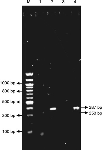
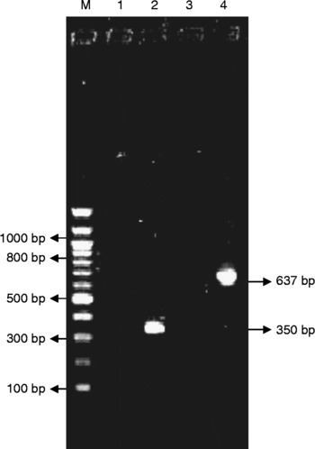
A transfection study was done in mammalian CHO cell lines to analyse the expression of BmALT-2 or BmVAH to validate their in vivo expression as DNA vaccines. Transfection of cell lines with pBUDALT-2 or VAH yielded a specific transcript of 387 bp for BmALT-2 (fig. 1) and 637 bp for BmVAH (fig. 2) in a semiquantitative real time polymerase chain reaction (RT-PCR) experiment with gene-specific primers. Transfection with control plasmid (vector) did not show any transcript.
RT-PCR amplification of the ALT-2 gene. Lanes: M, 100 bp ladder; 1, cDNA without GAPDH primers; 2, cDNA with GAPDH primers; 3, cDNA without gene-specific primers; 4, cDNA with gene-specific primers (BmALT-2). The expected size of the PCR product is 387 bp.

RT-PCR amplification of the VAH gene. Lanes: M, 100 bp ladder; 1, cDNA without glyceraldehyde-3-phosphate dehydrogenase (GAPDH) primers; 2, cDNA with GAPDH primers (positive control); 3, cDNA without gene-specific primers; 4, cDNA with gene-specific primer (BmVAH). The expected size of the PCR product is 637 bp.

Antibody response to immunized jirds
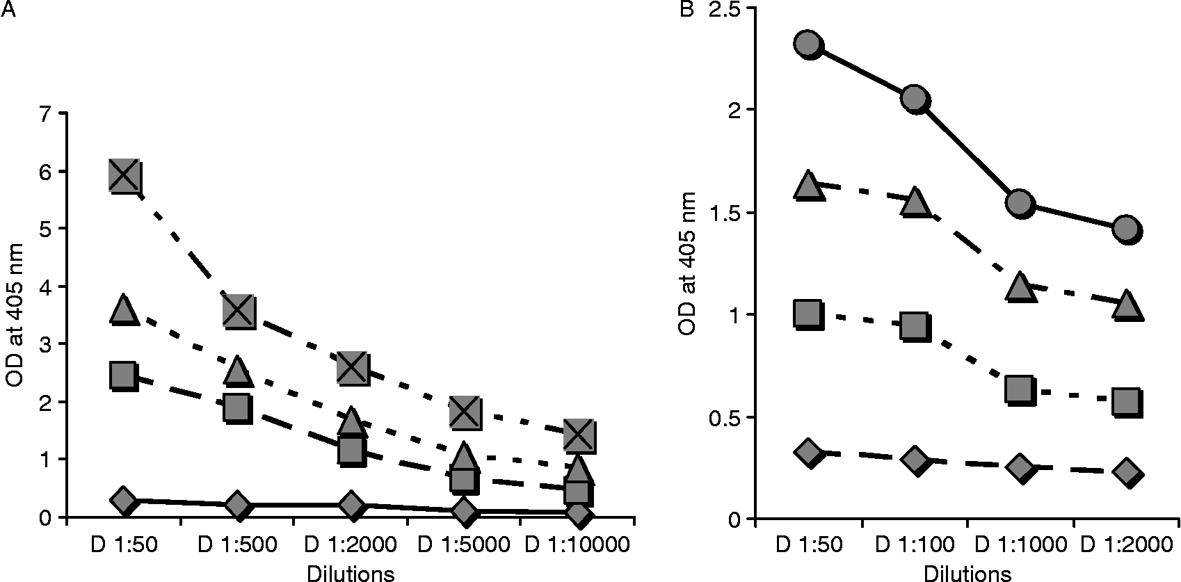
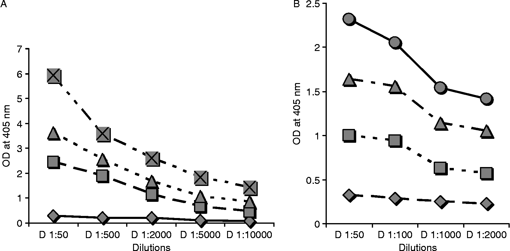
A recombinant DNA bicistronic construct (pBUDALT-2+VAH) was immunized in jirds as a cocktail of DNA vaccine constructs and also as a single DNA vaccine construct (pBUDALT-2 or pBUDVAH). The resulting antibody responses were measured 2 weeks after the fourth immunization. There was significant antigen-specific antibody production in those receiving DNA vaccines compared with the control groups. Significantly increased antibody levels were observed in those receiving the bicistronic DNA constructs compared with those receiving only a single DNA vaccine construct (fig. 3B). Similarly, following immunization with cocktail recombinant proteins (rALT-2+VAH), there were significant increases of antibody levels compared with those receiving individual protein immunizations (rALT-2 or rVAH) (fig. 3A).
Antigen-specific antibody titres (in thousands) in jirds immunized with (A) alum (diamonds), rALT-2 (squares), rVAH (triangles), rALT-2+ VAH (crosses within squares); (B) pBUD vector (diamonds), pBUDALT-2 (squares), pBUDVAH (triangles), pBUDALT-2+ VAH (circles) at different titre values. Results are expressed as the mean ± 3 SD of five jirds in each group, with control optical density (OD) values subtracted.
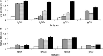
Since the relative IgG2a and IgG1 antibody level is a marker for overall T-cell phenotype, IgG2a and IgG1 titres were also determined for sera collected from these vaccinated animals. A Th2-type response favours the production of IgG1 antibodies, while IgG2a antibodies are associated with a Th1 response (Rizzo et al., Reference Rizzo, DeKruyff and Umetsu1992). For the recombinant rALT-2+VAH-vaccinated animals, the antibody titre showed an elevated IgG1, IgG2a and IgG2b isotype response (fig. 4A). While rALT-2 protein-vaccinated animals showed an elevated IgG1 and IgG3 isotype response, rVAH-vaccinated animals showed an elevated level of IgG1 and IgG2 isotype response. These results suggest that the cocktail protein vaccination induced a mixed Th1/Th2 type of response; while rALT-2 showed a Th2-type response and rVAH showed a Th1/Th2-type mixed response. Similarly, in the pBUDALT-2+VAH bicistronic DNA vaccination there was a significant increase in the levels of IgG2a and IgG2b and a very low level of IgG1. These results suggest that cocktail DNA vaccination induced a Th1 type of response (fig. 4B).
Antibody isotype profile of (A) alum (white bars), rALT-2 (grey bars), rVAH (striped bars) and rALT-2+VAH (black bars) immunized jirds; (B) pBUD vector (white bars), pBUDALT-2 (grey bars), pBUDVAH (striped bars) and pBUDALT-2+VAH (black bars) immunized jirds. Results are expressed as the mean ± 3 SD of five jirds in each group, with control optical density (OD) values subtracted. The results were statistically significant.

In vitro antibody-dependent cellular cytotoxicity assay
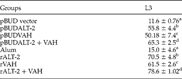
The serum of jirds immunized with BmALT-2 and VAH antigens promoted the adherence of peritoneal exudate cells to both L3 larvae and microfilariae. In the in vitro cytotoxicity assay, anti-pBUDALT-2 sera induced 55% cytotoxicity against infective larvae (L3), pBUDVAH sera induced 50% cytotoxicity against infective larvae (L3), and pBUDALT-2+VAH sera induced 65% cytotoxicity against infective larvae (L3). Similarly anti-rALT-2 sera induced 70% cytotoxicity against infective larvae (L3), anti-rVAH sera induced 61% cytotoxicity against infective larvae (L3) and anti-rALT-2+VAH sera induced 79% cytotoxicity against infective larvae (L3); the difference was statistically significant (table 1).
In vitro antibody-dependent cellular cytotoxicity (ADCC) against L3 larvae induced by sera of jirds immunized with rALT-2+VAH and pBUDALT-2+VAH. Five jirds were used/group.
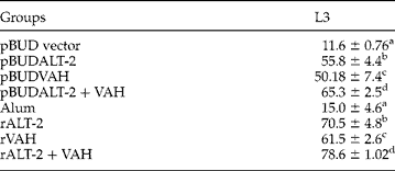
a versus b, significant, P < 0.001; a versus c, significant, P < 0.001; a versus d, significant, P < 0.001.
In situ cytotoxicity studies (micropore chamber technique)
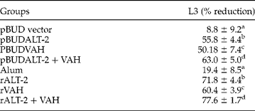
The in situ study in jirds immunized with pBUDALT-2 showed 55% cytotoxicity, pBUDVAH showed 50% cytotoxicity and with pBUDALT-2+VAH showed 63% cytotoxicity. The control group of jirds immunized with the pBUD vector showed 8.8% cytotoxicity. Jirds immunized with only alum shown 19% cytotoxicity and the difference in the test and control groups of animals was statistically significant (P < 0.001). The in situ study in jirds immunized with rALT-2 showed 72% cytotoxicity. Jirds immunized with rVAH showed 60% cytotoxicity and jirds immunized with rALT-2+VAH showed 78% cytotoxicity against L3 (table 2).
In situ cytotoxicity studies (micropore chamber technique) against L3 larvae induced by sera of jirds immunized with rALT-2+VAH and pBUDALT-2+VAH. Five jirds were used/group.
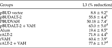
a versus b, significant, P < 0.001; a versus c, significant, P < 0.001; a versus d, significant, P < 0.001.
Parasite clearance test
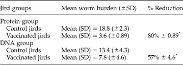
A mean (SD) of 3.6 ( ± 0.89) B. malayi larvae were recovered from rALT-2+VAH (cocktail vaccines) immunized jirds and a mean of 18.8 ( ± 2.3) were recovered from the alum control group of jirds. This represents an 80% reduction (table 3) in parasite recovery from rALT-2+VAH (cocktail vaccines) immunized jirds (P < 0.001). A mean (SD) of 7.8 ( ± 4.6) B. malayi larvae were recovered from pBUDALT-2+VAH (bicistronic vaccine) immunized jirds and a mean of 13.4 ( ± 4.3) were recovered from the vector control group of jirds. This represents a 57% reduction in parasite recovery from pBUDALT-2+VAH (bicistronic DNA vaccine construct) immunized jirds (P < 0.001).
The number of adult worms recovered from rALT-2+VAH (protein group) and pBUDALT-2+VAH (DNA vaccine group) and control group of jirds. Five jirds were used/group.
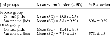
*Control versus vaccinated statistically significant, P < 0.001.
Antigen-specific cellular immune response
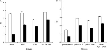
The SI of splenocyte proliferation was determined by the MTT assay (Cell Titre96® aqueous non-radioactive cell proliferation assay; Promega). Enumerated results showed a significantly higher level of proliferation in the pBUDALT-2-immunized group (GM SI = 2) (P < 0.001), pBUDVAH (GM SI = 1.5) (P < 0.02) and pBUDALT-2+VAH (P < 0.001) (GM SI = 2.5) compared with the pBUD vector (SI < 2) (fig. 5B). Similarly, the rALT-2 (GM SI = 2.2) (P < 0.001), rVAH (GM SI = 2) (P < 0.001) and rALT-2+VAH (GM SI = 2.85) (P < 0.001) immunized groups also showed significant proliferation compared with the alum control (SI < 2) (fig. 5A). Stimulation with ConA induced significant proliferation in both the control and vaccine-immunized cells.
Splenocyte cells (A) ConA (white bars) stimulated; rALT-2 vaccinated jirds stimulated with rALT-2 protein, rVAH vaccinated jirds stimulated with rVAH protein, rALT-2+VAH vaccinated jirds stimulated with rALT-2+VAH protein (black bars). (B) Splenocyte cells proliferated with ConA (white bars); pBUDALT vaccinated jirds stimulated with rALT-2 protein, pBUDVAH vaccinated jirds stimulated with rVAH protein, pBUDALT+VAH vaccinated jirds stimulated with rALT-2+VAH protein (black bars). The bars represent proliferation level of jird splenocytes stimulated with the vaccine compared to that of the control group cells. The data represent the mean stimulation index (SI) ± SD.

Cytokine response studies
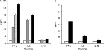
Immunized pBUDALT-2+VAH spleen cells from jirds when stimulated with BmALT-2+VAH resulted in higher levels of IFN-γ (34 pg/ml) (P < 0.05) compared with the control group (2.87 pg/ml) (P < 0.05), with diminished levels of IL-10 (11 pg/ml) and IL-5 (6.5 pg/ml). The levels of IFN-γ production were significantly elevated in vaccinated jirds, suggesting that the vaccination process induced a Th1 type of immune response (fig. 6B).
Cytokine-ELISA of jird spleen cells treated with (A) alum (white bars), rALT-2 (grey bars), rVAH (striped bars), rALT-2+VAH (black bars); and (B) pBUD vector (white bars) and pBUDALT-2+VAH (black bars). Data represent mean values ± SD.

Similarly, immunized rBmALT-2+VAH spleen cells from jirds when stimulated in vitro with BmALT-2+VAH resulted in a high level of IFN-γ (55 pg/ml) (P < 0.05) with significant levels of IL-5 (40 pg/ml) and a very low level of IL-10 (6 pg/ml) compared with the control group (IFN-γ, 4 pg/ml; IL-5, 4 pg/ml; IL-10, 6 pg/ml) (P < 0.05), suggesting that the vaccination process induced a mixed type of Th1/Th2 immune response. When rBmALT-2 spleen cells were stimulated in vitro with rALT-2 alone, a predominant increase of IL-5 was observed (32 pg/ml), with a significant level of IFN-γ (22 pg/ml) and a very low level of IL-10 (5 pg/ml) when compared with control cells (P < 0.05). This suggests that rBmALT-2-vaccinated jirds were biased towards a Th2 (IL-5) type of immune response. Similarly, when rBmVAH spleen cells were stimulated in vitro with rVAH alone it resulted in a predominant increase of IFN-γ (40 pg/ml) and lower levels of IL-5 (4 pg/ml) and IL-10 (4 pg/ml) compared with the control group (P < 0.05), which suggest that rBmVAH when vaccinated individually produces Th1 cytokines (fig. 6A). This result suggests that rBmALT-2+VAH vaccination induces high proliferation compared with single-antigen vaccination and the bicistronic DNA construct. Similarly, the cytokine level produced a mixed Th1- and Th2-type response.
Discussion
Although diethylcarbamazine against filariasis is currently proceeding as a drug of choice in combination with albendazole or ivermectin, these drugs are mainly microfilaricidial and fairly ineffective in killing worms, thereby providing only partial benefit to infected patients (Pokharel et al., Reference Pokharel, Rai, Nandakumar, Reddy and Rathaur2006). Thus there remains an urgent need to identify new candidate antigens for single use or as part of a protein or DNA vaccine or as a cocktail vaccine.
A previous study in our laboratory has shown that a stage-specific B. malayi abundant larval transcript-2 (BmALT-2) could offer 70% protection in jirds as a single protective antigen against filariasis. Similarly, Murray et al. (Reference Murray, Gregory, Gomez-Escobar, Atmadja and Maizels2001) have shown that B. malayi vespid allergen homologue (BmVAL or BmVAH) produced a mean reduction of 64% in parasite recovery against B. malayi in animal models, but the level of protection was not statistically significant. However, for multicellular parasites such as filarial nematodes, single antigen vaccination may not be strong enough to combat this complex life cycle and the use of multivalent vaccines may therefore be necessary to achieve a high level of protection (Doolan & Hoffman, Reference Doolan and Hoffman2001; Kumar et al., Reference Kumar, Villinger, Oakley, Aguiar, Jones, Hedstrom, Gowda, Chute, Stowers, Kaslow, Thomas, Tine, Klinman, Hoffman and Weiss2002; Burns et al., Reference Burns, Flaherty, Romero and Weidanz2003; Mendez et al., Reference Mendez, Zhan, Goud, Ghosh, Dobardzic, Wu, Liu, Deumic, Dobardzic, Liu, Bethony and Hotez2005).
In the present study, we have shown, using the in vitro ADCC assay and in situ micropore chambers, that the bicistronic DNA vaccine construct enhanced the protective efficacy against B. malayi infective larvae when compared with a single-DNA vaccine construct. Similarly, the recombinant protein cocktail vaccines enhanced the protective efficacy against B. malayi infective larvae when compared with single-protein vaccines. Our results, interestingly, have shown that cocktail protein vaccines induced higher protective efficacy when compared with a bicistronic DNA vaccine construct against B. malayi infective larvae. Also, in situ and in vitro experiments showed that the cocktail vaccine construct enhanced efficacy when compared with single vaccine constructs. Hence, in the further confirmatory study, only the cocktail vaccine constructs alone with appropriate controls were tested by a parasite clearance study. Jirds are slow to breed in animal houses, particularly when inbreeding or closed breeding is followed, and instances of cannibalism cannot be totally avoided. Hence, there are limitations to obtaining them in large number for experiments involving multiple groups.
In the present study we constructed DNA vaccines that express two antigens as a bicistronic construct (BmALT-2 and BmVAH); the resulting construct showed a slight increase of antibody level when compared with the monocistronic construct (BmALT-2 or BmVAH) in jirds. Similarly, we have formulated a cocktail of two filarial vaccine candidate recombinant antigens (BmALT-2 and BmVAH) and the resulting cocktail of recombinant antigens was shown to increase antibody titre level when compared with single recombinant antigens (BmALT-2 or BmVAH) in jirds.
ADCC is believed to be one of the principal immunological mechanisms that functions in animals and the disappearance of circulating parasites is mainly attributed to this phenomenon (Mehta et al., Reference Mehta, Sindhu, Subramanyam and Nelson1980; Chandrasekhar et al., Reference Chandrasekhar, Rao, Parab and Subramanyam1985a, Reference Chandrasekhar, Rao and Subramanyamb). In our study of the in vitro ADCC reaction, the antisera raised against both BmALT-2 and BmVAH in single and multiple antigen vaccination modes induced cytotoxicity to B. malayi infective larvae within 48 h. However, anti-rALT-2+VAH (79% cytotoxicity) serum induced stronger adherence of peritoneal cells, mainly macrophages, against B. malayi L3 larvae compared with the cellular adherence observed with either anti-rALT-2 (70% cytotoxicity) or anti-rVAH (61% cytotoxicity) as the single construct. Similarly, in the DNA vaccine construct anti-pBUDALT-2+VAH (65% cytotoxicity) serum showed better adherence of cells when compared with either pBUDALT-2 (55% cytotoxicity) or pBUDVAH (50% cytotoxicity) alone. Both antibody- and complement-mediated effector mechanisms have been shown to be involved in inducing cytotoxicity to the microfilaria and infective larvae in vitro (Sim et al., Reference Sim, Kwa and Mak1982). In vitro, there is a large body of evidence showing all mammalian stages of filarial parasites that can be killed by ADCC. Following engagement of cell surface FcR by antibodies attached to the parasite surface, cells recruited to the site of infection, such as macrophages, eosinophils and neutrophils, are all capable of extruding toxic mediators into the parasite surface. In vitro data show that a variety of cells and antibody isotypes are potentially capable of killing filarial parasites and the exact mechanism may vary according to the developmental stage of the parasite.
Like the ADCC assay, the micropore chamber experiment with the cocktail of antigens rALT-2+VAH showed 78% protective efficacy when compared with the single antigen rALT-2 (71% cytotoxicity) or rVAH (60% cytotoxicity). In the DNA vaccine construct pBUBALT-2+VAH, 63% protective efficacy was shown against Brugia infective stage L3 larvae when compared to either pBUDALT-2 (55% cytotoxicity) or pBUDVAH (50% cytotoxicity). The micropore chamber technique, in a closer physiological environment, can be used to study parasite growth and survival and also to assess the host effector mechanism. It has been used by many investigators to analyse whether a similar ADCC reaction could occur in vivo (Rajasekariah et al., Reference Rajasekariah, Monteiro, Nelto, Deshpande and Subrahmanyam1989). The involvement of macrophages in mediating cytotoxicity reactions has also been demonstrated against other filarial parasites, namely Dipitelonema setariosum adults (Worms & McLaren, Reference Worms and McLaren1982) and Litomosoides carinii microfilariae (Nelson et al., Reference Nelson, Subrahmanyam, Rao and Mehta1976). In the present study, our findings also demonstrated cytotoxicity results in situ (micropore chamber) which were similar to those of the ADCC reaction.
All the in vitro and in situ experiments were repeated twice, and from the obtained results, cocktail DNA vaccine or protein constructs were shown to have better protective efficacy when compared with single antigens. Hence, for further confirmation, an attempt was made to study the protective efficacy of protein and DNA vaccine constructs against B. malayi infection by the parasite clearance method. None of the jirds immunized with rBmALT-2+VAH and pBUDALT-2+VAH antigen showed microfilariae in circulation. The experiment was repeated twice.
In the present study, the group of jirds immunized with rBmALT-2+VAH showed a mean reduction of 80.4% in parasite recovery from the peritoneal cavity compared with the control group of jirds (P < 0.001), which is statistically significant. Earlier studies with rBmALT-2 as a single antigen vaccination (Gregory et al., Reference Gregory, Atmadija, Allen and Maizels2000) showed 70% protection in jirds and BmVAH showed a mean reduction of 64% in parasite recovery against B. malayi in animal models, but the level of protection was not statistically significant (Murray et al., Reference Murray, Gregory, Gomez-Escobar, Atmadja and Maizels2001). The present study thus confirms that cocktail antigens are substantially better than any previous filarial recombinant single antigen reported in the literature (Li et al., Reference Li, Chandrasekhar, Alvarez, Lifis and Weil1991; Peralta et al., Reference Peralta, Schmitz and Rajan1999; Gregory et al., Reference Gregory, Atmadija, Allen and Maizels2000; Murray et al., Reference Murray, Gregory, Gomez-Escobar, Atmadja and Maizels2001; Dabir et al., Reference Dabir, Krithika, Goswami and Reddy2006).
Interestingly, the new concept of delivery of DNA vaccination with multiple antigen vaccination enhanced the protection (up to 57%) and had a slight increase in immune response level compared with single antigen vaccination, but when compared with recombinant protein the protection level was low. Although these findings represent progress, we realize that much more work is needed before practical DNA vaccines for filariasis can be produced in reality. In addition, more work is needed to optimize vaccination protocols, many other candidate antigens need to be tested, and refinement of DNA vaccines is necessary for an effective prime protection immune response.
In the present study, the spleens from vaccinated animals were removed and spleen cells were stimulated with rALT-2 or rVAH or rALT-2+VAH. The results showed that the rALT-2+VAH-vaccinated group produced a significantly higher proliferation response (SI>3) (P < 0.05) compared with rALT-2 (SI < 2.2) and rVAH ( < 2). A similar observation has been reported from the studies on fusion of two malaria vaccine candidate antigens. The cellular response was investigated in lymphocytes proliferated with multiple antigens and a significant proliferation response was shown (SI>3) (Pan et al., Reference Pan, Huang, Zhang, Qu, Zhang, Zhang, Xue and Qian2004). Altogether, these findings demonstrated that multiple antigen vaccination may stimulate an increase in proliferative response compared with single or monocistronic vaccination.
The T-cell response that might be associated with high antibody titres was studied by analysis of IgG isotypes and cytokine profile, which revealed that animals immunized with rALT-2 single antigen have high concentrations of IgG1 and IgG3 in their sera, suggesting a Th2-type response. When cells from these animals were stimulated with recombinant BmALT-2 protein, significantly higher levels of IL-5 (Th2) and lower levels of IFN-γ (Th1) were induced, suggesting that a Th2-biased response may be important in the single antigen-mediated protective responses.
On the other hand, animals vaccinated with BmVAH showed high levels of IgG1 and IgG2 in their sera, suggesting a mixed-type Th1/Th2 response. However, this was not truly reflected when the antigen-specific cytokine profile was evaluated by ELISA. When cells from these animals were stimulated with recombinant BmVAH protein, a significant level of IFN-γ (Th1) was induced, suggesting a Th1-biased response. A similar discrepancy between isotype and cytokine has been reported previously (MacDonald et al., Reference MacDonald, Cao, He, Zhao, Jiang and Lustigman2005).
Analysis of the characteristics of protective immune responses has shown that in cocktail antigen-vaccinated animals there were high IgG1 and IgG2 isotypes in their sera, suggesting a mixed type of response. This was truly reflected when the antigen-specific cytokine profile was evaluated by ELISA. When cells from these animals were stimulated with mixed recombinant BmALT-2 and BmVAH protein, significant IFN-γ (Th1) and IL-5 (Th2) were induced and both Th1- and Th2-type responses were produced, which were important in the cocktail-mediated protective responses.
Some evidence has confirmed that Th2 responses are responsible for resistance to intestinal helminths; however, it is difficult to extrapolate such results to tissue-dwelling nematodes such as filariae. Several recent studies of different models of filarial diseases have shown that Th1-type responses are also involved in the immune protection to filarial infection. IFN-γ has been shown to be required for clearance of B. malayi in mice (Babu et al., Reference Babu, Ganley, Kliei, Shultz and Rajan2000). Cytokine profiles indicated that infection stimulates both Th1 and Th2 responses, and that the type of response is dependent on the time of post-infection.
Altogether, our results truly indicate that multiple antigen vaccination improves vaccine efficacy against B. malayi infection when compared with using single antigen vaccination. However, further study is warranted to improve the protective response.
Acknowledgements
This study was funded by the Department of Biotechnology and partly supported by the Indian Council of Medical Research and the University Grant Commission, Government of India. We sincerely thank Dr Thomas B. Nutman, NIH, Bethesda, USA for kindly providing clones for our study and for kindly critiquing the paper. S.B.A. is a recipient of a senior research fellowship from the Indian Council of Medical Research, Government of India.