Introduction
Belemnoid coleoids, with their well-known mineralized rostrum and characteristic arm hooks, represent widely distributed but often neglected Jurassic and Cretaceous microfossils. A belemnoid arm crown is composed of 10 arms, each equipped with up to 40 pairs of chitinous micro-hooks, that is, hooks rarely larger than 5 mm, resulting in 200–800 micro-hooks per specimen (Engeser Reference Engeser1987; Engeser and Clarke Reference Engeser and Clarke1988). Published records of belemnoid micro-hooks mainly refer to Jurassic deposits (e.g., Münster Reference Münster1834, Reference Münster1839; Owen Reference Owen1844; Quenstedt Reference Quenstedt1849; Engeser and Reitner Reference Engeser and Reitner1981; Reitner and Engeser Reference Reitner and Engeser1982; Reitner and Urlichs Reference Reitner and Urlichs1983; Engeser Reference Engeser1987; Urlichs et al. Reference Urlichs, Wild and Ziegler1994; Riegraf Reference Riegraf1996; Klug et al. Reference Klug, Schweigert, Fuchs and Dietl2010, Reference Klug, Schweigert, Fuchs, Kruta and Tischlinger2016; Fuchs et al. Reference Fuchs, Donovan and Keupp2013a,Reference Fuchs, Heyng and Keuppb), while Cretaceous findings were first reported by Riedel (Reference Riedel1936) and are rare in comparison (Engeser and Suthhof Reference Engeser and Suthhof1992; Reich and Frenzel Reference Reich and Frenzel1997; Reich Reference Reich2002; Mitta and Bogomolov Reference Mitta and Bogomolov2014). The rarity of reports on Cretaceous coleoid hooks may reflect a form of selection bias, as it is believed that they occur more abundantly but have often been overlooked, possibly due to lack of interest by researchers (Reich and Frenzel Reference Reich and Frenzel1997; Reich Reference Reich2002).
Complete or incomplete arm crowns associated with the remaining parts of the belemnoid specimen provide important paleobiological insights, particularly with respect to functional morphology, systematics, and taxonomy. Examples include the Late Triassic Phragmoteuthis (Rieber Reference Rieber1970; Doguzhaeva et al. Reference Doguzhaeva, Summesberger, Mutvei and Brandstaetter2007); Early Jurassic Sueviteuthis (Reitner and Engeser Reference Reitner and Engeser1982), Acrocoelites, Passaloteuthis (Reitner and Urlichs Reference Reitner and Urlichs1983), and Clarkeiteuthis (Quenstedt Reference Quenstedt1849; Riegraf Reference Riegraf1996; Fuchs et al. Reference Fuchs, Donovan and Keupp2013a,Reference Fuchs, Heyng and Keuppb; see Garassino and Donovan [Reference Garassino and Donovan2000] for an extended list); and Middle to Late Jurassic Belemnotheutis (Owen Reference Owen1844; Donovan and Crane Reference Donovan and Crane1992), Acanthoteuthis (Engeser and Reitner Reference Engeser and Reitner1981), Winkleriteuthis (Fuchs et al. Reference Fuchs, Donovan and Keupp2013a) and Hibolithes (Klug et al. Reference Klug, Schweigert, Fuchs and Dietl2010). Individuals of the above-mentioned species develop arms of similar length, and their arms are equipped with micro-hooks of similar shape. However, the hook shapes of those species show low interindividual and intraindividual variability but differ sufficiently between species and are abundant enough to be recognized as potential index fossils for biostratigraphic purposes (Engeser Reference Engeser1987). Conversely, in species in which hook shapes show intraspecific variation (e.g., Reitner and Urlichs Reference Reitner and Urlichs1983 for Acrocoelites, Garassino and Donovan Reference Garassino and Donovan2000 for Ostenoteuthis), morphological studies of their shapes offer the possibility for functional ecological interpretations of their roles in the living organisms. Consequently, different types of arm armature between species allow for speculations on the potential food resources.
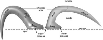
Kulicki and Szaniawski (Reference Kulicki and Szaniawski1972), who established the widely used terminology for micro-hook elements, divided the micro-hook into three morphological parts, namely the base, shaft, and the uncinus (Fig. 1). This terminology was adopted from earlier descriptions of mega-hooks (hooks>5 mm) by Quenstedt (Reference Quenstedt1856). Hook morphotypes were carefully characterized based on three traits: flat or concave base; straight or bent shaft; and strongly or weakly curved uncinus, with the same or different bending compared with the shaft. Using such techniques, it has been shown that hook morphologies normally vary only slightly within an individual arm crown (e.g., Engeser Reference Engeser1987; Fuchs Reference Fuchs2006). Alongside shape, hook size has been recognized to differ within individual belemnoid specimens. In general, micro-hooks are small when situated proximal (i.e., close to the mouth), becoming larger toward the middle section of the arm and decreasing in size when more distally situated (Reitner and Urlichs Reference Reitner and Urlichs1983).
General hook morphology showing the base, shaft, and uncinus. Left, micro-hook; right, mega-hook.

When Kulicki and Szaniawski (Reference Kulicki and Szaniawski1972) introduced their parataxonomy for isolated micro-hooks, the authors considered micro-hook morphotypes as species specific and therefore assumed the absence of distinct morphotypes within the same arm crown. However, the discovery of a pronounced morphological differentiation of micro-hooks observed in the arm crowns of Ostenoteuthis siroi (Garassino and Donovan Reference Garassino and Donovan2000) and Chondroteuthis wunnenbergi (Engeser Reference Engeser1987) made the parataxonomic system of Kulicki and Szaniawski (Reference Kulicki and Szaniawski1972) problematic. In addition, Reitner and Urlichs (Reference Reitner and Urlichs1983) and Reitner (Reference Reitner1986) recognized gradual shape changes of the hooks along the arm length of two belemnitids (Acrocoelites and Acanthoteuthis). The observation of gradual changes in hook morphology made sufficient descriptions for hook (para-)genera or species difficult. To overcome this limitation, a few studies supplied standard linear and angular measurements (Engeser Reference Engeser1987; Fuchs Reference Fuchs2006; Lehmann et al. Reference Lehmann, Solarczyk and Friedrich2012), including both maximum hook length and maximum hook height, length of the base, width and bending of the shaft, and hook curvature. From these primary parameters, ratios (e.g., total length/length of the base, total length/total height, total length/length of maximum height) can be inferred to quantitatively describe hook morphology. Going further, Fuchs (Reference Fuchs2006) used some univariate measures to quantify hook morphology for well-preserved hooks in which the inner and outer margin of the base (process) and the tip of the uncinus have been used as landmarks. These landmarks provide distinct coordinates to infer hook morphology. Because most animal hooks are shaped like a section of a logarithmic spiral, Hammer et al. (Reference Hammer, Hryniewicz, Hurum, Hoyberget, Knudsen and Nakrem2013) used the outer margin of the uncinus of hook of complete mega-hooks and fitted them to logarithmic spirals. The use of logarithmic spirals as morphological descriptors of hook shapes demonstrated a remarkably consistent morphology when comparing Upper Jurassic material from Greenland, northern Norway, and the North Sea. Given the short stratigraphic (Kimmeridgian–Volgian) and small biogeographic range, Hammer et al. (Reference Hammer, Hryniewicz, Hurum, Hoyberget, Knudsen and Nakrem2013) assumed that the hook morphotype they had analyzed and assigned to Onychites quenstedti indicates its connection with a single biological species or genus.
Despite those refinements, the use of belemnite micro-hook morphology for stratigraphic, phylogenetic, and ecological studies still faces the following problems: (1) parataxonomy of micro-hooks is in some cases based on minute morphological features that often neglect potential taphonomic bias; (2) gradual morphological changes in micro-hook morphology may cause an artificially high number of hook parataxa and require quantification of morphological variation; and (3) some hook morphotypes seem to represent stable forms showing limited morphological variation from the Carboniferous to the Upper Cretaceous, and can thus be expected to be found in several coleoid taxa.
Here we apply for the first time elliptic Fourier analysis (EFA) to objectively quantify and categorize belemnoid micro-hook morphology. This analysis technique allows a quantitative description of shape as a whole, thus allowing a more integrated analysis of hook shape than a multivariate dataset of several lengths and angles (e.g., Engeser Reference Engeser1987). Fourier analysis is a well-established method in paleobiological studies to quantify shapes and distinguish different groups (e.g., species) within a set of specimens or to reconstruct phylogenetic trajectories (Kaesler and Waters Reference Kaesler and Waters1972; Anstey and Delmet Reference Anstey and Delmet1973; Christopher and Waters Reference Christopher and Waters1974; Healy-Williams and Williams Reference Healy-Williams and Williams1981; Renaud et al. Reference Renaud, Michaux, Jaeger and Auffray1996; Renaud and Schmidt Reference Renaud and Schmidt2003) and is also used for comparable applications in other fields, such as geology (Ehrlich and Weinberg Reference Ehrlich and Weinberg1970). EFA is a particular form of Fourier analysis and was chosen for this study because it is well established (Rohlf and Archie Reference Rohlf and Archie1984) and has been shown to yield reliable results with shapes lacking pronounced features and having only few homologous points along their outlines (Crampton Reference Crampton1995; Van Bocxlaer and Schultheiß Reference Van Bocxlaer and Schultheiß2010).
On the basis of isolated hook assemblages, Engeser (Reference Engeser1987) assumed that Chondroteuthis wunnenbergi was equipped with four different hook morphotypes. This hook differentiation is obvious in new specimens with excellently preserved in situ arm hooks and thus ideal for the first morphological quantification of belemnoid arm hooks. It is the aim of our contribution (1) to demonstrate the possibility of objectively classifying micro-hooks in distinct morphological groups, (2) to quantify morphological variation of these morphotypes, (3) to document the distribution of the hook morphotypes within the arm crown and along the arms, and (4) to discuss arm differentiation based on our analysis of hook distribution. Such analyses allow establishing a homogenous and broadly applicable morphotype system for belemnoid hooks, which is essential for quantitative studies based on hook morphology. Practically, EFA can be supplemented with descriptions of other morphological features not seen in the outline (e.g., ornamentation, shape of the basal opening) to further enhance the system in the future.
Materials and Methods
Materials.—We analyzed the micro-hooks of arm crowns of three specimens of Chondroteuthis wunnenbergi from the lower Toarcian Posidonia shales (falciferum Zone, mainly boreal nodule layer) of northern Germany. All three specimens are housed at the Bundesanstalt für Geowissenschaften und Rohstoffe in Hanover (NW Germany) with the registration nos. BGR MA 13435, 13436, and 13437. The monotypic genus Chondroteuthis is characterized by the absence of a solid and calcitic rostrum proper and by a narrow, long, spatulate proostracum, which is slightly longer than the orthoconic phragmocone (ratio phragmocone length:total shell length ~0.4). With respect to shell morphologies, Chondroteuthis is closest to the coeval genus Clarkeiteuthis, but differs significantly in hook morphology (Fuchs et al. Reference Fuchs, Donovan and Keupp2013a). Jeletzky (Reference Jeletzky1966) and other workers regarded Chondroteuthis as a member of the belemnitid suborder Belemnotheutidina, but owing to the presence of a conspicuously narrow proostracum, we follow Engeser (Reference Engeser1995), who first placed Chondroteuthis within the order Diplobelida.
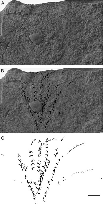
Our specimens exhibit shell (hence mantle) lengths between 12.5 and 8.2 cm, while the holotype is 15.0 cm long (Table 1). This suggests that all three examined specimens belong to similar (subadult to adult) growth stages. The arm crowns (the holotype has no arm crown preserved) are respectively located anterior to the narrow proostracum and visible thanks to aligned carbonized hooks, which are laterally embedded in the surrounding carbonaceous matrix (Fig. 2). The arms are more or less outspread and of comparable length (about 60 mm, indicating a moderate arm length). For the best-preserved arm crown (specimen 1) we counted approximately 208 hooks, not all suitable for the subsequent analyses but traced in Figure 2 for a better impression of the hook-morphotype distribution.
Best-preserved arm crown of Chondroteuthis wunnenbergi and its reconstruction (specimen 1). Scale bar, 10 mm.

Dimensions (mantle length, arm length, and total length), number of hooks analyzed per specimen, and morphotype distribution of the three examined specimens.

Methods.—A total of 87 micro-hooks (55 of specimen 1, 20 of specimen 2, and 12 of specimen 3; see Table 1 for details) have been morphologically analyzed in R (Version 3.3.1; R Development Core Team 2016), using code from Claude (Reference Claude2008). Micro-hook images were taken with a Zeiss binocular microscope and were manually traced as black-on-white silhouettes, because the contrast between the micro-hooks and the surrounding rock was insufficient for a fully automated object recognition. The outlines of the individual micro-hooks were then extracted and standardized for 70 curvilinear equally spaced outline points (Supplementary Fig. 1). The tip of the uncinus was chosen as a morphologically homologous starting point for the outline extraction. The outline was subsequently smoothed (one iteration) according to methods described by Haines and Crampton (Reference Haines and Crampton2000) to reduce the amount of arbitrary outline irregularities. To estimate the relative measurement error (Yezerinac et al. Reference Yezerinac, Lougheed and Handford1992) during the extraction step, the process of outline extraction was replicated. We also replicated the process of manually tracing the micro-hooks in the images, but because this step is very time-consuming, we only did this in a stratified random subsample of 24 hooks (Supplementary Fig. 2). During the outline extraction, the size of the micro-hook, as cross-sectional area, was calculated as an additional morphological parameter. To avoid the influence of growth-related interspecimen differences in hook size, intra-individually standardized size measurements were used for the ensuing analyses. Additionally, for specimen 1, which preserved the most micro-hooks, the position of the mouth was estimated, and the distance between this position and each individual micro-hook was measured to analyze the morphotypical distribution of hooks along the arms. While this procedure likely introduced a certain error due to the fact that the position of the mouth as well as the curvature of the arms had to be estimated, this error can be ignored here, because it affects all measurement values alike and would only be relevant when comparing such measurements among several specimens.
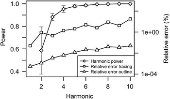
The outline coordinates were then subjected to a normalized EFA (Giardina and Kuhl Reference Giardina and Kuhl1977; Kuhl and Giardina Reference Kuhl and Giardina1982), calculating the first 10 harmonics, so that the harmonic amplitudes (excluding the first harmonic) sum up to a cumulative descriptive power of at least 99% in each individual micro-hook (Fig. 3). During the normalization, the micro-hook outlines were normalized for orientation and size (Rohlf and Archie Reference Rohlf and Archie1984), preserving the starting point of the outline (tip of the hook), because it can be considered to be mutually homologous among individual micro-hook morphotypes. The orientation was standardized according to the longest axis of the first harmonic. We also tested a normalization of orientation based on a manually measured baseline stretching from the outer process to the tip of the uncinus. This normalization introduced a higher degree of orientation variation while at the same time leading to nearly identical results, and we therefore used the normalization according to the first harmonic ellipse, because it is more objective and not influenced by human errors. For all ensuing analytical steps, both the size measurements and the results of the EFA were averaged over the two replications applied during the extraction process. The harmonic coefficients were further tested for their signal-to-noise ratio using methods described by Yezerinac et al. (Reference Yezerinac, Lougheed and Handford1992), discarding all harmonics that carry more error variation than explained variation. As expected, the relative error increases with harmonic number (Fig. 3). All raw data necessary to replicate this work and a visualization of number of outline points, smoothing, and number of harmonics have been stored on DataDryad (doi:10.5061/dryad.5q5m1).
Mean cumulative power of the first 10 harmonics (disregarding the first harmonic) for the shape reconstruction; relative error of the manual tracing of the hooks as a black-and-white form; and relative error of extracting the hook outline per harmonic (calculated according to Yezerinac et al. Reference Yezerinac, Lougheed and Handford1992, based on two replicates). Error bars for harmonic power represent the standard deviation of the cumulative power across individual hooks. Note the log-scaling of the relative error axis.

For all subsequent analyses of the EFA results, the first three coefficients of the first harmonic were excluded. We retained the fourth coefficient of the first harmonic, however, because the elongation of the hook could contain valuable shape information (Claude Reference Claude2008). We also tested the micro-hook shape for ontogenetic allometry between specimens, based on their body size, to ensure that any observed morphological differences would not be the result of a systematic change of hook shape with the age of the individual. We did this by multivariate regression of the EFA harmonics against the size parameters shell length and total body length of the three Chondroteuthis specimens (using Pillai trace as the test statistic), as suggested by Zelditch et al. (Reference Zelditch, Swiderski and Sheets2012). The same technique was used to test for a relationship between hook size and hook shape, which could also be indicative of allometric effects.
The results of the EFA were visualized using a principal components analysis (PCA; Hotelling Reference Hotelling1933) on the covariance matrix, in which theoretical mean forms of micro-hooks were reconstructed on the basis of the calculated harmonics representative for the respective centroid of squares along an equidistant grid in the plane of the first two principal components (PCs). The existence of a hypothetical grouping of morphological types was visually analyzed using a complete-linkage cluster analysis on the basis of the Euclidian distances between harmonic configurations in individual micro-hooks.
The optimal number of clusters supported by the data was subsequently determined using the gap statistic (Tibshirani et al. Reference Tibshirani, Walther and Hastie2001), as implemented in the R-package ‘cluster’ (Version 2.0.4), to determine whether the data support a subdivision into clusters and that any observed clustering is not an artifact of the clustering algorithm (Legendre and Legendre Reference Legendre and Legendre2012). As further support, the confidence for the nodes separating our chosen morphotypes were calculated by multistep–multiscale bootstrap resampling using the R-package ‘pvclust’ (Version 2.0-0; Shimodaira Reference Shimodaira2002, Reference Shimodaira2004). A canonical variate analysis (CVA; Fisher Reference Fisher1936) was used to test whether this morphological classification of the hooks can be related to specimens, that is, whether the observed clusters can be attributed to the fact that hooks among belemnite specimens are significantly different.
For data distribution, we used a Shapiro-Wilk test (Shapiro and Wilk Reference Shapiro and Wilk1965) to test for the normality of distribution and a Fligner-Killeen test (Fligner and Killeen Reference Fligner and Killeen1976) to test for homoscedasticity wherever necessary. Further characteristics of the morphotypes (size, position on the arm) were compared using a Kruskal-Wallis test (Kruskal and Wallis Reference Kruskal and Wallis1952), followed by pairwise Mann-Whitney U-tests (Mann and Whitney Reference Mann and Whitney1947), for which the p-values have been adjusted for the false discovery rate according to Benjamini and Hochberg (Reference Benjamini and Hochberg1995). The relative position of the individual micro-hook morphotypes was additionally subjected to a multinomial logistic regression, predicting the morphotype based on the distance to the mouth of the belemnite.
Results
Our material allowed the reconstruction of the arm crown in specimen 1 (Fig. 2) and a reasonable analysis of hook morphology in all three specimens. The reconstructed arm crown clearly indicates the existence of 10 arms of equal lengths in specimen 1. The hooks are arranged in a single longitudinal row (uniserial). Individual hooks can in most cases be attributed to a specific arm, allowing us to semiquantitatively analyze the distribution of hook morphotypes across the arms of the three arm crowns.
The replication of the extraction process of hook outlines reveals a high reproducibility of the parameters extracted from the images of micro-hooks. For the size measurements, the mean squares within the same micro-hook per replicate is only MSS within=0.00026, while the mean squares between micro-hooks is MSS between=0.13115. With MSS between >> MSS within and a relative error per individual of only 0.096%, the measurement error introduced by providing the image scale manually in each replicate of the outline extraction based on the scale bar in the image can therefore be neglected.
All 10 harmonics initially extracted carry more explained variation than error variation and therefore provide meaningful shape information. For the replicated outline extraction, the maximum relative measurement error per individual during the EFA was only 0.148% (median: 0.036%; 3rd quartile: 0.057%; Fig. 3), with the interindividual variation always being much larger than the intra-individual variation (p max<0.001). The error between replications was never significant (p min=0.967), and variation within replicates was always much larger than between replicates. The error introduced by manually tracing the micro-hooks was significantly larger, with a median value of 7.171% (3rd quartile: 13.36%; maximum: 33.34%; Fig. 3). However, this is still in an acceptable range (compare with Claude Reference Claude2008); the within-replicate variation was always larger than the between-replicate variation (p min=0.362), and the variation between specimens was always larger than the variation within the same specimen over the replicates (p max<0.001). We thus conclude that our data are reliable enough for the intended studies. However, we note that the relative error for the manual tracing of hooks drastically increased with the tenth harmonic (from ∼8% before to >17% in harmonic 10; Fig. 3). We therefore limited our shape analyses to the first nine harmonics extracted from the hooks, which still captures more than 98.8% of the available shape information.
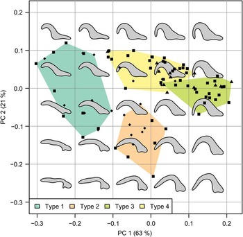
A PCA of the results of the EFA shows the existence of a large, homogenous morphospace within Chondroteuthis micro-hooks, without any immediately visible grouping (Fig. 4). The first PC explains 63.0%, and the second PC 21.4% of the observed morphological variation, so that the first PCs together already capture nearly 85% of the morphological variation of the micro-hooks. We tested the hook shape for allometry linked to a shape change of the hooks during the lifetime of individual Chondroteuthis specimens, under the assumption that larger specimens are older. We are not able to detect any systematic change of hook shape with individual age, neither when approximating age by using the shell length (p=0.389) nor when using the total body length, including the arm crown (p=0.120). We thus conclude that our data are not complicated by any ontogenetic effect within the studied specimens.
PCA of the results of an elliptic Fourier analysis of hooks using nine harmonics (excluding the first three components of the first harmonic in the ordination process). The first two axes of the PCA together explain more than 84% of the total morphological variation of the hooks. Symbols represent the individual hooks (squares: specimen 1; diamonds: specimen 2; triangles: specimen 3). The gray silhouettes represent the theoretical mean form of the centroid of the respective 0.1×0.1 square. Not all geometrically possible forms, especially the more extreme morphotypes in the lower left corner, can be found in nature. The position of the morphotypes subsequently distinguished using a cluster analysis (Fig. 5) are indicated by convex hulls including all specimens of the respective morphotype. All morphotypes occupy clearly different areas of the morphospace, with only a small overlap between morphotypes 3 and 4.

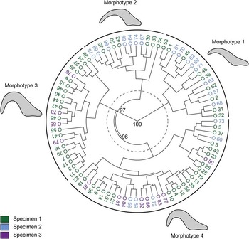
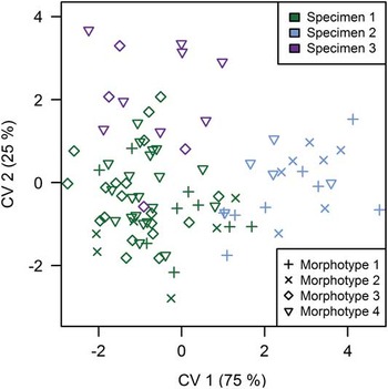
The lack of visible groups in a PCA does not necessarily imply the nonexistence of such morphological groups, since a PCA spreads data along the axes of greatest variation rather than those of strongest separation. A cluster analysis (complete linkage) of the EFA results using Euclidian distances as similarity index implies the possible existence of four morphotypes at a homogenous cutoff value of 0.3 (Fig. 5). The gap statistic supports this by calculating the optimal number of clusters within the data to be four (999 replications). We further note that when the four identified clusters are projected into the PCA space (Fig. 4), they occupy different parts of the morphospace, with only morphotypes 3 and 4 showing a minor overlap. Both are indicators that our shape data can indeed be clustered into these morphotypes and that the observed clusters are not an artifact of the clustering algorithm. To further validate the results, we calculated a CVA using the identity of the belemnite specimen that contained the respective hook as grouping factor (Fig. 6). Should such an analysis show a significant difference between hooks of the different specimens, this would mean that hook shape is already too variable across individuals for our analysis to be reliable. We found no significant difference between hooks of the different specimens (p=0.150), indicating that the distinguished morphotypes described here do not result from inherently different hook shapes among the investigated specimens.
Cluster analysis (complete linkage) of results of an elliptic Fourier analysis of hooks using Euclidian distances. At a homogenous distance level of 0.3 (indicated by the gray, dashed line) the existence of four different morphotypes is implied; these are indicated by the solid black lines in the outer ring. The approximately unbiased confidence (Shimodaira Reference Shimodaira2002, Reference Shimodaira2004) of the nodes delineating those clusters is indicated by numbers. The gray silhouettes represent the reconstructed mean shape of hooks in each cluster. Morphotype 1 has a long base and a small, slightly curved uncinus. Morphotype 2 has a narrow base and a nearly horizontal uncinus with very little curvature. Morphotype 3 is characterized by a strong curvature of the uncinus, the tip of which nearly reaches base level, and a narrow base. Morphotype 4 has a broader base than morphotype 3 and a medium curvature of the uncinus, the tip of which does not reach the base level. All morphotypes are well distributed across the three specimens investigated (indicated by the color of the specimen label).

CVA of an elliptic Fourier analysis of belemnite hooks using the three investigated belemnite specimens as grouping factor. The CVA is insignificant at p=0.150, and the different specimens show considerable overlap in the CVA space, with no clear groups emerging.

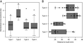
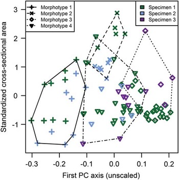
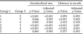
The morphotypes, as defined on the basis of the cluster analysis, were also tested with respect to other intermorphotype differences. For the size of the micro-hooks, we cannot assume that the data in all groups are normally distributed (p min=0.001), although the variances are equal (p=0.120). We therefore applied the Kruskal-Wallis test for the extracted size parameter, which revealed that not all micro-hook morphotypes are of the same size (χ2=26.935, df=3, p<0.001). A pairwise Mann-Whitney U-test (Table 2) shows that hooks of morphotype 2 are significantly larger than those of any other morphotype, while the three other morphotypes do not differ significantly in size (Fig. 7A). Plotting the individual hook sizes against the unscaled first axis of the PCA as a shape parameter (Fig. 8) further shows that this size difference is not linked to morphology or specimens but that hooks of morphotype 2 are consistently larger than those belonging to any other morphotype.
Box plots depicting size differences and differences in the distance to the belemnite mouth within four different hook morphotypes supported by a cluster analysis (Fig. 5). The assignment of morphotypes to particular groups, supported by a pairwise Mann-Whitney U-test, is indicated by lowercase letters on the boxes (compare with Table 2). A, Size data have been standardized per belemnite specimen to eliminate ontogenetic influences on hook size. The width of the boxes is proportional to the square root of the number of within-group observations. Thick lines indicate the median, boxes cover the interquartile range (IQR), whiskers extend to 1.5 × IQR, outliers are plotted as black dots. Hooks of morphotype 2 are significantly larger than those of any other morphotypes. B, Distance data were extracted from specimen 1. The width of the boxes is proportional to the square root of the number of within-group observations. Thick lines indicate the median, boxes cover the interquartile range, whiskers extend over the full range of the data, original data points have been plotted as black diamonds. The order of hook morphotypes along the belemnite arm from mouth to tip is 1+2 → 3+4.

Cross-plot of standardized hook size against the score along the first PC axis as shape descriptor. The size of the hooks does not systematically change with shape or across specimens. Rather, hooks of morphotype 2 are larger than those of any other morphotype. Convex hulls including all hooks of the same morphotype are indicated. Note that the overlap between morphotype 2 and morphotypes 3 and 4 is mainly caused by a few outliers.

Results of a pairwise Mann-Whitney U-test concerning standardized hook size and distance of the hooks to the supposed mouth of the belemnite between the respective morphotypes listed in the group 1 and 2 columns (compare with Fig. 5). Both the original p-values and the p-values adjusted for the false discovery rate (Benjamini and Hochberg Reference Benjamini and Hochberg1995) are listed. Size values are based on the whole data set, and the size difference of morphotype 2 compared with all other morphotypes is significant (Fig. 7A). Values for the distance to the mouth are based on specimen 1 only, and morphotypes 1+2 and 3+4 differ significantly in their distance to the mouth of the belemnite (Fig. 7B).

For the distance to the mouth for the individual micro-hooks, we again found the data deviated from normal distribution (p min=0.043), although they showed equal variances (p=0.698). We tested the distances of the micro-hook morphotypes to the presumed mouth of the diplobelid animal using a Kruskal-Wallis test. The test confirms that the group-dependent distances differ at χ2=20.407, df=3, p<0.001. The ensuing pairwise Mann-Whitney U-test (Table 2) shows there are two morphological groups of hooks that occur at different distances from the mouth of the diplobelid animal (Fig. 7B). A multinomial logistic regression analysis (significant at p<0.001, with a McFadden R 2=0.209) confirms that observation. Both morphotypes 3 (p=0.007) and 4 (p=0.001) occur based on the distance of the micro-hook to the mouth of the diplobelid (compared with morphotype 1 as base level). Morphotype 2, however, shows no such dependence (p=0.692). This confirms the results of the Mann-Whitney U-test and indicates that the distance to the mouth does have predictive (and thus interpretational) value for the expected hook morphotype.
An investigation of the distribution of different hook morphotypes across the arms of specimen 1 (Fig. 2) reveals that morphotype 2 seems to be restricted to four arms.
Discussion
The results of our analyses of the micro-hook composition and distribution in Chondroteuthis provided new insights into the paleobiology of this taxon. First, we provide for the first time direct evidence for the existence of 10 arms of equal length in Chondroteuthis. Second, we observed that hooks in Chondroteuthis are arranged in a single row, which is in contrast to the biserial arrangement in all other known belemnoid taxa with articulated arm crowns (e.g., compare with Engeser and Clarke Reference Engeser and Clarke1988; Fuchs et al. Reference Fuchs, Boletzky and Tischlinger2010, Reference Fuchs, Donovan and Keupp2013a,Reference Fuchs, Heyng and Keuppb). According to the uniserial arrangement of hooks and the assumed maximum number of hooks per arms (i.e., about 20–40), in Chondroteuthis the maximum number of hooks per arm crown ranges between 200 and 400 micro-hooks. None of the analyzed hooks shows a spur (Fig. 1), which is assumed to occur exclusively in true belemnites (Engeser Reference Engeser1987). Therefore, our analyses confirm the exclusion of Chondroteuthis from the order Belemnitida. Some of the recognized hook shapes are unknown among belemnoid orthotaxa; however, these shapes resemble those of established hook parataxa. Third, our analyses provide evidence for the existence of four different micro-hook morphotypes within individuals of Chondroteuthis based on a cluster analysis with associated validity tests (Legendre and Legendre Reference Legendre and Legendre2012). However, it remains highly speculative whether the observed hook differentiation in Chondroteuthis compensates for a reduced (uniserial instead of biserial arrangement) hook number. We must further admit that a multivariate regression between hook shape and hook size is significant (p<0.001). This leaves the possibility that the observed differences in shape could be the result of allometry of individual hooks within the same individual during growth, assuming that hooks are built consecutively and are thus not in the same ontogenetic stage within the individual. We note that it is not possible to be certain about either of these interpretations. However, the distribution of hook morphotypes along the arm (with co-occurrence of different morphotypes and their restriction to certain parts of the arm) seems more indicative of a functional separation than an allometric growth trend, comparable to the functionally adapted distribution of teeth in the jaws of mammals.
Hook Morphotypes 1–4 and Their Positions in the Arm Crown.—Using the best-preserved Chondroteuthis arm crown of specimen 1 (Fig. 2; for additional specimens see Supplementary Fig. 3), we can gain important insights into the distribution of the hook morphotypes along individual and across different arms.
Our study suggests that the four distinguished hook morphotypes are not occurring in a strict, monotypic sequence along the arms, as assumed by Engeser (Reference Engeser1987). In contrast, morphotypes 1, 2, and 4 all occur in proximal and median positions and thus show considerable overlap in their distribution along the arms. Only morphotype 3 occurs exclusively in the distal position, but morphotype 4 can also be present in this position. A clear trend of size increase toward the midarm and a subsequent size decrease of a distinct micro-hook type could not be observed. However, the largest micro-hooks (morphotype 2) occur only in proximal to median positions and are restricted to four arms in the middle of the arm crown. While rows of morphotype 1 are thus arranged next to the medium-sized morphotypes 3 and 4 on the same arms, hooks of morphotype 2 show that not all hook morphotypes are homogenously distributed across the arm crown. This type of micro-hook distribution, different hook morphotypes on different arms at similar distance to the mouth, is reported here for the first time and indicates an arm-wise functional differentiation according to the specific hook morphotype.
Hook Morphotypes 1–4 in Comparison with Engeser (1987) and Hook Parataxa.—Kulicki and Szaniawski (Reference Kulicki and Szaniawski1972) established a parataxonomy for micro-hooks that is still widely used but based on partly subjective or hard to define criteria. Fitting our morphotypes within the framework of this artificial system can provide further insight into its validity and applicability. With its long base, morphotype 1 is probably equivalent to the hook of Chondroteuthis wunnenbergi figured by Engeser (Reference Engeser1987: Fig. 1A), which is similar to the paragenus Paraglycerites. Morphotype 2 is the largest and is characterized by a long, nearly straight to slightly curved uncinus, sometimes with a small hump at the outer margin of the shaft–uncinus boundary. That hook morphology represents a new, so far unnamed, morphotype, which is similar to the hook figured by Engeser (Reference Engeser1987: Fig. 1C). Morphotype 3 has a strongly curved uncinus, with its tip reaching the baseline, that is, the horizontal extension of the base. This morphotype is closest to Engeser (Reference Engeser1987: Fig. 1D) and is referred to the paragenus Arites. Morphotype 4 resembles morphotype 3 but does not show such a strong curvature of the uncinus and has a broader base. Because uncinus curvature was recognized as highly variable in some hooks (e.g., Lehmann et al. [Reference Lehmann, Solarczyk and Friedrich2012] for Arites), morphotype 4 should also be referred to Arites but may belong to a different paraspecies. Although we did not reach the anticipated goal of a fully established morphotypical distinction of all different hook types, we can state that the currently applied parataxonomic system is too rough to distinguish between objectively existing morphotypes and does not capture the entire morphospace of micro-hooks. We thus advocate deprecating this practice in favor of more neutral terminology complemented by a more robust typological concept of hook morphology in the future.
Hook Armament of Chondroteuthis Compared with Other Belemnoid Orthotaxa.—The presence of different hook types within the same specimen appears to be rare among belemnoid coleoids. Genera such as Acanthoteuthis, Belemnoteuthis, Clarkeiteuthis, Phragmoteuthis, Uncinoteuthis, and Winkleriteuthis are typified by low intra- and interindividual variation of the hook morphology. A set of various hook morphotypes within an individual arm crown has so far been reported by Reitner and Urlichs (Reference Reitner and Urlichs1983) for Acrocoelites, by Reitner (Reference Reitner1986) for Acanthoteuthis, and by Garassino and Donovan (Reference Garassino and Donovan2000). The lower Sinemurian Ostenoteuthis siroi from northern Italy has a pronounced intra-individual change in hook morphology, while the co-occurring Uncinoteuthis is characterized by arms equipped with uniform hooks (Garassino and Donovan Reference Garassino and Donovan2000). Surprisingly, Garassino and Donovan (Reference Garassino and Donovan2000) also reported four different hook morphotypes based on line drawings for Ostenoteuthis. Without a detailed comparison of the Ostenoteuthis material, it is difficult to judge whether both genera, Ostenoteuthis and Chondroteuthis, possessed similar hook morphotypes or not. Regardless, a fundamental difference is established by the uniserial hook arrangment in Chondroteuthis. A functional interpretation of hook morphotypes and their distribution was not provided by Garassino and Donovan (Reference Garassino and Donovan2000), but a functional differentiation of the arms was assumed by Fuchs (Reference Fuchs2006).
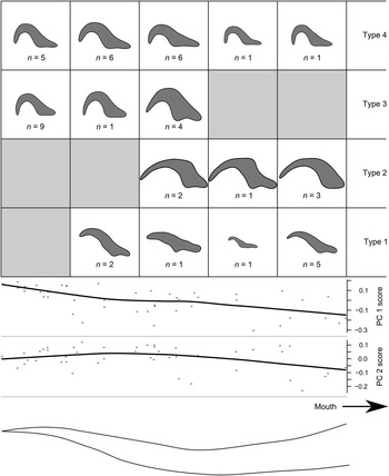
Based on our simplified model for Chondroteuthis, which shows the development of hook morphology and size from the proximal to the distal part of the arm (Fig. 9), we will thus try to infer the potential function of the four different hook types and the hook-bearing arms.
Reconstruction of hook morphology of the four morphotypes identified by a cluster analysis (Fig. 5) within five intervals of equal length along the arm of a belemnite (indicated at the bottom). The reconstructions are based on the mean form of hooks of any particular morphotype found in the respective interval in specimen 1. The number (n) of hooks averaged for each reconstruction is indicated. The hooks are scaled relative to one another to show size differences; scaling was performed according to a log10(x) scale to enhance visibility. The scores along the first two components of a PCA of the elliptic Fourier coefficients used as shape descriptors are plotted at the bottom (lines based on local polynomial regression fitting (LOESS) smoothing). Higher values within both scores toward the end of the arm coincide with the distribution of morphotypes in the PCA space (Fig. 4): morphotypes 1 and 2 predominate close to the mouth and reduce the score along PC 1 and PC 2, respectively, at that position along the arm.

Speculations on Functional and Ecological Adaptations of Belemnoid Hooks.—When compared with the best-fitting functional analogous structures, namely the hooks and suckers of extant coleoids, it is important to note that these structures are most likely not homologous to belemnoid hooks (Engeser and Clarke Reference Engeser and Clarke1988; Young et al. Reference Young, Vecchione and Donovan1998; Fuchs et al. Reference Fuchs, Boletzky and Tischlinger2010, Reference Fuchs, Donovan and Keupp2013a,Reference Fuchs, Heyng and Keuppb). Ordinary hooks aid in latching onto prey items during the initial strike. We assume that the hooks in belemnoids, analogous to some types of suckers, were not stalked (sessile) but could be moved, which would allow a generally increased flexibility in arm movement.
Due to the similar function of hooks and suckers, that is, to grab and hold prey, both are primarily hunting weapons. It is interesting to note that tiny hooklike (<5 mm) structures can be found in the suckers of oegopsid Decabrachians (Nixon Reference Nixon2011), in which tentacular sucker rings can bear relatively large hooks (>5 mm). Those hooks are positioned curving inward, with the tip pointing in the direction of desired prey movement toward the mouth. The sucker size distribution in modern coleoids is similar to the belemnoid hook size distribution, with smaller suckers around the mouth (due to less space?), the largest suckers in the center, and smaller suckers at the distal end of the arms. The size reduction toward the distal end could be because the arms thin out and thus less space is available, or because maximum power cannot be exerted distally, or because large distal hooks would increase the risk of losing arms.
Nixon and Dilly (Reference Nixon and Dilly1977) postulated that the differences in diameter, size, and shape of sucker components depend on physiology and behavior. We assume that belemnoid hooks, due to their comparable function, can also be separated into functional groups. This allows us to compare our results with observations from other taxa to arrive at an integrated functional interpretation of hook armament in Chondroteuthis.
Mattheck and Reuss (Reference Mattheck and Reuss1991) discussed the nearly perfect adaptation of vertebrate claws to the logarithmic spiral, in which no localized stress peaks occur, and suggested that the logarithmic spiral is a highly shape-optimized design for short hooks leading to a nearly constant stress state at the surface of the claw (first mentioned by Thompson [Reference Thompson1917]). The main advantage of a spirally shaped hook seems to be that the hook can penetrate the surface of the prey at a constant angle, avoiding lateral wiggling, while maintaining a constant force direction (Hammer et al. Reference Hammer, Hryniewicz, Hurum, Hoyberget, Knudsen and Nakrem2013). If the prey tries to escape distally, it will only cause the hook to dig in farther. Therefore, it is interesting to note that not all Chondroteuthis hooks (i.e., morphotypes 1 and 2) match a logarithmic spiral. This could result from a relaxation of physical constraints, because belemnoid hooks are not attached to a solid base as in vertebrates, but could also mean that not all Chondroteuthis hooks are designed for the same function.
It seems likely that hooks with a broader base were fixed within the arm tissue more securely due to a greater area of attachment. This is based on the assumption that the base provided a “handle” for the muscles to hold. “Perhaps where the handle was long it gave some fulcrum for leverage and when it was laterally expanded it provided a means for the muscles to move the hook laterally” (Engeser and Clarke Reference Engeser and Clarke1988: p. 140).
For an extended discussion of similar hook-shaped microstructures and their functions, the reader is referred to Murdock et al. (Reference Murdock, Sansom and Donoghue2013) for conodonts, to Kasatkina (Reference Kasatkina1982) and Bieri (Reference Bieri1983) for the spines and teeth of chaetognaths, and to Leatham (Reference Leatham1985) and Merz and Woodin (Reference Merz and Woodin2006) for the chitinous trematode haptors and polychaetae chaetae.
Toward a Functional Model of Chondroteuthis Hook Armament.—Morphotypes 3 and 4 show stronger curvature of the uncinus than morphotypes 1 and 2 and occur at the distal end of the arm (though morphotype 4 also occurs less abundantly in proximal positions). A stronger curvature would conceivably be better suited to catch prey, limiting possibilities of escape. Micro-hooks of morphotype 4 have a broader base, indicating that they are more securely fixed within the tissue, but are otherwise similar in morphology to morphotype 3. It seems likely that morphotype 3 was mainly used for first grasping the prey, allowing for easy release by means of ripping some hooks out of the arm tissue in cases in which the prey was too strong and thus a threat to the predator. If that was not the case, the coleoid animal could then proceed by further entangling the prey using hooks of morphotypes 3 and 4, which would then allow it to hold and fix the prey with the arms. It might also be that hooks of morphotype 3 were designed to be more flexible than those of morphotype 4, allowing a better entangling of the prey item. While not as securely fixed within the arm tissue as hooks of morphotype 4, they could act in combination with those (both morphotypes occur at the same positions along the arm) to deliver the optimal combination of secure yet rigid and highly flexible but loose grasp.
Morphotypes 1 and 2 occur closer to the mouth, and it is therefore likely that they participated in dissecting and transporting the prey toward the mouth. The large size makes hooks of morphotype 2 more likely to either completely surround and fix smaller prey items or to be rammed into the tissue of larger prey and hold it during a first attack. The lack of morphotype 2 in specimen 3 (smallest or youngest, subadult specimen examined) could represent a preservational artifact, or indicate a shift in prey selection during ontogeny, with differently shaped hooks in use. It could also indicate that this specimen represents a female, since such large hooks may have played a role during mating, with males holding females with such large hooks, as was discussed for fossil finds of Passaloteuthis (Stevens Reference Stevens2010 and references therein), or in male-to-male agonistic behavior (as is known from modern coleoids; see Jackson and O’Shea Reference Jackson and O’Shea2003). In modern squids large hooks are developed as a function of maturity, and their presence only in males suggests that their principal role is reproductive rather than predatory, so it seems likely that fossil mega-hooks fulfilled a similar role (Stevens Reference Stevens2010).
Conclusion
We analyzed the arm hooks of three specimens of Chondroteuthis (belemnoid) with respect to their morphology and distribution along and across different arms.
The application of elliptic Fourier shape analysis for the quantification of belemnite hook morphology successfully demonstrates the presence of four distinct morphotypes within the arm crown of the diplobelid coleoid Chondroteuthis wunnenbergi. Our data support a pronounced intra-individual morphological variation of micro-hooks, which can be interpreted on grounds of functional adaptation. We recommend applying similar techniques to other belemnoid taxa, and we advise against the parataxonomic approach to hook-shape description, because it appears to be both too rigid and subjective to capture the entire possible morphospace.
Acknowledgments
R.H. acknowledges K. Wiedenroth (Hanover), who brought two Chondroteuthis wunnenbergi arm crowns, deposited at the Bundesanstalt für Geowissenschaften und Rohstoffe (BGR) in Hanover, Germany, to our attention. R. Lemanis and K. Stevens contributed to the discussion about form and function of hook-shaped structures. We thank C. Klug and one anonymous reviewer for helpful comments on the manuscript. Further, we acknowledge the editor, S. Xiao, and the associate editor, J. Crampton.
Supplementary Material
Data available from the Dryad Digital Repository: https://doi.org/10.5061/dryad.5q5m1