INTRODUCTION
Mesocestodes corti possesses particular characteristics such as the requirement for three different hosts in its life cycle (Soldatova, Reference Soldatova1944; Etges and Marinakis, Reference Etges and Marinakis1991; Loos-Frank, Reference Loos-Frank1991; Rausch, Reference Rausch, Khalil, Jones and Bray1994), asexual reproduction of the tetrathyridial (larval) form by longitudinal fission in both laboratory isolates as well as in isolates from domestic dogs and coyotes (Crosbie et al. Reference Crosbie, Nadler, Platzer, Kerner, Mariaux and Boyce2000) and development from larva to adult worm in appropriate culture conditions (Barrett et al. Reference Barrett, Smyth and Ong1982; Thompson et al. Reference Thompson, Sue and Buckley1982). This last property led to the use of this parasite as an experimental model system for studying cestode development, host-parasite relationships and antihelminthic drug activity in some other human disease-causing cestodes, such as Echinococcus spp. and Taenia spp. (Thompson et al. Reference Thompson, Sue and Buckley1982; Ong and Smyth, Reference Ong and Smyth1986; Siles-Lucas and Hemphill, Reference Siles-Lucas and Hemphill2002). The effect of drugs on the developing stages of M. corti has also been investigated (Britos et al. Reference Britos, Dominguez, Ehrlich and Marin2000; Saldaña et al. Reference Saldaña, Marin, Fernández and Dominguez2001; Markosky et al. Reference Markoski, Trindade, Cabrera, Laschuk, Galanti, Zaha, Nader and Ferreira2006).
Our previous studies have focused on defining cellular and molecular aspects that are involved in the process of M. corti differentiation from larva to adult worm induced by trypsin in vitro. It was found that this protease, present in the intestinal juice of the vertebrate hosts, makes the larvae permeable to growth factors, inducing dynamic reorganization of the tegument, activation of cell proliferation and induction of differentiation to adult worms in vitro (Espinoza et al. Reference Espinoza, Galindo, Bizarro, Ferreira, Zaha and Galanti2005). During this process two waves of DNA synthesis were identified that are tightly associated with two waves of transcription of the parasite histone H4 gene and with the acquisition of the phenotypic characteristics of each stage of development from larva to adult (Espinoza et al. Reference Espinoza, Gómez, Galindo and Galanti2007). On the other hand, the presence, activity, localization and molecular forms of cholinesterases in the nervous system of M. corti were assessed in its different developmental stages and related to the achievement of mature forms of the parasite (Kemmerling et al. Reference Kemmerling, Cabrera, Campos, Inestrosa and Galanti2006).
In this paper, we describe morphological features of developmental stages of M. corti obtained in vitro after trypsin and serum stimulation of tetrathyridia, showing unambiguously that sexually mature worms are obtained as a result of this process of differentiation. As with the protoscolex of Echinococcus granulosus (Galindo et al. Reference Galindo, Schadebrodt and Galanti2008), cellular territories can be identified in the M. corti larvae which are maintained throughout the process of development to adult worm. Additionally, we report the presence and distribution of neutral polysaccharides in M. corti larva encapsulated in the mouse liver and in free larvae, as well as glycogen mobilization in different stages of larval development to adult worm. We propose that degradation of polysaccharides in the transition from segmented to adult worm may be related to energy needs necessary for the cellular processes leading to the mature specimen.
MATERIALS AND METHODS
Parasite maintenance
Female Balb/c mice (3 months old) were infected with 100 μl of fresh Mesocestoides corti tetrathyridia in PBS solution by intraperitoneal inoculation. After 3 months, the animals were killed by cervical dislocation after chloroform anaesthesia and the parasites were recovered from the peritoneal cavity using standard aseptic techniques. Tetrathyridia were sedimented by gravity and washed twice with sterile phosphate buffered saline (PBS) pH 7·2 and supplemented with penicillin (75 U/ml) and streptomycin (75 μg/ml).
Induction of development of tetrathyridia to adult worms
Tetrathyridia were incubated in RPMI medium 1640 culture medium (GIBCO BRL, Life Technologies, Grand Island, NY) supplemented with 20% inactivated foetal bovine serum (FBS), 75 U/ml penicillin and 75 ug/ml streptomycin containing 1×10⁵ BAEE (N-benzoil-L-arginine ethyl ester) units of trypsin for 24 hr at 37° C under 5% CO2 partial pressure. The trypsin-containing medium was then substituted with the same medium without trypsin. The medium was changed every 2 days to avoid acidification.
Following previous protocols (Espinoza, Reference Espinoza2002; Espinoza et al. Reference Espinoza, Galindo, Bizarro, Ferreira, Zaha and Galanti2005), tetrathyridia treated for 24 h with trypsin were considered as stimulated larvae while parasites between 1 to 4 days post-treatment with trypsin were taken as elongated non-segmented larvae. From day 5 to day 9 after trypsin induction of development, the process of segmentation is evident but reproductive apparatus is lacking; these parasites were considered segmented. Finally, parasites from day 10 after trypsin induction were considered adult worms (Kemmerling et al. Reference Kemmerling, Cabrera, Campos, Inestrosa and Galanti2006).
Histological techniques
Tetrathyridia obtained from the peritoneum of mice, larvae stimulated to differentiate with trypsin, elongated larvae, segmented worms, adult worms and livers from infected mice were fixed in 1% (w/v) paraformaldehyde in PBS pH 7·2 at 4° C for 24 h, and then embedded in paraffin (all reagents from Merck KGaA, Darmstadt, Germany). Five μm thick sections were stained with hematoxylin and eosin following standard procedures.
Identification and localization of glycogen
Tetrathyridia obtained from mice and different developmental stages of the parasite after trypsin and serum stimulation of larvae were fixed and sectioned as indicated above. For detection of neutral polysaccharides, sections 5 μm thick were treated with the periodic acid-Shiff reagent (PAS) (McManus, Reference McManus1946) and counter-stained with hematoxylin following standard procedures. Polysaccharides were defined by a red-violet colour. As a control of glycogen detection, 5 μm thick sections were incubated with a 4 μg/ml solution of α-amylase (Nutritional Biochemical Corporation) in PBS pH 6·0, for 30 minutes or for 2 hours at 37°C prior to the PAS-hematoxylin reaction. A decrease in the intensity of the PAS colour reaction after enzyme treatment was considered as evidence of the presence of α1-4 neutral polysaccharide (mainly glycogen) (Kiernan, Reference Kiernan1981). The samples were analyzed with a Nikon Optiphot microscope; the images were taken with a Kodak Digital Science DC 120 camera and processed with Corel Draw 11 software.
Arteta's histochemical reaction
Tetrathyridia, as well as segmented and adult worms, were fixed in alcoholic Bouin and embedded in paraffin. Sections of 5 μm were sequentially stained with Harris haematoxylin and erythrosine-Orange G. After treatment with phosphotungstic acid, slices were stained with aniline blue. Collagen from the basal lamina is identified by a blue colour (Humason, Reference Humason1979; Galindo et al. Reference Galindo, Schadebrodt and Galanti2008).
RESULTS
Morphological features of different stages of differentiation of M. corti from larvae to mature worms
Fig. 1 shows in vivo images of different forms of the parasite as observed with an inverted microscope. In 1A a larva obtained from the mouse peritoneal cavity is shown. Suckers (S), tegument (T) and parenchyma (P) are evident. In 1B, a larva in a process of asexual reproduction by longitudinal fission is shown. Fig. 1C shows a segmented worm 7 days after trypsin stimulation where strobila invaginations (St) and genital pores (G) are evident. In Fig. 1D an adult worm obtained from day 10 on after trypsin stimulation is shown, with all morphological features typical of this final stage of development, particularly proglottids (Pg). In the same worm, an oncosphere (Fig. 1E, O) and cirrus (Fig. 1F, C) are shown as evidence of parasite maturity.
In vivo images of M. corti observed with an inverted microscope. A and B: tetrathyridia obtained from the abdominal cavity of an infected mouse. In B asexual reproduction by longitudinal fission; C: Segmented worm obtained 7 days after trypsin and serum in vitro stimulation of tetrathyridia; D: Adult, mature worm; E: Proglottid of an adult worm showing an oncosphere (O); F: A cirrus (Ci) from an adult worm. Bars: 100 μm. S: suckers; T: tegument; P: parenchyma; G: genital pore; St: strobilar invaginations; Pg: proglottids.

Fig. 2 A shows a longitudinal section of a tetrathyridium obtained from the mouse peritoneal cavity. Anterior (AR), medial (MR) and posterior (PR) regions are shown in brackets. Tegument (T) is well defined in the three regions (Figs. 2A to 2D). In Fig. 2B the anterior region is shown amplified, showing a high cellular concentration as compared with the other regions. Suckers (S) are well defined as cellular regions separated from other structures by a basal lamina (BL). A cellular region anterior to the suckers, named the apical massif (AM) (Hess, Reference Hess1980) is shown. Another cellular region below the suckers corresponds to the cerebral ganglia (CG). Though clearly defined, the apical massif and the cerebral ganglia are not surrounded by a basal lamina. Nerve cords (NC) that originate in the cerebral ganglion (CG, 2B) are observed all along the larva (2A to 2D). The cellular concentration in the parenchyma diminishes from the AR to the MR being still lower in the PR.
Tetrathyridium obtained from the mouse peritoneal cavity. A: Longitudinal section of the whole larva; B: Anterior region; C: Medial region; D: Posterior region. AR: anterior region; MR: medial region; PR: posterior region; S: suckers; BL: basal lamina; AM: apical massif; CG: cerebral ganglium; T: tegument; NC: nerve cords. Bars: 100 μm.

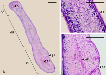
Fig. 3A shows a longitudinal section of a tetrathyridium 24 hours after stimulation with trypsin and serum. The same regions as described for tetrathyridia obtained from the mouse peritoneal cavity are shown. The excretory pore (EP) is evident in the posterior region (PR). In some areas of the larva, particularly in the anterior region (Fig. 3B, arrows) a ‘hairy’ appearance of the tegument (T) corresponds to alterations in the structure of the tegument produced by trypsin. Acidophilic fibres (AF) probably corresponding to collagen or smooth muscle fibres are present in the parenchyma (Fig. 3A and 3C); these structures were not found in non-stimulated tetrathyridia. In Fig. 3C the excretory pore (EP) and channel are shown together with the acidophilic fibres (AF). Parenchyma is vacuolized (Fig. 3B) and shows less contrast to the hematoxylin-eosine staining than tetrathyridia obtained from the mouse peritoneal cavity.
Tetrathyridium 24 h after stimulation with trypsin and serum. A: Longitudinal section of the whole parasite; B: Tegument showing erosions produced by trypsin (arrow); C: An area of the posterior region. AR: anterior region; MR: medial region; PR: posterior region. S: Suckers; AF: acidophilic fibres; EP: excretory pore; T: tegument. Bars: 100 μm.

Fig. 4A shows the longitudinal section of an elongated larva 4 days after trypsin stimulation. Clusters of cells are evident in the parenchyma probably related to the differentiation processes leading to the formation of genital pores (GP, Fig. 4B). The tegument is well defined showing regeneration after the initial trypsin effect.
Elongated M. corti larvae 4 days after trypsin and serum stimulation. A: Longitudinal section of a whole parasite. B: Medial region. C: Posterior region. S: suckers; GP: genital pore; NC: nerve cords; T: tegument. Bars: 100 μm.

Fig. 5A shows the longitudinal section of a worm showing full segmentation (St). Testis are present (Te) in this stage (Fig. 5B) as well as rudimentary female genitalia (RFG) (Fig. 5C). In Fig. 6A the section of a fully developed, mature worm is shown. Proglottids (Pg) derived from tegument invaginations are present. In Fig. 6B ovaries (Ov), a sigmoid region of the uterus (SigU), testis (Te) and a genital pore (GP) are shown. The parauterine organ (PUO) with ovaries (Ov) is also shown (Fig. 6C).
Segmented worm 7 days after trypsn and serum stimulation. A: Full longitudinal section of a whole parasite. B and C: Medial region. S. suckers; St: strobilar invaginations; RFG: rudimentary female genitals; Te: testis; NC: nerve cords. Bars: 100 μm.

Adult worm 10 days after trypsin and serum stimulation. A: Full longitudinal section of a whole parasite. B and C: Medial region. S: suckers; Pg: proglottids; Ov: ovary; Te: testis; GP: genital pore; PUO: parauterine organ; SigU: uterus sigmoid region; NC: nerve cords. Bars: 100 μm.

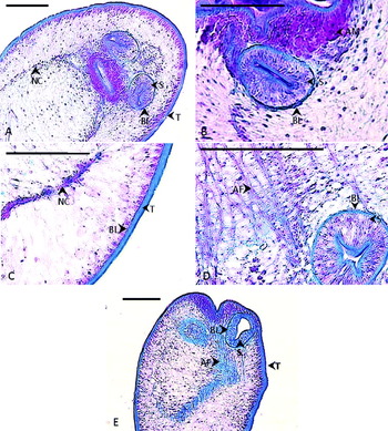
The basal lamina was identified surrounding the suckers and all along the internal side of the tegument (Fig. 7A to E). Other structures from the larva, such as apical massif and cerebral ganglia, do not present basal lamina. The tegument and acidophilic fibres (AF) are stained with the Arteta staining (Fig. 7D and E), indicating presence of collagen in these structures.
Basal lamina in tetrathyridia obtained from the mouse peritoneal cavity (A, B and C) and from segmented (D) or adult (E) worms. S: suckers; BL: basal lamina; AM: apical massif; T: tegument; AF: acidophilic fibres; NC: nerve cords. Bars: 100 μm.

Presence and mobilization of glycogen during larva to adult worm development
Fig. 8 shows the detection of neutral polysaccharides in histological sections of a liver-encapsulated tetrathyridium (A, frontal view) and in a tetrathyridium obtained from the peritoneum of infected mice (B, frontal view). In C, a longitudinal section of a tetrathyridium is shown. Neutral polysaccharides detected by the periodic-Schiff (PAS) reaction are concentrated in the cephalic region of the parasite parenchyma, principally among the nerve cords (NC), the tegument (T) and the suckers (S). No evident differences in glycogen location or mobilization were observed between encapsulated and free tetrathiridia.
Neutral polysaccharides (violet red) visualized by PAS-hematoxylin in A: Histological section (frontal view) of a liver encapsulated tetrathyridium; B: Tetrathyridium obtained from the peritoneum of infected mice (frontal view); C: Tetrathyridium obtained from the peritoneum of infected mice (longitudinal view); D: Tetrathyridium stimulated to differentiate with trypsin and serum for 24 h; E: Tetrathyridium one day after stimulation to differentiate with trypsin and serum; F: Elongated worm four days after stimulation to differentiate with trypsin and serum; G: Segmented worm; H: Mature, fully differentiated worm. P: parenchyma; NC: nervous cords, S: suckers, T: tegument. Bars: 100 μm.

After 24 h of trypsin stimulation to differentiate, neutral polysaccharides in the larvae change drastically to a homogeneous distribution in the entire parasite parenchyma (Fig. 8D). However, an important concentration of polysaccharides is still evident along the nerve cords (NC) and suckers (S).
In larvae at day 1 after trypsin stimulation (Fig. 8E), neutral polysaccharides remain distributed throughout the whole parenchyma as well as concentrated in cells surrounding the nerve cords (NC) and suckers (S). Elongated worms (Fig. 8F) show a clear increase in the staining of neutral polysaccharides, maintaining the distribution observed in the previous stage of development. Segmented worms (Fig. 8G) show a high concentration of neutral polysaccharides all along the parasite body while remaining concentrated in the basal lamina defining the suckers (S) and in nerve cords (NC). In Fig. 8H a fully mature worm is shown after the PAS reaction. Clearly, neutral polysaccharide concentration as defined by the intensity of the PAS reaction shows a sharp decrease though these macromolecules are still concentrated mostly at the level of nerve cords (NC).
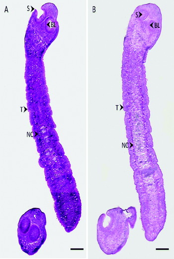
Specificity for the detection of neutral polysaccharides was achieved by treatment of the larvae and different stages of worm development with α-amylase previous to the PAS staining. As an example, Fig. 9A shows a segmented worm after PAS staining. Fig. 9B shows a serial section of the same worm that was treated for 2 h with α-amylase before PAS staining. A clear decrease in the reactivity to the PAS reagent after the enzyme incubation is observed (compare Fig. 9 A and B), confirming the specificity of the reaction for the detection of neutral polysaccharides.
Specificity of the PAS reaction. A: Neutral polysaccharides visualized by PAS-hematoxylin in M. corti segmented worms. B: The same segmented worms treated with α-amylase for 30 minutes previous PAS-hematoxylin staining. NC: nervous cords; S: suckers; T: tegument; NC: nerve cords; BL: basal lamina Bars: 100 μm.

DISCUSSION
Mesocestoides corti was described from the intestine of mice (Hoeppli, Reference Hoeppli1925) and its tetrathyridium form from the body cavity of lizards and small mammals (Specht and Voge, Reference Specht and Voge1965). In addition, it was reported that tetrathyridia were able to multiply asexually in the peritoneal cavity of mice and could be maintained indefinitely by serial passage in these animals (Specht and Voge, Reference Specht and Voge1965; Hart, Reference Hart1968). Furthermore, it was reported that different proteases had the capacity to induce cestode development (Berntzen and Mueller, Reference Berntzen and Mueller1964; Smyth and Davies, Reference Smyth and Davies1974; Esch and Smyth, Reference Esch and Smyth1976). Later, it was described that incubation of M. corti tetrathyridia with trypsin induced the development of these larval forms to adult worms (Kawamoto et al. Reference Kawamoto, Fujioka and Kumada1986a, Reference Kawamoto, Fujioka, Kumada and Kojimab) and that tetrathyridial development was synergistically stimulated by diacylglycerol and phorbol esters, probably by Ca ions and the activation of protein kinase C (Kawamoto et al. Reference Kawamoto, Fujioka, Mizuno, Kumada and Voge1986c). These studies were extended and refined conditions of incubation as well as mechanisms of inducing development by trypsin treatment were described (Espinoza, Reference Espinoza2002; Markoski et al. Reference Markoski, Bizarro, Farias, Espinoza, Galanti, Zaha and Ferreira2003; Espinoza et al. Reference Espinoza, Galindo, Bizarro, Ferreira, Zaha and Galanti2005). As a consequence, M. corti is recognized as an ideal research model for experimental studies in cestode biology.
Several authors have described the morphological features of M. corti larvae and the different stages of development of the parasite obtained from the abdominal cavity of infected mice and other hosts (Specht and Voge, Reference Specht and Voge1965; Hart, Reference Hart1968; Novak, Reference Novak1972; Hess and Guggenheim, Reference Hess and Guggenheim1977; Hess, Reference Hess1980; Thompson et al. Reference Thompson, Sue and Buckley1982; Hrckova et al. Reference Hrckova, Halton, Maule, Brennan, Shaw and Johnston1993, Reference Hrckova, Halton, Maule, Shaw and Johnston1994; Terenina et al. Reference Terenina, Gustafsson and Reuter1995), in larvae stimulated to develop spontaneously to the adult worm in vitro (Barrett et al. Reference Barrett, Smyth and Ong1982) or after stimulation with trypsin (Kawamoto et al. Reference Kawamoto, Fujioka, Kumada and Kojima1986b, Reference Kawamoto, Fujioka, Mizuno, Kumada and Vogec; Ong and Smyth, Reference Ong and Smyth1986; Markoski et al. 2003, Reference Markoski, Trindade, Cabrera, Laschuk, Galanti, Zaha, Nader and Ferreira2006; Espinoza et al. Reference Espinoza, Galindo, Bizarro, Ferreira, Zaha and Galanti2005, Reference Espinoza, Gómez, Galindo and Galanti2007; Kemmerling et al. Reference Kemmerling, Cabrera, Campos, Inestrosa and Galanti2006). A systematic description of each developmental stage of M. corti (larvae, stimulated larvae, elongated larvae, segmented worm and adult, sexually mature worm) in one experimental model is missing. This lack of information has led to contradictory data. Thus, on one hand, sexual differentiation in vitro was reported (Ong and Smith, Reference Ong and Smyth1986) with the parauterine organ developing from 8 days after segmentation. Moreover, even the designation of M. corti was considered as uncertain because a fully grown adult had never been obtained experimentally (Etges and Marinakis, Reference Etges and Marinakis1991).
In accordance with previous reports, we divided the body of the larvae into three regions corresponding to a gradient in cellular concentration, from the anterior region, showing a high number of cells, to the posterior region with a low cell concentration. High cellularity in the anterior region corresponds to defined cellular regions: the four suckers, the cellular massif, the cerebral ganglia and cellular columns surrounding the nerve cords. These internal cellular regions do not disorganize during the process of worm development and maintain their disposition and location. Interestingly, only the suckers and the tegument present a basal lamina. As a consequence of parasite development, from the segmented stage on, new cellular territories appear mostly related with the organization of the genital apparatus. Cells in the posterior region are mainly located in the tegument.
Larvae stimulated with trypsin for 24 h showing lacerations in the tegument of the anterior region and vacuolization in the parenchyma are evident. The discontinuities and ‘hairy’ appearance of the tegument produced by the enzyme were thought to be related with activation of cell proliferation induced by serum in the incubation media, which in turn is the initial process of differentiation and development to mature worms (Espinoza et al. Reference Espinoza, Galindo, Bizarro, Ferreira, Zaha and Galanti2005). The lacerations in the tegument disappear in the elongated larvae; this stage of development shows grouping of cells in the centre of the parenchyma probably corresponding to a process of differentiation, and formation of the male and female genital apparatus.
Segmented worms show periodic invaginations of the tegument and a more developed genital apparatus (ovary and uterine structures, genital pores and testis in lateral areas in the parenchyma). In mature worms invaginations are more pronounced defining proglottides; the genitalia are fully developed, showing oncospheres and cirrus. Contrary to previous reports (Etges and Marinakis, Reference Etges and Marinakis1991), this description is a clear demonstration that full developed mature worms may be obtained by in vitro stimulation of larvae with trypsin and serum.
An interesting observation is the presence of different cellular regions in the parasite that are maintained over the entire development process from larva to adult worm. The sucker and the tegument are surrounded by a basal lamina, which provides both a physical barrier and a regulatory means of communication with other cellular regions and with the external medium. It should be taken into account that these two regions are the only ones in direct contact with the host. The other internal regions are clusters of cells most probably playing specific functions. Thus, the apical massif and the cells underlying the tegument are regions concerned with stimulation of DNA synthesis after trypsin and serum stimulation, probably leading to asexual reproduction by binary fission (Espinoza et al. Reference Espinoza, Galindo, Bizarro, Ferreira, Zaha and Galanti2005). The concept of cellular regions in the organization of the parenchyma in cestodes that was proposed for the Echinococcus granulosus protoscolex (Galindo et al. Reference Galindo, Schadebrodt and Galanti2008) provides a new point of view for understanding the physiology of these parasites, through synthesis and processing of signals in different areas of the body. Indeed, the presence of defined cellular regions in all stages of development from the larva to the mature worm point to a highly structural organization with specific roles during differentiation and growth.
Cestodes require glucose from the host, as reported for Hymenolepis nana (Smyth and McManus, Reference Smyth and McManus1989). Concomitantly, studies on enzymes involved in the carbohydrate metabolism were reported in M. corti and Heterakis spumosa (Dubinsky et al. Reference Dubinsky, Ruscinova, Hetmanski, Arme, Turcekova and Rybos1991). Tapeworms incorporate large amounts of glucose from the host through an extensive tegumentary syncytium and store it as cytoplasmic glycogen (Bush et al. Reference Bush, Fernandez, Esch and Seed2001).
Glycogen has been isolated and its molecular structure established in the cestode Monezia expansa (Orpin et al. Reference Orpin, Huskisson and Ward1976). Glycogen-containing vesicles and glycogen granules have been described in the proglottides of mature Taenia solium presenting a gradient of concentration from the scolex to the mature proglottides (Willms et al. Reference Willms, Roberts and Caro2003). Since the scolex is the part of the worm attached to the intestine of the host, it has been proposed that the large stores of glycogen in the scolex were related to the uptake of glucose at this host-parasite interface, representing the main source of energy in these parasites (Willms et al. Reference Willms, Roberts and Caro2003, Reference Willms, Fernández Presas, Jiménez, Landa, Zurabián, Juarez Ugarte and Robert2005).
In a related system, the glycogen content in Echinococcus granulosus hydatid cyst wall was measured before and after drug treatments and it was proposed that a decrease in glycogen may be related to the effectiveness of anti-parasitic treatments (García-Llamazares et al. Reference García-Llamazares, Merino-Pelaez, Prieto-Fernández and Alvarez de Felipe2002). Similarly, different developmental stages of Taenia crassiceps cysticerci treated with sub-lethal doses of praziquantel or albendazole decrease the excretion of lactate and induce aerobic energy production while maintaining the internal concentration of glucose. This suggests impairment of glucose uptake, particularly by praziquantel. These results led to the proposal that the blockage of glucose uptake is one of the main modes of action of these drugs (Vinaud et al. Reference Vinaud, Ferreira, Lino Junior and Bezerra2008).
The findings presented in those studies are important contributions to our knowledge of the metabolic pathways of sugar metabolism and in defining the importance of glycogen and glucose uptake in the parasite-host interface, in addition to suggesting specific drug action. However, no specific references are available on the participation of glycogen as well as of energy requirements during the differentiation process starting in larvae and leading to mature worms.
Clearly, there is an increase in the concentration of glycogen in the larva stimulated with trypsin and serum for 24 h when compared to liver encapsulated or free larvae, as well as a redistribution of this polysaccharide that is now found all over the body of the parasite. The fact that glycogen is still concentrated in the suckers and nerve cords excludes a simple re-distribution of glycogen in the larval body. The increase in glycogen in the stimulated larva could be a consequence of the lacerations in the tegument produced by trypsin, increasing permeability and accessibility to glucose in the culture medium.
The distribution of glycogen in the entire body of the parasite is conserved during all stages of development, from the stimulated larva up to the segmented worm. However, a sharp decrease in glycogen content in the parenchyma of mature worms is observed that cannot be explained by its increase in length considering the high concentration of this polysaccharide observed in the nerve cords. The most plausible explanation is that in the last step of development glycogen present in the parenchyma is hydrolyzed and its products are used for energy requirements derived from the important differentiation process observed between segmented and sexually mature adult worms. This proposal agrees well with previous observations on the increase in acetylcholinesterase activity and with the presence of a more complex molecular form of this enzyme in the adult worm related to the development of a more complex nervous system in this stage of the parasite (Kemmerling et al. Reference Kemmerling, Cabrera, Campos, Inestrosa and Galanti2006).
In summary, we report in this paper: (1) structural changes in different stages of development of M. corti; (2) the achievement of full sexually mature worms in culture; (3) the presence of cellular territories which are maintained during development; and (4) the mobilization of glycogen in connection with stages of development from larva to adult worm.
ACKNOWLEDGMENTS
This work was supported by grants 1010817 and 1050135 from FONDECYT, Chile and the RTPD Network (SIDA/SAREC). Contract grant sponsor: Fondecyt (to N.G.); Contract grant numbers: 1010817 and 1050135. Contract grant sponsor: Network for Research and Training in Parasitic Diseases, SIDA/SAREC (to N.G); Contract grant number: 2003/2005. We thank Dr. Catherine Connelly for helpful suggestions and corrections of the manuscript.