Introduction
Among the many recent natural challenges, numerous countries on the African continent have been severely impacted by the latest upsurge of desert locusts (Orthoptera: Acrididae). Due to their high fecundity and ability to gregarise and form highly mobile swarms, these insects cause widespread damage to crops and endanger the food supplies of entire countries (Pener and Simpson Reference Pener and Simpson2009). Migratory flight of the desert locust, Schistocerca gregaria (Forskål), and the closely related migratory locust, Locusta migratoria Linnaeus, is fuelled by lipids stored in their fat body. The use of these lipids depends on various lipid-binding proteins that carry the hydrophobic lipids to their target tissues, such as the haemolymph lipoprotein, lipophorin (van der Horst and Rodenburg Reference Van der Horst and Rodenburg2010), and the intracellular fatty acid–binding protein (Rajapakse et al. Reference Rajapakse, Qu, Syed Ahmed, Rickers-Haunerland and Haunerland2019). Similarly, reproduction relies on efficient nutrient storage and mobilisation; here, vitellogenin is the protein that transports nutrients through the haemolymph into the ovaries and is the main source of the molecules needed for offspring development (Hagedorn and Kunkel Reference Hagedorn and Kunkel1979).
Vitellogenins belong to the superfamily of large lipid–transfer proteins, members of which can be found throughout the animal kingdom (Babin et al. Reference Babin, Bogerd, Kooiman, Van Marrewijk and Van der Horst1999). In insects, the protein was first detected in the middle of the last century (Telfer Reference Telfer1954), and vitellogenins have subsequently been isolated and characterised from locusts and other insect orders (Sappington and Raikhel Reference Sappington and Raikhel1998). Vitellogenins are the most abundant haemolymph proteins in mature female insects but cannot be found in males or immature females. In studies on the regulation of vitellogenesis by juvenile hormone in Locusta migratoria 35 years ago, Locke et al. (Reference Locke, White and Wyatt1987) and Wyatt (Reference Wyatt1988) identified two separate mRNAs that encode two nonidentical proteins called vitellogenin A and vitellogenin B (Vg-A and Vg-B). Although the approximate size and short amino–terminal sequences of these proteins were obtained, the complete primary structure of any locust vitellogenin had not been determined. In contrast, over the past 20 years, the sequences of vitellogenins from most insect orders other than Orthoptera have been elucidated (Tufail and Takeda Reference Tufail and Takeda2008; Tufail et al. Reference Tufail, Nagaba, Elgendy and Takeda2014). Even when the first draft of the genome of L. migratoria was published (Wang et al. Reference Wang, Fang, Yang, Jiang, Jiang and Zhao2014), vitellogenin sequences were not found in the automatic annotation process. Subsequently, partial sequences for two vitellogenins were derived from genome and transcriptome sequencing data, but both missed several hundred amino terminal residues, including the parts originally identified in 1987 by Locke et al. (Reference Locke, White and Wyatt1987) and Wyatt (Reference Wyatt1988).
The size of the genome of the desert locust, S. gregaria, is estimated to be 8.55 Gb, and thus it is 50% larger than that of the migratory locust, making the sequencing of these genes from genomic sources even more challenging (Camacho et al. Reference Camacho, Ruiz-Ruano, Martín-Blázquez, López-León, Cabrero and Lorite2015). However, a transcriptome analysis of the central nervous systems of various developmental stages of the desert locust (Badisco et al. Reference Badisco, Huybrechts, Simonet, Verlinden, Marchal and Huybrechts2011a) and, more recently, transcriptome shotgun analysis from various Schistocerca species (Texas A&M University 2020) also contain partial sequences that bear similarity to the large lipid–transfer protein superfamily.
The present study was carried out to sequence the complete complementary DNA (cDNA) of both Vg-A and Vg-B from the fat body tissue of the desert locust and to measure the proteins’ expression levels during adult development. The findings are compared to the information that is available for the closely related L. migratoria and help to consolidate the fragmentary knowledge of the structure, function, and gene regulation of these proteins in locusts.
Materials and methods
Insects
Gregarious locusts (Schistocerca gregaria Forskål) were reared in the insectary of Simon Fraser University, Burnaby, British Columbia, Canada on a diet of rye grass and wheat bran (Poaceae) at a temperature of 30 °C and with a 16-hour:8-hour light:dark cycle. Before vitellogenin measurements or treatment, freshly emerged female adults were removed from the cages, marked on the ventral side of the thorax with a permanent-marking pen, and subsequently reared separately until used at the age specified. Locusts between 0 and 12 hours after adult moulting are designated as day-0 adults, and each subsequent “day” represents an additional 24-hour period.
For haemolymph collection, one hind leg was cut off of each female adult, and the haemolymph was flushed out into a precooled Eppendorf tube by injection of phosphate-buffered saline, pH 7.4, into the body cavity, as previously described by Chino et al. (Reference Chino, Hirayama, Kiyomoto, Downer and Takahashi1987). The tubes were centrifuged at 4 °C for 10 minutes at 3500 × g to remove the haemocytes before storage at –20 °C. Fat body tissue was dissected from the same insects into RNase-free tubes and either was used immediately for RNA isolation or was rapidly cooled in liquid nitrogen and stored at –80 °C for later use.
Sodium dodecyl sulfate–polyacrylamide gel electrophoresis
Analysis of haemolymph proteins was carried out by sodium dodecyl sulfate–polyacrylamide gel electrophoresis (SDS–PAGE). Proteins were separated on an 8% separating gel and 5% stacking gel. The electrophoresis was timed for 1 hour at 200 mA and 130 V in a mini-slab electrophoresis unit. A total protein concentration of 50 mg of each sample was mixed with the double volume of the Laemmli Sample Buffer (Bio-Rad, Mississauga, Ontario, Canada) and boiled for 90 seconds before loading on the gel. The gel was stained overnight with EZBlueTM staining reagent (Sigma-Aldrich, Oakville, Ontario, Canada).
Treatment and sample collection
To knockdown the juvenile hormone receptor, methoprene-tolerant (Met) transcript, met double-stranded RNA (10 µg in 10 µL water) was injected with a 10-µL Hamilton syringe into the haemolymph of adult female locusts within 12 hours of adult eclosion and again at days 7 and 13. To exclude the possibility of off-target effects of the double-stranded RNA injection, control female locusts were injected in the same manner with double-stranded RNA from the green fluorescent protein (GFP) pZgreen1-DR vector (gfp double-stranded RNA) with an equivalent volume of diethyl pyrocarbonate–treated water.
Collection and preparation of RNA
Total RNA extraction was carried out using the RNeasy Plus Universal Mini Kit (Qiagen, Toronto, Ontario, Canada), following the manufacturer’s protocol. The fat body was homogenised in 900 µL of QIAzol Lysis Reagent, and the homogenate was left at room temperature for 5 minutes to promote the complete dissociation of nucleic acid and protein complexes. To reduce genomic DNA contamination, 100 µL of gDNA eliminator solution (Qiagen) was added to the homogenate. After the addition of chloroform (180 µL), the sample was centrifuged at 16 000 × g for 15 minutes at 4 °C. The upper aqueous phase was mixed with an equal volume of chilled 70% ethanol and then was separated on an RNeasy spin column, as described by the manufacturer. The concentration and purity of RNA were determined photometrically on a Nanodrop 2000C visible-ultraviolet spectrophotometer (ThermoScientific, Burlington, Ontario, Canada).
Synthesis of cDNA
Synthesis of cDNA from total RNA (1 µg) was carried out with the Bio-Rad iScript™ cDNA synthesis kit in an MJ Mini thermal cycler (Bio-Rad; cDNA-synthesis programme: 5 minutes at 25 °C, 30 minutes at 42 °C, 5 minutes at 85 °C, cooled down to 4 °C).
Polymerase chain reaction amplification
Polymerase chain reaction amplification of vitellogenin fragments was carried out using the Fermentas 2× PCR master mix (ThermoFisher Scientific, Waltham, Massachusetts, United States of America). Polymerase chain reaction was carried out at denaturation for 3 minutes at 98 °C, followed by 40 cycles of: denaturation for 30 seconds at 98 °C, annealing for 30 seconds, elongation for 1 minute at 72 °C, and eventually a final elongation step for 15 minutes at 72 °C.
3′ and 5′ RACE library construction
Libraries of the rapid amplification of cDNA ends (RACE) were constructed using the SMARTer™ RACE cDNA Amplification kit from Clontech (Mountain View, California, United States of America). For the preparation of 5′ RACE cDNA, 1 µg of total RNA was mixed with 1 µL of 5′-CDS Primer A (5′-(T)25VN-3′, 12 µM) in total volume of 11 µL. For the 3′ RACE library, 1 µg of total RNA was mixed with 3′-CDS Primer A (5′-AAGCAGTGGTATCAACGCAGAGTAC(T)30VN-3′, 12 µM) and sterile water in a total volume of 12 µL. The contents were mixed and incubated in a thermocycler for 3 minutes at 72 °C and cooled for 2 minutes at 42 °C. The SMARTER II A Oligo (1 µl) with a sequence (5′-AAGCAGTGGTATCAACGCAGAGTACXXXXX-3′) was added to the 5′ RACE reaction. A master mix (4 µL 5× first strand buffer, 0.5 µL dithiothreitol (100 mM), and 1 µL deoxynucleotide triphosphates), 0.5 µL RNase inhibitors (40 U/µL), and 2 µL SMARTScribe Reverse transcriptase (100 U) were added to the 3′ RACE and 5′ RACE reactions to a total volume of 20 µL. Each reaction was incubated for 90 minutes at 42 °C, followed by 10 minutes at 70 °C, diluted with 90 µL of tricine–ethylenediaminetetraacetic acid buffer, and stored at –20 °C.
An optimised polymerase chain reaction kit SeqAmp™ DNA polymerase from Clontech was used to amplify the 3′ and 5′ ends of the vitellogenin transcripts. Different internal gene-specific primers were used along with universal primer mix to amplify and sequence the entire cDNA, inclusive of the 3′ and 5′ untranslated regions, transcription start site, and the polyadenylation signal.
Quantitative real-time polymerase chain reaction
Quantitative real-time polymerase chain reaction was performed as described in Rajapakse et al. (Reference Rajapakse, Qu, Syed Ahmed, Rickers-Haunerland and Haunerland2019), with the following thermal cycling profile: 95 °C for 3 minutes, followed by 40 cycles of 95 °C for 10 seconds and 60 °C for 30 seconds. Reactions were carried out in triplicate, each with 2 µL cDNA, 0.5 µL each of forward and reverse primers (10 µM), 5 µL 2× SSO Fast EvaGreen Supermix (Bio-Rad), and 2 µL water. All amplification products exhibited a single melting profile peak.
Gene-specific quantitative real-time polymerase chain reaction primers were designed from the obtained sequences of Vg-A (forward: 5′-CGCTGAACATCACTGCAATAC-3′, reverse: 5′-CACCCAGCAAGTCTTCATAGT-3′), Vg-B (forward: 5′-ACTGCTCTTCTGGGATTTGG-3′, reverse: 5′-GGACTGTCTAACTTGCCGTATAA-3′), and Met (forward: 5′-CATGCAGCGACAGGAAACAC-3′, reverse: 5′-TGTGTGAGGTTTGAGCGGAA-3′), along with primers targeting the reference gene EF1α (GenBank accession JG665860.1; forward: 5′-GATGCTCCAGGCCACAGAGA-3′, reverse: 5′-TGTGTGAGGTTTGAGCGGAA-3′). For quantification, the amplification efficiency was considered to be 100% because the experimentally determined values for all primer pairs ranged between 93% and 100%. In each sample, the transcript levels were normalised against the reference gene EF1α.
The 2−ΔΔCt method described by Livak and Schmittgen (Reference Livak and Schmittgen2001) was used to evaluate the expression of vitellogenin after the knockdown of the Met nuclear receptor. For each data point, the average threshold cycle value of three polymerase chain reaction replicates with 10 ng of cDNA template for the reference gene and the target genes was determined. The expression of each target gene was normalised against the reference gene, and the statistical significance was evaluated by paired sample t-tests.
Synthesis of double-stranded RNA
The double-stranded RNA constructs were prepared using T7 RiboMAX Express RNAi System (Promega, Madison, Wisconsin, United States of America). A 944-bp fragment of met cDNA (GenBank accession MK855050) was amplified by polymerase chain reaction (forward: 5′-CTCATTCGGGAACTACGGAG-3′, reverse: 5′-CTCTTCCTTCCTGGCATGTATC-3′). For the synthesis of single-stranded sense and antisense RNA, T7 promoter sequence (5′-GGATCCTAATACGACTCACTATAGG-3′) was added to the 5′ end of the forward or reverse primers, respectively. Separate polymerase chain reactions with each of these primers and their respective unmodified reverse or forward primer (95 °C for 3 minutes, followed by 10 cycles of 95 °C for 30 seconds, 53 °C for 45 seconds, and 72 °C for 45 seconds, and then 35 cycles of 95 °C for 30 seconds, 65 °C for 45 seconds, and 72 °C for 45 seconds, and eventually 7 minutes at 72°C) resulted in two separate single-promoter templates that were then directly used in a single high-yield in vitro transcription reaction. After nuclease treatment to remove any remaining single-stranded nucleic acids, the double-stranded RNA was further purified following the manufacturer’s instructions. Double-stranded RNA was diluted to 1 µg/µL in diethyl pyrocarbonate–treated water, analysed on a 1% agarose gel, and aliquoted for storage at –20 °C. For the nontargeting negative control, single-stranded sense and antisense RNA corresponding to a 153-bp region of the GFP-expression vector pZsGreen1-DR (Clontech) was amplified in a single in vitro transcription reaction and was processed as described above.
Results
Vitellogenin-protein analysis in the haemolymph
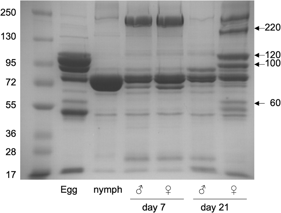
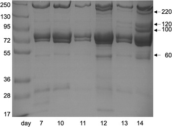
The haemolymph was extracted and analysed by SDS–PAGE to reveal the presence of vitellogenin proteins. By comparing the protein bands from females with those from males, four strong bands (∼200 kDa, 120 kDa, 100 kDa, and 60 kDa) were visible in the haemolymph of mature adult females but not in younger adult females, adult males, or nymphs (Fig. 1). Except for the largest band, these protein bands are also prominent in extracts of developing oocytes that contain the yolk protein, vitellin. In adult females, these bands are not detectable during the first 12 days after adult eclosion but become prominent in the following two days (Fig. 2). Because the appearance of these protein bands coincides with the timing of vitellogenesis, it is likely that these bands represent components of the mature vitellogenins after their processing by endoproteases. Therefore, messenger RNA was extracted from the fat body of 14-day-old females, and cDNA was prepared as the starting material for the amplification of the vitellogenin DNA fragments.
Sodium dodecyl sulfate–polyacrylamide gel electrophoresis (SDS–PAGE) of Schistocerca gregaria haemolymph. Aliquots of haemolymph from nymphs and adult male and female insects (7 and 21 days after adult ecdysis), as well as egg extracts were analysed by SDS–PAGE, as described in the “Materials and methods” section. First lane: marker proteins, with molecular weight in kilodaltons. The arrows at the right point to the four bands originating from vitellogenin.

Sodium dodecyl sulfate–polyacrylamide gel electrophoresis (SDS–PAGE) of haemolymph from adult female Schistocerca gregaria. Haemolymph samples were taken from adult females 7–14 days after adult ecdysis, as indicated, and separated by SDS–PAGE, as described in the “Materials and methods” section. First lane: marker proteins, with molecular weight in kilodaltons. The arrows indicate the putative vitellogenin proteins bands.

Complementary-DNA sequences of Vg-A and Vg-B
At the onset of our study, the only sequence information on orthopteran vitellogenins was the short 5′ sequences identified by Locke et al. (Reference Locke, White and Wyatt1987) and a partial vitellogenin sequence from the Lubber grasshopper, Romalea microptera (Palisot de Beauvois) (Orthoptera: Romaleidae) (Borst et al. Reference Borst, Eskew, Wagner, Shores, Hunter and Luker2000; GenBank accession AF135420.1). Blast searches of this 733-bp nucleotide sequence revealed one significant match from S. gregaria, an expressed sequence tag from a central nervous system cDNA library (GenBank accession LC01027B2G10.f1). This sequence was used to generate specific polymerase chain reaction primers, which resulted in prominent amplification products in the cDNA from mature females. Gene-specific primers obtained from this sequence were used to amplify the entire cDNA using the 3′ and 5′ RACE techniques in multiple and overlapping sequencing runs. The 5′ end of the mRNA is almost identical to the exon in the Vg-A clone identified by Locke et al. (Reference Locke, White and Wyatt1987; 55 out of 56 nucleotides), and therefore, it was concluded that the cDNA encodes Vg-A. The resulting sequence contains an open reading frame of 5455 nucleotides, followed by untranslated sequence with a polyadenylation signal. The conceptually translated protein is composed of 1806 amino acids (GenBank accession MK206969), yielding a calculated molecular weight of 203 kDa and an estimated isoelectric point of 6.73.
The publication of the first draft of the complete genome of the migratory locust, L. migratoria (Wang et al. Reference Wang, Fang, Yang, Jiang, Jiang and Zhao2014), provided another means for obtaining partial sequences of potential vitellogenins that could be used for RACE-primer design. Searches via T-BLASTX of the S. gregaria Vg-A showed strong similarities with translated genome sequences from two adjacent contigs (GenBank accession AVCP010784604 and AVCP010784605); in addition, much weaker similarities were detected for two shorter parts of the sequence (residues 571–715 and 943–1002) on contig AVCP010784615, suggesting the possibility that this may represent an additional vitellogenin sequence. We were able to amplify the homologous sequences in S. gregaria and obtained the full-length sequence of Vg-B through 3′ RACE and 5′ RACE.
The cDNA of S. gregaria Vg-B is 5591 bp in length, with an open reading frame of 5439 bp. Its 5′ end matches the sequence of the Vg-B clone identified by Locke et al. (Reference Locke, White and Wyatt1987; 60 out of 66 nucleotides). The open reading frame translates to a protein with 1812 amino acid residues, a molecular weight of approximately 202 kDa, and an isoelectric point of 7.28 (GenBank accession MK206970).
Analysis of the Vg-A and Vg-B cDNA sequences showed untranslated 5′ regions of 33 and 35 bp, respectively, that precede the start codon ATG. The following 45 bp (Vg-A) or 54 bp (Vg-B) encodes the N-terminal 15 or 18 amino acid residues that match the criteria for classical signal peptides that direct the nascent proteins to the endoplasmic reticulum. Vitellogenin A cDNA has the stop codon TGA followed by 87 bp, which contains the polyadenylation signal AATAAA before the codon’s poly A tail, whereas in Vg-B cDNA, the same polyadenylation signal is located 67 bp after a TAA stop codon.
Vitellogenin expression in adult females
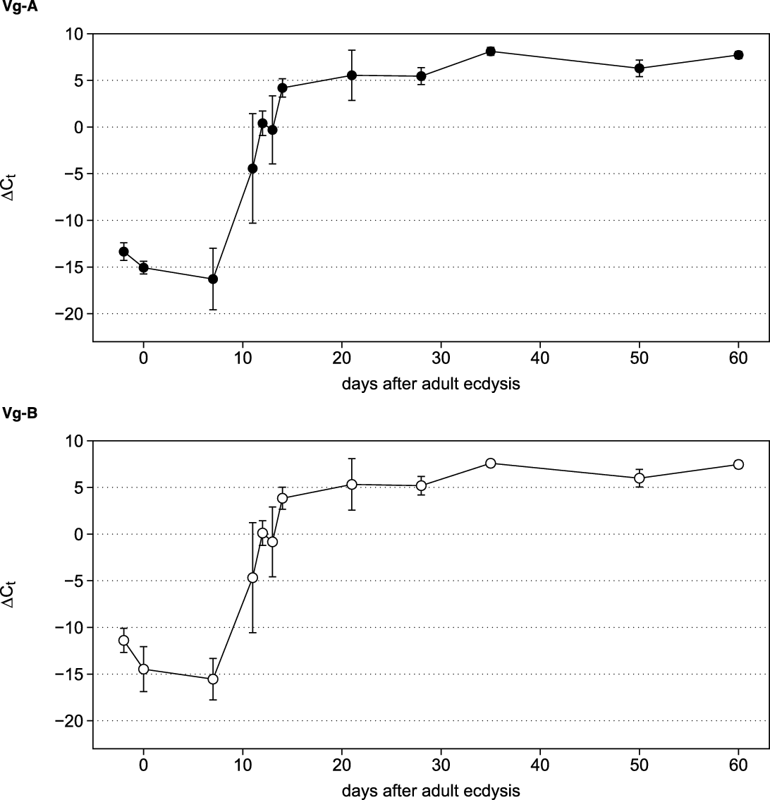
Quantitative real-time polymerase chain reaction was used to measure the expression levels of these genes in the fat bodies of adult female locusts. In nymphs and over the first 10 days after adult eclosion, vitellogenin mRNA was below the detection limit, more than 10 cycles later than in the internal control. The first detectable signal for both Vg-A and Vg-B occurred in adult females 11 days after adult eclosion, followed by a steep increase over the following three days. At day 14, both vitellogenins were very strongly expressed, more than five cycles earlier than the internal control threshold cycle values, and their transcript levels remained at similar levels throughout the entire lifespan (Fig. 3). Vitellogenins A and B were expressed at similar amounts. The difference between their ΔCt values was generally less than one cycle, and the ratio of these transcripts did not change significantly during the adult stage.
Expression of vitellogenin in adult female Schistocerca gregaria. Total RNA was extracted from the fat body of female S. gregaria at various times after adult ecdysis, and the expression of Vg-A and Vg-B was determined by quantitative real-time polymerase chain reaction, as described in the “Materials and methods” section. The data points show the difference in the threshold cycle (Ct) values for each sample relative to the internal housekeeping gene (ΔCt ± standard deviation; n = 3–5). Each Ct value measured up to day 7 is statistically highly significantly different from each value measured from day 14 onward (P < 0.001 for all pairings).

Vitellogenin expression after knockdown of Met
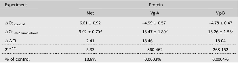
A hallmark of insect vitellogenins is that their expression depends on the intracellular juvenile hormone receptor, methoprene-tolerant (Met). To confirm that the sequenced proteins are indeed vitellogenins, double-stranded RNA targeting the Met mRNA was injected into adult locusts. Double-stranded RNA (10 µg in 10 µL) was administered within 12 hours of adult eclosion to precede the first gonadotropic cycle and again at day 7 and day 13. On day 14, the fat body and haemolymph were collected from treated and control insects to examine the effect of met double-stranded RNA on its corresponding transcript and met double-stranded RNA’s influence on vitellogenin expression at the RNA and protein levels. The met double-stranded RNA treatment reduced met RNA by more than 80% without any noticeable phenotypical effects on the insects. The knockdown of met almost completely eliminated the mRNA for both Vg-A and Vg-B (Table 1).
Changes in expression after met double-stranded RNA treatment in adult female Schistocerca gregaria. Expression levels were determined by the ΔΔCt method, with normalisation against the housekeeping gene EF1α (ΔCt ± standard deviation).

Different from control: a P = 7.6 × 10⁻⁶; b P = 2.7 × 10⁻¹⁴; and c P = 1.2 × 10⁻¹⁵.
Met, methoprene-tolerant; Vg, vitellogenin; Ct, threshold cycle.
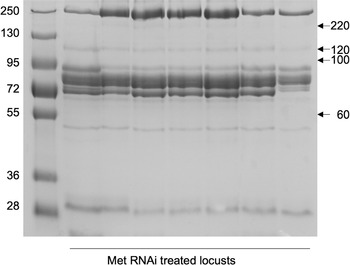
Haemolymph from the same insects was collected along with the fat body at day 14. Comparison of the protein pattern by SDS–PAGE (Fig. 4) shows that the characteristic vitellogenin bands (200, 120, 100, and 60 kDa) disappeared in all of the met double-stranded RNA–treated insects. A faint band at 120 kDa, normally the most prominent in control insects, remained visible, possibly originating from an unrelated protein with the same molecular mass.
Sodium dodecyl sulfate–polyacrylamide gel electrophoresis (SDS–PAGE) of haemolymph proteins after knockdown of met. Adult Schistocerca gregaria females were treated with met double-stranded RNA, as described in the “Materials and methods” section. Haemolymph samples were taken at day 14 after adult ecdysis and separated by SDS–PAGE. First lane: marker proteins, with molecular weight in kilodaltons. The arrows indicate the putative vitellogenin bands found in untreated locusts (see Fig. 1).

Discussion
Vitellogenin sequence analysis
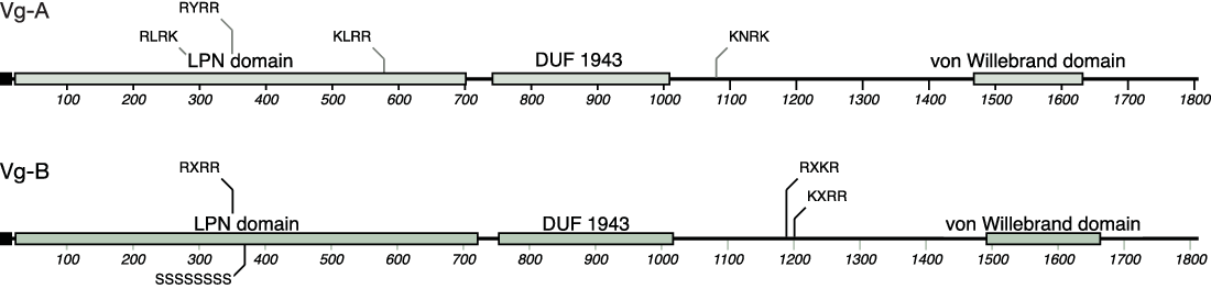
The deduced amino acid sequences for Vg-A and Vg-B contain a number of conserved motifs common to members of the vitellogenin gene family (Fig. 5): a vitellogenin N-terminal domain, also called a lipoprotein N-terminal domain because of its presence in most other members of the large lipid–transfer protein superfamily (Anderson et al. Reference Anderson, Levitt and Banaszak1998), spans the region from position 22 to position 770 (Vg-A) and from position 23 to position 790 (Vg-B); towards the C-terminus (residues 1469–1685, Vg-A; 1498–1717, Vg-B), a von Willebrand factor D domain is present that is believed to be involved in multimerisation and secretion of extracellular proteins and in the interactions with other ligands.
Protein domains of Vg-A and Vg-B A from Schistocerca gregaria. The sequences of Vg-A and Vg-B start with an N-terminal signal peptide (black box) and contain three domains conserved in the vitellogenin family: a lipoprotein N-terminal domain (LPN), a domain of unknown function (DUF 1943), and a von Willebrand domain towards the C-terminus. Also indicated are potential multibasic cleavage sites for proteases of the subtilisin family and the polyserine domain in Vg-B.

Other motifs often found in insect vitellogenin proteins include a highly conserved GL/ICG site at the C-terminus in both Vg-A (1613) and Vg-B (1645), with a DGXR site close by (1595 for Vg-A; 1616 Vg-B). Noticeably, a polycysteine sequence that follows the GL/ICG motif in other insects is absent. Also, a polyserine sequence that is common to many vitellogenins is found only in Vg-B (369–376). Finally, a domain of unknown function (DUF 1943) that can be found in many vertebrate and invertebrate vitellogenins is identified in Vg-A (745–1002) and Vg-B (754–1017).
There are seven potential N-glycosylation sites in Vg-A and four in Vg-B, as expected for vitellogenins. The proteins are glycolipoproteins because they are responsible for transferring lipids and carbohydrates along with their amino acids and other minerals to the ovaries. The glycosylation happens as a co- or posttranslational modification in the fat body and before excretion of vitellogenin into the haemolymph (Dhadialla and Raikhel Reference Dhadialla and Raikhel1990).
All insect vitellogenins sequenced to date have a cleavage site for proteases of the subtilisin family (Barr Reference Barr1991; Chen and Raikhel Reference Chen and Raikhel1996) in the lipoprotein N-terminal domain, and an additional cleavage site in the centre or the C-terminal domain was identified in hemimetabolous insects in which the vitellogenin precursor is cleaved into several fragments (Tufail and Takeda Reference Tufail and Takeda2008). The amino acid sequences of Vg-A and Vg-B both have the consensus cleavage site RXR/KR in the N-terminal domain (Vg-A: RYRR, 349–352; Vg-B: RPRR, 351–354), and Vg-B contains an additional potential cleavage site closer to the C-terminal end (RSKR, 1190–1193). Cleavage in these positions would result in a smaller N-terminal fragment (∼40 kDa) and large and small subunits for Vg-B of approximately 94 kDa and 69 kDa. There are additional similar polybasic motifs, in which one or more R has been replaced by a K, that may also be possible cleavage sites (Fig. 5). The exact sizes of the resulting fragments depend on glycosylation or other posttranslational modifications. Sodium dodecyl sulfate–gel electrophoresis of haemolymph proteins from mature reproductive females showed a similar banding pattern for those proteins absent in the haemolymph of males and young adult females (∼200 kDa, 120 kDa, 100 kDa, and 60 kDa). The three smaller polypeptides correspond to the polypeptides from the egg extract, in which vitellin is the primary component. The highest band may represent vitellogenins without further cleavage other than the removal of the small signal peptide at the N-terminus. However, because this band is fairly weak, it appears that most of the protein has been cleaved by endoproteases.
Comparative analysis
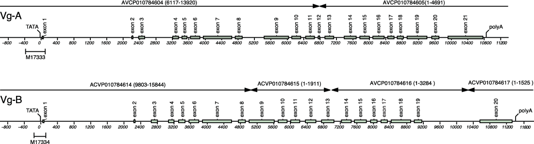
Although the automatic annotation of the genome of L. migratoria originally did not identify vitellogenin, Song et al. (Reference Song, Guo, Jiang, Kang and Zhou2013) identified a partial sequence for the 3′ end of Vg-A cDNA, coding for the amino-terminal part of Vg-A (GenBank accession KF171066). The translated protein misses the first 465 amino acid residues that include the original sequences found by Locke et al. (Reference Locke, White and Wyatt1987), but of the remaining 1346 residues, 69% are identical to S. gregaria Vg-A. The sequence of Vg-B was not identified by Song et al. (Reference Song, Guo, Jiang, Kang and Zhou2013), but later Wang and Zhou deposited the sequence of a small fragment of Vg-B from L. migratoria to GenBank (accession ASU09687) that is highly similar to S. gregaria Vg-B (84% identity over the 239-bp overlapping sequence). With the knowledge of the full-length cDNA sequences for Vg-A and Vg-B from S. gregaria, we were able to assemble the likely gene structure for the homologous proteins in L. migratoria (Fig. 6). The gene for L. migratoria Vg-A contains 21 exons, spanning over a 10.9-kb sequence found on assembly contigs AVCP010784604 and AVCP010784605, yielding a protein that is 71% identical to the homologue from S. gregaria. The 20 exons of the VG-B gene are spread over 11.4 kb on assembly contigs AVCP010784614, AVCP010784615, AVCP010784616, and AVCP010784617, and the protein sequences for Vg-B are 72% identical between the two locust species. The most recent annotation of the L. migratoria genome by Insect Base 2.0 (Mei et al. Reference Mei, Jing, Tang, Chen, Chen and Duanmu2022) identified the genes for both Vg-A (Lmig001370) and Vg-B (Lmig001371) as vitellogenins, albeit missing some parts that we identified based on the S. gregaria sequences: for Vg-A, the annotation specifies a start codon upstream of exon 3, thus missing 19 residues, including the N-terminus detected by Locke et al. (Reference Locke, White and Wyatt1987), and omits 122 residues (78% identity) that are coded in exon 11, exon 12, and part of exon 13 (78% identity with S. gregaria). For Vg-B, exon 9 (71% identical to S. gregaria) is missing, as is a large part of exon 13 (76% identical to S. gregaria).
Predicted gene structure for Vg-A and Vg-B from L. migratoria. Homologous sequences in the genome of Locusta migratoria were identified by BLAST searched with the protein sequences of Vg-A and Vg-B from Schistocerca gregaria. The entire sequence of Vg-A is found on 21 exons on the adjacent contigs AVCP010784804 and 010784805, whereas Vg-B is encoded by 20 exons on the contigs AVCP010784614-010784617. The 3′ end of the genes contains the sequences of proximal promoter regions, 3′ untranslated sequence, first exon, and part of the first intron published by Locke et al. (Reference Locke, White and Wyatt1987) for Vg-A and V-B (GenBank accession M17333 and M17334, respectively).

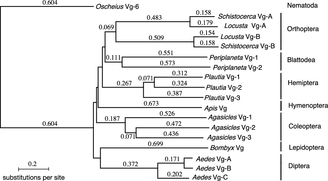
Although the respective orthologues in L. migratoria and S. gregaria are highly similar, with the characteristic domains (signal peptide, lipoprotein N-terminal domain, DUF 1943, and von Willebrand domain) conserved, Vg-A and Vg-B within either species are far less similar (only ∼28% identity), a fact that indicates that gene duplication of an ancestor vitellogenin gene has occurred long before these two species separated. Phylogenetic analysis (Fig. 7) shows that all insect vitellogenins seem to have originated from a common ancestor and that any gene duplications occurred later in the various orders. A similar relatively low degree of sequence identity is observed for many other insect vitellogenins (Comas et al. Reference Comas, Piulachs and Bellés2000; Tufail et al. Reference Tufail, Hatakeyama and Takeda2001). The number of different vitellogenin paralogues identified varies; most insects seem to possess only one, but two or three genes have been found in some species (Tufail and Takeda Reference Tufail and Takeda2008). It has been proposed that the amino acid composition, rather than the amino acid sequence, is conserved between these paralogues (Hughes Reference Hughes2010). Indeed, the amino acid compositions of Vg-A and Vg-B from S. gregaria are highly similar to each other and to other members of the vitellogenin family, despite their low degree of sequence conservation.
Phylogram of insect vitellogenins from seven orders. The phylogenetic tree was constructed from vitellogenin protein sequences by the neighbour-joining method (uncorrected, gaps distributed proportionally) with MacVector 17.0 software (MacVector Inc., Apex, North Carolina, United States of America). Vitellogenin 6 from the nematode Oscheius tipulae was used as an outgroup. The sequences and their GenBank accession numbers are Oscheius tipulae Vg-6 (Q94637) (Nematoda: Rhabditidae); Schistocerca gregaria Vg-A (QGR25457); Locusta migratoria Vg-A (TPA: BK059897); Locusta migratoria Vg-B (TPA: BK059898); Schistocerca gregaria Vg-B (QGR25458); Periplaneta americana Vg-1 (Q9U8M0) (Blattodea: Blattidae); Periplaneta americana Vg-2 (Q9BPS0); Plautia stali Vg-1 (BAA88075) (Hemiptera: Pentatomidae); Plautia stali Vg-2 (BAA88076); Apis mellifera Vg (NP_001011578) (Hymenoptera: Apidae); Agasicles hygrophila Vg-1 (QDP42282) (Coleoptera: Chrysomelidae); Agasicles hygrophila Vg-1 (QDP42283); Agasicles hygrophila Vg-1 (QDP42284); Bombyx mori Vg (NP_001037309) (Lepidoptera: Bombycidae); Aedes aegypti Vg-A (Q16927) (Diptera: Culicidae); Aedes aegypti Vg-B (AAQ92367); Aedes aegypti Vg-C (AAQ92366).

Expression
Vitellogenin is the most abundant transcript in adult females because producing enormous amounts of this protein is necessary before oviposition expression (Chen et al. Reference Chen, Couble, Abu-Hakima and Wyatt1979; Chinzei and Wyatt Reference Chinzei and Wyatt1985; Glinka and Wyatt Reference Glinka and Wyatt1996; Wyatt and Davey Reference Wyatt and Davey1996). The presence of two genes possibly may help in meeting these demands. Tufail et al. (Reference Tufail, Hatakeyama and Takeda2001) pointed out that the existence of multiple vitellogenins in other insect species may be advantageous in the provision of the required amounts of yolk protein to the developing oocytes.
The expression of vitellogenin in adult females is transcriptionally regulated, and it has been shown to be under the control of various endocrine factors (Wyatt and Davey Reference Wyatt and Davey1996; Badisco et al. Reference Badisco, Marchal, Van Wielendaele, Verlinden, Vleugels and Vanden Broeck2011b). In orthopteran insects, it is well established that juvenile hormone acts as a primary regulating hormone during the adult fertility stage, controlling reproduction mechanisms, including the transcription of several vitellogenesis-related genes. In Locusta migratoria, juvenile hormone was found to induce the expression of vitellogenin in a tissue-, stage-, and sex-dependent manner (Chen et al. Reference Chen, Couble, Abu-Hakima and Wyatt1979; Chinzei et al. Reference Chinzei, White and Wyatt1982; Dhadialla and Wyatt Reference Dhadialla and Wyatt1983).
A growing body of evidence from various species, including L. migratoria (Jindra et al. Reference Jindra, Palli and Riddiford2013; Wu et al. Reference Wu, Yang, He and Zhou2021), suggests that the transcriptional regulator Met acts as an intracellular receptor for juvenile hormone. The action of juvenile hormone is known to lead to dramatic changes in the fat body at the cellular level, including inducing extensive DNA replication. During the maturation of the adult female, fat body cells become polyploid, which is believed to contribute to the rapid synthesis of the large amounts of vitellogenin that are required (Nair et al. Reference Nair, Chen and Wyatt1981; Oishi et al. Reference Oishi, Locke and Wyatt1985). More recent studies by Guo et al. (Reference Guo, Wu, Song, Jiang, Wang and Deng2014) showed that Mcm4 and Mcm7, the two genes that induce DNA replication and polyploidisation, are also under the control of juvenile hormone. However, this alone cannot explain the several thousand-fold reduction in vitellogenin transcripts found in Met-knockdown locusts. Our results therefore support a direct action of Met on the transcription of the two vitellogenin genes in the desert locust and are in agreement with previous studies in both hemi- and holometabolous insects that showed the involvement of Met in this pathway.
Acknowledgements
This research was supported by a grant from Simon Fraser University, Burnaby, British Columbia, Canada. A.S.A. contributed methodology, investigation, writing – original draft; and N.H.H. contributed conceptualisation, writing – review and editing.
Competing interests
The authors declare that there are no competing interests.
Data availability
The nucleotide and protein sequences for S. gregaria vitellogenin have been deposited with GenBank (Vg-A: MK206969; QGR25457; Vg-B: MK206970; QGR25458). Nucleotide sequence data reported for the L. migratoria vitellogenin are available in the Third-Party Annotation Section of the DDBJ/ENA/GenBank databases under the accession numbers TPA: BK059897–BK059898.