INTRODUCTION
Leishmania are protozoan parasites transmitted by blood-sucking sandflies. The flagellated promastigotes introduced into the mammalian host during a bloodmeal are internalized by host cells into the phagolysosomal compartment, where they transform into obligatory intracellular amastigotes (Locksley and Louis, Reference Locksley and Louis1992; Handman, Reference Handman1999). The most virulent and protective factors in this hostile environment are surface glycoconjugates. The surface glycoconjugates of Leishmania include lipophosphoglycan (LPG), the glycoinositolphospholipids (GIPL), and GPI-anchored proteins, all tethered to the membrane with a GPI anchor. They differ in composition, structure and size of their glycans and lipid moieties (Mc Conville et al. Reference McConville, Homans, Thomas-Oates, Dell and Bacic1990, Reference McConville, Schnur, Jaffe and Schneider1995).
PPG is another major phosphoglycan which is produced and secreted by Leishmania species. This phosphoglycan, unlike LPG, contains a protein backbone and forms filamentous structures in the centre of promastigote rosettes and in culture medium. More recently, it was found that L. major promastigote PPG exists in 2 forms; a water-soluble secreted form and a structurally distinct form, which is tethered to the membrane with a glycosylphosphatidyl anchor. L. major PPG has a repetitive amino acid backbone rich in serine, alanine and proline. Most of the serine residues in PPG are glycosylated via a phospho-diester linkage and the sugars are remarkably similar to those present in LPG, where they have been shown to play a role in the attachment to the host cells (Ilg, Reference Ilg2000).
The innate immune system senses the presence of invading microbes by the use of specific receptors that recognize pathogen-associated molecular patterns (PAMPs), and activation of these receptors initiate host responses. Sensing has been shown to be mediated by an ancient family of membrane proteins known as the Toll-like receptors (TLR), which recruit and activate signalling molecules involved in innate immunity. Members of the TLR family are responsible for the recognition of pathogen-associated molecular patterns expressed by a wide spectrum of infectious agents. To date, over 13 TLRs have been reported in human and mouse. TLRs activate the NF-κB pathway, which regulates cytokine expression, nitric oxide and reactive oxygen species production through several adaptor molecules including MyD88. Activation of the NF-κB pathway links innate and adaptive immune response by production of inflammatory cytokines such as IL-1, IL-6, IL-8, TNF-α, IL-12, chemokines and induction of co-stimulatory molecules such as CD80, CD86, and CD40 (Aderem and Ulevitch, Reference Aderem and Ulevitch2000; Takeda et al. Reference Takeda, Kaisho and Akira2002; Van Der Kleij et al. Reference Van Der Kleij, Latz, Brouwers, Kruize, Schmitz, Kurt-Jones, Espevik, de Jong, Kapsenberg, Golenbock, Tielens and Yazdanbakhsh2002; Pasare and Medzhitov, Reference Pasare and Medzhitov2003). We already assessed functional properties of parasite membrane isolated LPG (mLPG) and culture supernatant LPG (sLPG) in addition to PPG (Kavoosi et al. Reference Kavoosi, Ardestani, Kariminia, Abolhassani and Turco2006, Reference Kavoosi, Ardestani, Kariminia, Zeinali and Alimohammadian2008). It was shown that L. major PGs are able to induce IL-12 and IL-10; however, only sLPG is able to induce IFN-γ secretion by PBMCs. In the present study we showed for the first time that TLR2 recognition of LPG promotes differential cytokine secretion by PBMCs of healthy individuals. The pathological and immunological implications will be discussed.
MATERIALS AND METHODS
Parasite culture
The strain of L. major used in this study was the vaccine strain (MRHO/IR/75/ER), kindly provided by Dr Ali Mohammadian (Department of Immunology, Pasteur Institute of Iran). Infectivity was maintained by regular passage in susceptible BALB/c mice. The parasites were cultured in the RPMI-1640 medium (Sigma, Chemical Co., St Louis, USA) supplemented with 10% FBS (Sigma), 292 μg/ml L-glutamine (Sigma) and 4·5 mg/ml glucose. The starting parasite inoculation was 1×10⁶/ml. Under these culture conditions, the stationary phase of parasite growth was obtained in 6 days as determined by peanut agglutination assay.
Leishmania major LPG and PPG purification
Membrane LPG (mLPG) was extracted from 1011 metacyclic promastigotes and then purified by hydrophobic interaction chromatography and cold methanol precipitation (Turco et al. Reference Turco, Wilkerson and Clawsons1984). Soluble LPG (sLPG) and PPG from the culture medium were purified with ion-exchange chromatography, hydrophobic interaction chromatography, ultrafiltration and cold methanol precipitation (Ilg et al. Reference Ilg, Stierhof, Craik, Simpson, Handman and Bacic1996). Then purified LPGs and PPG were analysed by immunoblotting with WIC97.3 monoclonal antibody that recognizes the galactosylated Gal-Man repeat.
Immunoblotting of Leishmania major LPG and PPG
The purified LPG, as described above, was subjected to electrophoresis on an SDS–PAGE gel using 5% and 12% polyacrylamide for the stacking and separating gel, respectively. The purified PPG was subjected to electrophoresis on the agarose gel using 1% DNA grade agarose. The transfer was done at 12 V for 1 h onto nitrocellulose membrane. The membrane was blocked with 5% milk in phosphate-buffered saline. The primary mouse monoclonal antibody WIC79.3 (kindly provided by E. Handman, The Walter and Eliza Hal Institute of Medical Research, Melbourne, Victoria 3050, Australia) that recognizes the galactosylated Gal-Man-P repeat units of LPG, was used at 1:1000 dilution (Greenblatt et al. Reference Greenblatt, Slutzky, De Ibarra and Snary1983). A goat anti-mouse horseradish peroxidase secondary antibody (Pierce, USA) was used to detect the LPG and PPG at a 1:10 000 dilution.
LAL test
In order to indicate the presence of gram-negative bacterial endotoxin (LPS), the Limulus amoebocyte lysate (LAL) assay was performed based on the manufacturer's instruction (LAL Kit, Charles Riever Endosafe, T2092 CTK7, USA).
Peripheral blood mononuclear cell (PBMC) culture
Healthy individuals from a non-endemic area without prior travel to an endemic area or a history of any parasitic infection were included in the study. Peripheral blood samples were isolated after venipuncture on a Ficoll–Hypaque gradient (d=1·077). The cells at the inter-phase were washed twice in cold RPMI-1640 and cultured in 24-well plates (106 cells/ml, 1 ml/well) in RPMI-1640 supplemented with 10% FCS, 2 mM L-glutamine, 100 U/ml penicillin and 100 μg/ml streptomycin at 37°C and 5% CO2. For TLR blocking, PBMC were pre-treated for 2 h with anti-TLR2 (10 μg/ml, anti-human TLR2, TL2.3 clone, CD282, IgG2a, ebioscience, USA) and anti-TLR4 (10 μg/ml, anti-human TLR4, CD284, HTA125 clone, IgG2a, ebioscience, USA). Pre-treated and untreated PBMC were stimulated with purified L. major LPG and PPG (1 μg/ml).
Reactive oxygen species (ROS) measurement
Reactive oxygen species production was analysed by a previously described method (Conour et al. Reference Conour, Graham and Gaskins2004). Briefly, PBMCs were cultured in 24-well plates (106 cells/ml, 1 ml/well) in RPMI-1640 and 10% FCS at 37°C with 5% CO2, and pre-treated with anti-TLRs and then stimulated with LPG or PPG (1 μg/ml). The plates were incubated for an additional 24 h. At the end of the incubation, a final concentration of 25 μM of dichlorodihydrofluorescein diacetate (H2DCFDA) was added and the cells were incubated in 37°C, with 5% CO2 in a humidified incubator for 30 min to allow loading of the dye. After 30 min, the cells were washed 3 times with phosphate-buffered saline (PBS) and the conversion of non-fluorescent H2DCFDA into the highly fluorescent dichlorofluorescein (DCF) was measured using a flow cytometer (FACScan, Becton Dickinson, USA). In order to define the background auto-fluorescence, non-stained cells were used. In addition, non-treated cells were used as control.
Determination of IFN-γ, IL-12 and IL-10
Treated and untreated PBMCs were cultured in 24-well plates (106 cells/ml, 1 ml/well) in RPMI-1640 and 10% FCS for 24 h at 37°C and 5% CO2 in the presence of sLPG, mLPG or PPG (1 μg/ml). At the end of the incubation, the culture supernatants were harvested and kept frozen until used. IFN-γ, IL-12 and IL-10 were determined by sandwich ELISA with IFN-γ Kit (Quantikine HS, R&D Systems, USA), IL-10 Kit (Quantikine HS, R&D Systems, USA) and IL-12 Kit (Quantikine, R&D Systems, USA). The manufacturer's instructions were followed.
Statistical analysis
Statistical significance (P<0·05) was analysed by Student's t-test using SPSS version 10.
RESULTS
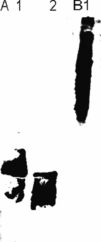
The L. major LPG was extracted and purified by solvent extraction, hydrophobic interaction chromatography and cold methanol precipitation. The efficiency of purification was analysed by immunoblotting. As seen in Western blotting of mLPG and sLPG purified from metacyclic stages of L. major promastigotes, both the membrane-bound and excretory LPGs are highly heterogeneous in size (Fig. 1, Lane A1, 2). The antibody used in this Western blot was WIC79.3 which recognizes galactosylated side-chains that branch off the Gal-Man-PO4 backbone of LPG. Thus, it is likely that the mLPGs and sLPGs are very similar in structure but their molecular weights are different due to the special repeat units of the glycan portion. Soluble PPG was purified by ion-exchange, hydrophobic interaction and gel filtration chromatography. The efficiency of purification was analysed by immunoblotting. The results indicate that PPG is a heterogenous molecule and its glycan portion is similar to LPG (Fig. 1, Lane B1). The results of the LAL test showed that these samples contain less than 300 pg endotoxin/mg phosphoglycan.
Immunoblotting of purified mLPG, sLPG and PPG from metacyclic Leishmania major promastigotes. mLPG, sLPG and PPG (10 μg per lane), isolated and purified from promastigotes of L. major, were subjected to Western blotting and probed with the monoclonal antibody WIC79.3, which recognizes the galactosylated–PO4-6Gal-Man repeat units of LPG. sLPG has a higher molecular weight than mLPG and PPG is a heterogeneous molecule with high molecular weight. Lane A1, sLPG; Lane A2, mLPG; Lane B1, PPG.

ROS production
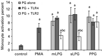
The cells were cultured in RPMI medium in the presence or absence of anti-TLR2 and/or anti-TLR4. The cells were then stimulated with LPG and PPG (1 μg/ml). After 30 min, ROS production was determined by flow cytometry. We observed that LPG and PPG induced ROS production (P<0·05); however, in the presence of anti-TLR2 the ROS production induced by mLPG and sLPG was reduced to the baseline. The specificity of treatment was confirmed by having no change in the presence of anti-TLR4 (Fig. 2).
Anti-TLR2 inhibits ROS production in human peripheral blood mononuclear cells. PBMCs were cultured (106 cell/ml) in RPMI medium in the presence of mLPG, sLPG and PPG (1 μg/ml), anti-TLR2 (10 μg/ml) and anti-TLR4 (10 μg/ml). After 24 h ROS production was analysed by flow cytometry. mLPG, sLPG and PPG induced ROS production significantly (P<0·05) but in the presence of anti-TLR2, ROS production was reduced. In the presence of anti-TLR4, ROS production was not affected. The bars represent mean +/− S.D. for 5 different experiments. Letters indicate significant differences at P<0·05. (a) Compared with untreated cells (control), (b) compared with PG alone and (c) PG+TLR2 compared with PG+TLR4.

Cytokine study
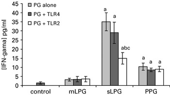
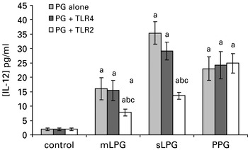
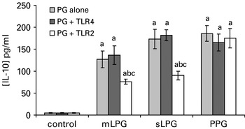
PPG and sLPG induced a significantly higher amount of IFN-γ (P<0·05); however, sLPG was far stronger than PPG. In addition, partial inhibition was only demonstrated in the presence of anti-TLR2 for sLPG (Fig. 3). No inhibition was seen in PPG-induced ROS production in the presence of anti-TLR2 or anti-TLR4. It was shown that all purified PGs including mLPG, sLPG and PPG induced IL-12 production (P<0·05) and, consistent with the IFN-γ results, sLPG had a significantly higher stimulatory effect than mLPG and PPG, which was also blocked by anti-TLR2 but not by anti-TLR4. In the presence of either anti-TLR2 or anti-TLR4, IL-12 stimulated by PPG was not affected (P<0·05, Fig. 4). On the other hand, all PGs profoundly stimulated secretion of IL-10 but PPG and sLPG were more potent than mLPG. Anti-TLR-2 partially diminished the IL-10 production induced by sLPG and mLPG but not by PPG (P<0·05, Fig. 5).
Anti-TLR2 inhibits IFN-γ production in human peripheral blood mononuclear cells. PBMCs were cultured (106 cell/ml) in RPMI medium in the presence of mLPG, sLPG and PPG (1 μg/ml), anti-TLR2 (10 μg/ml) and anti-TLR4 (10 μg/ml). After 24 h IFN-γ production was analysed by ELISA. Only sLPG induced IFN-γ production in a significant manner (P<0·05). In the presence of anti-TLR2 IFN-γ production was reduced. The bars represent mean +/− S.D. for 5 different experiments. Letters indicate significant differences at P<0·05. (a) Compared with untreated cells (control), (b) compared with PG alone and (c) PG+TLR2 compared with PG+TLR4.

Anti-TLR2 inhibits IL-12 production in human peripheral blood mononuclear cells. PBMCs were cultured (106 cell/ml) in RPMI medium in the presence of mLPG, sLPG and PPG (1 μg/ml), anti-TLR2 (10 μg/ml) and anti-TLR4 (10 μg/ml). After 24 h cytokine production was analysed by ELISA. Both mLPGs and sLPG induced IL-12 production (P<0·05) but in the presence of anti-TLR2, IL-12 production was reduced. PPG induced IL-12 (P<0·05) and the presence of anti-TLR did not affect its stimulatory effect. The bars represent mean +/− S.D. for 5 different experiments. Letters indicate significant differences at P<0·05. (a) Compared with untreated cells (control), (b) compared with PG alone and (c) PG+TLR2 compared with PG+TLR4.

Anti-TLR-2 inhibits IL-10 production in human peripheral blood mononuclear cells. PBMCs were cultured (106 cell/ml) in RPMI medium in the presence of mLPG, sLPG and PPG (1 μg/ml), anti-TLR2 (10 μg/ml) and anti-TLR4 (10 μg/ml). After 24 h cytokine production was analysed by ELISA. Both mLPGs and sLPG induced IL-10 production (P<0·05) but in the presence of anti-TLR2, IL-10 production was reduced. PPG induced IL-10 and the presence of anti-TLR did not change its stimulatory effect. The bars represent mean +/− S.D. for 5 different experiments. Letters indicate significant differences at P<0·05. (a) Compared with untreated cells (control), (b) compared with PG alone and (c) PG+TLR2 compared with PG+TLR4.

DISCUSSION
LPG is the main promastigote molecule and is composed of repetitive units of disaccharide and phosphate, which are linked to the external surface of the plasma membrane by the GPIL anchor. The structure of LPG is different in different species of Leishmania and also in different promastigotes of the same species. LPG is significantly more common in the metacyclic form and is rare in the amastigotes (McConville et al. Reference McConville, Turco, Ferguson and Sacks1992). The roles of the LPG and gp63 pathogen-associated molecular patterns appear to complement each other, and these molecules appear to be the trigger of the immune response against Leishmania (Zambrano-villa et al. Reference Zambrano-Villa, Rosales-Borjas and Ortiz-Ortiz2002). Some studies have demonstrated that LPG might inhibit the fusion of the phagosome with the lysosomes. There is evidence that the interaction of the parasitophorous vacuoles with the endocytic organelles is very limited. In contrast, vacuoles formed around a Leishmania mutant lacking the cell surface LPG fused extensively with endosomes and lysosomes, promoting complete destruction of the parasite (Dermine et al. Reference Dermine, Scianimanico, Prive, Descoteaux and Desjardins2000). Activated macrophages produce tumor necrosis factor alpha (TNF-α) that synergizes with IFN-γ in the induction of inducible nitric oxide synthase (iNOS) production and further Leishmania destruction (Amato et al. Reference Amato, Tuon, Bacha, Neto and Nicodemo2008).
Several studies have demonstrated that different receptors mediate the uptake and phagocytosis of Leishmania sp. and there are several studies showing interaction of LPG with TLR2 and induction of an immune response in mice. The first report showed that interaction of LPG with TLR2 activates cytokine production and innate immune responses (de Veer et al. Reference De Veer, Curtis, Baldwin, Di Donato, Sexton, McConville, Handman and Schofield2003). Additionally, RNA interference study showed that TLR2 and TLR3 are involved in LPG recognition of L. donovani by IFN-γ primed mouse macrophages; however, naive macrophages barely express those TLRs (Flandin et al. Reference Flandin, Chano and Descoteaux2006). The level of TLR2 mRNA is reported to be significantly higher in resistant C57BL/6 mice than susceptible BALB/c mice (Charmoy et al. Reference Charmoy, Megnekou, Allenbach, Zweifel, Perez, Monnat, Breton, Ronet, Launois and Tacchini-Cottier2007). More recently it has been shown that, depending on the genetic background of the mice, mast cells are activated by LPG and direct the outcome of the disease (Villasenor-Cardoso et al. Reference Villasenor-Cardoso, Salaiza, Delgado, Gutierrez-Kobeh, Perez-Torres and Becker2008). However, there is only one report on TLR2 involvement with Leishmania infection showing that human NK cells are directly activated by purified L. major LPG leading to IFN-γ and TNF-α secretion (Becker et al. Reference Becker, Salaiza, Aguirre, Delgado, Carrillo-Carrasco, Kobeh, Ruiz, Cervantes, Torres, Cabrera, Gonzalez, Maldonado and Isibasi2003).
We previously demonstrated that purified PGs from L. major are able to induce IFN-γ, IL-10, IL-12 and ROS production in PBMCs of naive healthy individuals who did not have any history of Leishmaniasis or of travelling to an endemic area. It was found that the most effective inducer of Th1 cytokines was sLPG (Kavoosi et al. Reference Kavoosi, Ardestani, Kariminia, Abolhassani and Turco2006, Reference Kavoosi, Ardestani, Kariminia, Zeinali and Alimohammadian2008). Similar to our previous report, in the present study sLPG was shown to be the potent inducer of cytokine production compared to mLPG and PPG; however, IFN-γ production was only induced by sLPG. Interestingly, anti-TLR2 mAb was able to partially yet significantly block secretion of cytokines which is consistent with the previous report showing that L. donovani LPG recognition is mediated with TLR2 and TLR3 in IFN-γ primed mouse macrophages (Flandin et al. Reference Flandin, Chano and Descoteaux2006). Therefore, in our study TLR2 and TLR3 might also synergistically be involved in L. major LPG recognition and cytokine secretion.
Recently, TLR2 targeted vaccines have been designed and used in different immunization studies in the mouse model. A herpes simplex virus type 2 (HSV-2) CD8+ T-cell peptide epitope extended by a palmitic acid moiety (a TLR2 agonist) was synthesized and applied intravaginally to B6 mice. HSV-2-specific memory CD8+ cytotoxic T cells were generated both locally in the genital tract draining lymph nodes and systemically in the spleen. Moreover, lipopeptide-immunized TLR2(−/−) and MyD88(−/−) mice developed a significantly reduced HSV-specific CD8+ T-cell response, earlier death, faster disease progression, and higher vaginal HSV-2 titres compared to lipopeptide-immunized wild-type B6 mice (Zhang et al. Reference Zhang, Chentoufi, Dasgupta, Nesburn, Wu, Zhu, Carpenter, Wechsler, You and BenMohamed2009). This report introduces the next generation of needle-free mucosal vaccines using TLR2 agonist peptide as adjuvant.
TLR2 agonists covalently conjugated to antigenic peptides specific for CD8+ T cells were successfully used in another mouse immunization model (Khan et al. Reference Khan, Weterings, Britten, de Jong, Graafland, Melief, van der Burg, van der Marel, Overkleeft, Filippov and Ossendorp2009). In addition, a Toll-like Receptor2 directed fusion protein vaccine against tuberculosis has been shown to promote a Th1 response and control the disease in mice (Wang et al. Reference Wang, Henao-Tamayo, Harton, Ordway, Shanley, Basaraba and Orme2007). These pioneering experimental vaccines implicate the valuable adjuvant impact of TLR-2. Therefore, our data propose sLPG as a strong adjuvant against L. major infection; however, its antigenicity remains to be shown. Covalent conjugation of sLPG with potential L. major-protective peptides or a mixture of protein antigens with sLPG might be a novel multi-valent vaccine (Pinheiro et al. Reference Pinheiro, Pinto, Guedes, Filho, de Mattos, Saraiva, de Mendonça and Rossi-Bergmann2007). Interestingly, cytokine production induced by PPG which has biochemical similarity with LPG was not affected with anti-TLR2. This might be due to the lack of the lipid moiety of PPG since ligand-receptor interaction studies with Pam3CSK4, a synthetic lipopeptide specific for TLR1/2 heterodimer inducing signal transduction, proposed a model in which 3 acyl chains of Pam3CSK4 promote heterodimerization of TLR-1 and TLR2 (Buwitt-Bechmann et al. Reference Buwitt-Beckmann, Heine, Wiesmuller, Jung, Brock and Ulmer2005; Brodsky and Medzhitov, Reference Brodsky and Medzhitov2007). It is noteworthy that PPG does not bear any lipid component making it unrecognizable by TLR2. However, PPG interaction with other TLRs such as TLR3 is not ruled out.
In conclusion, LPG recognition is partially mediated via TLR2 in humans, leading to cytokine and ROS production. However, TLR2 and TLR4 are not involved in PPG recognition. In addition, we introduce sLPG as a TLR2 agonist promoting strong Th1 cytokine production by human PBMCs.
The authors would like to thank Mrs Vaziri for her excellent technical assistance in providing large-scale Leishmania promastigotes.