INTRODUCTION
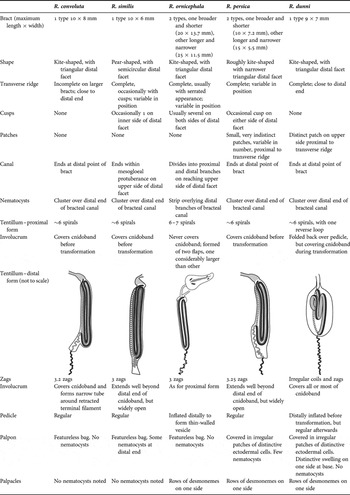
The genus Resomia, a replacement name for Moseria Totton, Reference Totton1965, which is multiply pre-occupied, has been reviewed recently by Pugh (Reference Pugh2006). The genus was there characterized by the heart-shaped mature nectophores, and by tentacles that bore tentilla of two forms, the more proximal one transforming into the more distal one as the tentacle grew. The genus was considered to include two species, R. convoluta (Moser, 1925) and R. similis (Margulis, 1977). Because of the uniqueness of the tentacles and tentilla, Pugh considered that the genus should be placed in a separate family, the Resomiidae.
Pugh (Reference Pugh2006) also considered the systematic position of all physonect genera based on recent molecular data (Dunn et al., Reference Dunn, Pugh and Haddock2005a) and on certain morphological characters. Although no molecular data were available for the Resomia species at that time, the morphological characters indicated that, because the species bore gonophores of both sexes, it belonged amongst the ‘Monoecious’ group of physonects; further verified by the presence of a descending pallial canal on the surface of the nectophores. Within this grouping the family Agalmatidae, including the genera Agalma, Athorybia, Halistemma, Melophysa and Nanomia, was distinctly divided off from the other ‘Monoecious’ genera by the fact that the nectophores were budded off on the dorsal side of the nectosome, while in the other genera they were budded off on the ventral side. Although this distinction cannot apply to the species of the genus Athorybia, as they do not possess a nectosome, nonetheless the molecular data clearly indicated that they should be included within the Agalmatidae.
Because the family Agalmatidae now had to be restricted to the genera mentioned above, this meant that several genera previously included within that catch-all category (see Totton, Reference Totton1965; Pugh, Reference Pugh2006), but which have a ventral nectosome, were now not attributable to any extant family. These genera included Cordagalma, Frillagalma, Lychnagalma, Resomia and Marrus, with the genus Erenna having already been removed to a separate family, the Erennidae (Pugh, Reference Pugh2001). Morphological data indicated that the first four of these genera belonged within the ‘Monoecious’ grouping of physonects, while Marrus and Erenna, belonged to the ‘Dioecious’ grade, despite the fact that M. orthocanna (Kramp, 1942) appears independently to have become monoecious. Of the above genera only Cordagalma was included in the molecular study by Dunn et al. (Reference Dunn, Pugh and Haddock2005a), but it was found difficult to define its exact phylogenetic position most likely due to problems of long-branch attraction. In a subsequent analysis (Dunn, Reference Dunn2005), Lychnagalma was included and, as expected, fell within the ‘Monoecious’ group of physonects. However, the exact relationships of the first four genera mentioned above remain uncertain and it is to be hoped that further molecular studies will clarify the situation.
In this paper three new resomiid species are described.
TYPE MATERIAL
All collected specimens were fixed in 5% borax-buffered formalin and later tranferred to Steedman's preserving fluid.
Holotype: specimen collected during the ROV ‘Tiburon’ Dive 845 from a depth of 233 m (9 April 2005; 35°28.76′N 123°51.72′W). The specimen has been deposited at the United States National Museum (Smithsonian Institution), Washington, DC (USNM 1121482).
Paratype: specimen collected during the ROV ‘Tiburon’ Dive 847 from a depth of 241 m (11 April 2005; 36°41.92′N 122°03.41′W). The specimen has been deposited at the United States National Museum (Smithsonian Institution), Washington, DC (USNM 1121483).
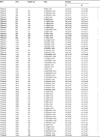
Other material: the station data for the other material examined are shown emboldened in Table 1. These remain in the collections of P.R.P. The remaining data refer to specimens identified from in situ photographs but not collected.
List of specimens of Resomia ornicephala sp. nov. examined (emboldened) and/or identified from in situ frame grabs (*).

DIAGNOSIS
Nectophores with upper and lower lateral ridges, the former not quite reaching ostium, the latter reaching it. A pair of lateral ridges, extending from the lateral sides of the ostium toward, but not reaching, the upper laterals, also present underlain by long narrow lateral ectodermal tracts extending from the ostium. Young nectophores with distinctive lateral flaps.
Two types of bract both roughly kite-shaped with complete transverse ridge, which in the younger bracts at least has a distinctly serrated appearance. Bracteal canal divides distally with the branches running in the mid-line proximally and distally on the upper surface of the distal facet, and overlain by a strip of nematocysts.
Involucrum of tentillum not enclosing cnidoband on either spiralled–coiled or zigzagged forms, but forming two asymmetrical flaps.
DESCRIPTION
The whole animal, and particularly the nectosome, is extremely transparent (Figure 1) and very difficult to photograph in situ. The siphosomal zooids, apart from the bracts, have a milky white appearance, and the only noticeable pigmentation is the yellow coloration of the cnidobands of the tentilla, although the involucrum of each tentillum also contains pigmentation of a variety of colours (see Figures 7 & 8). These characters, together with the characteristic arc of nematocysts at the distal end of the bracts, make this species easy to identify in situ and easy to distinguish from the commonest physonect encountered in the same depth range, namely Nanomia bijuga.
Resomia ornicephala sp. nov. In situ photographs from (A) ‘Tiburon' Dive 1075 and (B) ‘Tiburon’ Dive 1042. Specimens up to ~30 cm in length.

Pneumatophore: the pneumatophores of the specimens examined measured 2.0–2.2 mm in length and 0.72–0.79 mm in diameter. They showed no characteristic features and were without any obvious pigmentation.
Nectosome: the nectophores were budded off on the ventral side of the nectosome. No pigmentation was observed within the nectosomal region, and the nectophores themselves were very transparent in life.
Nectophore: there were ~27 nectophores associated with the type specimen, together with some young buds attached to the nectosome. The nectophores measured up to 14 mm in length and 15 mm in width; the largest, in their preserved state, being very flimsy, particularly in the region of the axial wings.
Four detached young nectophores were found with one of the ‘Tiburon’ Dive 847 specimens (Figure 2). They all showed the presence of small axial wings and extensive lateral flaps whose edges were demarcated by the upper lateral ridges. These ridges ran from their proximal connection with the lower lateral ridges to end just proximal to the ostium (see Figure 3). The lower laterals reached the ostium closer to the points of insertion of the lateral radial canals onto the ostial ring canal than to that of the lower canal. A pair of lateral ridges extended out and up from the lateral margins of the ostium, toward the upper lateral ridges but petered out before reaching them. On the lower side of these ridges, for approximately two-thirds of their length, there was a narrow tract of distinctive ectodermal cells. There were also clusters of large ectodermal cells, probably photocytes, in the vicinity of the ostial ring canal where the upper and lateral radial canals were inserted. The ostium itself opened obliquely on the lower side of the nectophore. There were no signs of a thrust block and the upper and lower pallial canals, of approximately equal length, appeared only as scars on the surface of the nectophore. The pedicular canal ran straight to the top of the nectosac and directly gave rise to the four straight radial canals. There was no muscle-free zone on the nectosac.
Resomia ornicephala sp. nov. Lower views of very young nectophores from ‘Tiburon' Dive 847. Scale bar: 1 mm.

Resomia ornicephala sp. nov. Lateral (left) and upper (right) views of developing nectophore from type specimen. pc, pallial canal; ped, pedicular canal; rl, lateral ridge; rll, lower lateral ridge; rul, upper lateral ridge; tec, tract of ectodermal cells. Scale bar: 1 mm.

Although only slightly larger than those shown in Figure 2, a nectophore from the type specimen was at a further stage of development (Figure 3). It showed the same basic arrangement of ridges as the young nectophores, with the upper lateral ridges (Figure 3, rul) not reaching the ostium, the lateral ridges (rl) petering out before reaching the upper lateral ones, and the lower lateral ridges (rll) extending from their proximal junction with the upper laterals to the ostium, ending close to where the laterals terminated. The axial wings had increased in size and the lateral flaps, demarcated by the upper lateral ridges, had folded inwards to define a broad, deep median furrow on the upper surface of the nectophore. The tracts of ectodermal cells (tec) that paralleled the lateral ridges were very distinctive. The upper pallial canal was now seen to be about twice the length of the lower one.
The largest nectophores of the type specimen generally had extensive axial wings (Figure 4, aw), but still a minute, almost indistinguishable, thrust block (tb). However, one or two nectophores had relatively small axial wings and appeared much more rectangular in outline. Nonetheless, the same basic arrangement of the ridges was maintained, and the ostium (ost) opened obliquely onto the lower side of the nectophore.
Resomia ornicephala sp. nov. Upper (left) and lower (right) views of mature nectophores from type specimen. aw, axial wing; ne, nectosac; ost, ostium; tb, thrust block. For other annotations see legend to Figure 3. Scale bar: (A) 2 mm, (B) 5 mm.

There was much greater variation in the shape of the thirteen nectophores found with the young ‘Ventana’ Dive 2613 specimen (Figure 5), all of which were relatively small in comparison with those of the type specimen. The two types of nectophores found in the type specimen, with extensive or reduced axial wings, were also found with the ‘Ventana’ 2613 specimen, with six of the former (Figure 5A), measuring up to 6.6 mm in length and 5.75 mm in width, and two of the latter (~7 × 7.4 mm) (Figure 5B). The remaining five, which were roughly the same size as the others, had no axial wings (Figure 5C, D), the upper ridges being comparatively straight and only turning in toward the ostium close to the distal end of the nectophore. Nonetheless, the basic arrangement of the ridges and the canals was the same, while the shape of the nectosac was affected to a lesser or greater extent. It is presumed that these variations in the general shape of the nectophores represent an ontogenetic series and reflect their position on the nectosome, with the ones with distinct axial wings most posterior. However, this could not be proven as all but the youngest nectophores had become detached from the nectosome.
Resomia ornicephala sp. nov. Upper views of larger nectophores from ‘Ventana’ Dive 2613. Scale bar: 1 mm.

The nectophores of one of the ‘Tiburon’ Dive 847 specimens also showed some variability, with twelve having more or less extensive axial wings (cf. Figure 5A) and twelve having these wings somewhat reduced (cf. Figure 5B). The remaining two nectophores had no axial wings and resembled those shown in Figure 5D.
Siphosome: all the in situ observations indicate that the siphosome was permanently contracted and rigid. It occupied approximately half of the total length, estimated as up to ~30 cm, of the mature specimens. As noted above, the gastrozooids and palpons had a milky white appearance, while each mature tentillum had a yellow pigmented cnidoband and an orange and green pigmented involucrum.
Because the siphosome was highly contracted and bore a large number of zooids it was difficult to discern the arrangement of these zooids within each cormidium. The ratio of the number of bracts and palpons to the number of gastrozooids was quite variable between specimens, ranging from 4 to 10 palpons and 19 to 60 bracts per gastrozooid.
Bract: although the younger bracts (Figure 6A) tended to be long and narrow, as they matured some expanded to a greater extent longitudinally (Figure 6B), while others expanded to a greater extent laterally to become roughly kite shaped (Figure 6C). The former measured up to 25 mm long by 11.5 mm wide, and the latter 20 mm by 13.7 mm. On the upper side of the bract a transverse ridge (Figure 6, tr) separated off a distal facet (df) that had a median mesogloeal swelling (ms) extending proximally from its distal tip toward that ridge. The transverse ridge was almost invariably serrated in the younger bracts, but in the laterally expanded mature bracts the serrations were usually less obvious such that in some cases only two small cusps were present. Small spherical ectodermal cells were scattered along the transverse ridge and the edges of the distal facet, with concentrations of them being found on the tips of the serrations and cusps. They appeared to be too small to be nematocysts, and no devaginated threads could be seen arising from them, and so their function remains unknown. In the preserved state the transverse ridge always overhung the distal facet, the depth of which was considerably less than that in the proximal part of the bract. Lateral cusps (lc) were also usually present on one or both sides of the distal facet, and again these were more pronounced in the younger bracts. The bracts were packed tightly together on the intact siphosome, and each bore several rows of minute cilia, only visible under oblique illumination, along their surfaces. No patches of distinctive ectodermal cells were found on the bracts.
Resomia ornicephala sp. nov. Upper views of bracts. Proximal end at bottom, distal at top. (A) Young bracts; (B) elongated mature bracts; (C) kite-shaped mature bracts. bc, thickened region of bracteal canal within attachment zone; df, distal facet; lc, lateral cusps; ms, median mesogloeal process; ne, tract of nematocysts overlying two distal branched of bracteal canal; pbc, point at which bracteal canal leaves lower surface and penetrates into mesogloea; tr, transverse ridge. Scale bar: (A&B) 1 mm; (C) 5 mm.

Proximally the bracteal canal always extended for a short distance from its point of origin on to the upper side of the bract. On the lower side of the bract, in the proximal region where it was attached to the stem (Figure 6, bc), the canal remained in contact with the lower wall and had distinctly thickened walls. This portion of the canal decreased proportionally as the bract increased in size. The canal walls abruptly thinned as the canal left the lower wall (pbc) and penetrated obliquely through the mesogloea toward the upper distal end of the bract. Approximately at the mid-length of the mesogloeal swelling on the distal facet, the canal curved sharply up toward the upper surface of the bract. However, it did not end when it reached that surface but divided into two median branches, one running distally and one proximally below the surface of the upper median swelling, for the whole of its length. Overlying these superficial canals was a strip of nematocysts (ne), the arc of which gave the bracts a characteristic appearance when observed in situ (Figure 1). These nematocysts appeared to be microbasic mastigophores. The intact nematocysts measured ~120 × 35 µm, while the capsules of the devaginated ones were slightly smaller (~106 × 32 µm), with the thread having a ~145 µm long shaft that bore distinct spines at its distal end. On the youngest bracts there were only about 30 of these nematocysts, but as the bracts enlarged the number increased to ~300. However, for the largest bracts the number decreased again, probably as a result of abrasion or devagination and subsequent disposal. These losses were more noticeable over the proximal branch of the bracteal canal.
Gastrozooid and tentacle: although 6–7 tentacles could be seen on intact specimens in situ, their fragility meant that only a few mature gastrozooids, with a fully formed tentacle having its full complement of tentilla, were found with each preserved specimen. Typically the gastrozooids were variable in shape, dependent on their degree of contraction or extension, particularly with regard to the distal proboscis and mouth. They measured up to 7.8 mm in length and 3.7 mm in diameter and were attached directly to the stem. In their preserved state they usually consisted of a large cupulate basigaster that occupied about one-third their total length; a more or less swollen stomach region; and a proboscis, often long and drawn out and bearing internal longitudinal hepatic stripes. In life the basigaster was less evident, while the stomach region and proboscis where often drawn out into long tubes (Figure 7A).
Resomia ornicephala sp. nov. (A) Gastrozooid and tentacle from the type specimen, before preservation; (B–I) stages in the development of the tentilla (see text for details). Proximal end at top, distal at bottom, with the exception of the right-hand tentillum in (E). cn, cnidoband; eb, elastic band; in, involucrum; llin, larger lobe of involucrum; pe, pedicle; slin, smaller lobe of involucrum; spe, swelling at distal end of pedicle; tf, terminal filament; z1,2,3, first, second and third zags of distal tentillum. Scale bar: (A) 3 mm; (B, C, D & F) 0.5 mm; (E, G, H & I) 1 mm.

The tentacle, attached to the base of the gastrozooid, bore marked annulations, with the tentilla arising at the nodes. The youngest tentilla on the tentacle were simple, undifferentiated, straight tubes (Figure 7B). Gradually, as they enlarged, the three main regions, pedicle (Figure 7B, pe), cnidoband (cn) and terminal filament (tf), became demarcated by slight constrictions between them. With further development the terminal filament began to coil up into a tight irregular spiral (Figure 7B). Then the cnidoband became loosely coiled and large nematocysts (mastigophores) could be seen on the sides of its proximal third. The regular coiling of the cnidoband became more defined and ultimately comprised six or seven spiral turns. By then the pedicle had lengthened and its distal end had begun to enlarge, forming a thin-walled vesicle (Figure 7C, spe). At this stage the involucrum began to appear (Figure 7C, in). It did not entirely encircle the base of the cnidoband but was split into two asymmetric parts, with small gaps between them. The larger part of the involucrum continued to enlarge and formed a structure like a cupped hand or a leaf, broad at the base with a curved taper to a distal point (Figure 6E, F, llin). It was covered in large, roughly hexagonal vacuolar cells, and included the first signs of pigmentation, visible in the live specimens but quickly lost on preservation. Two yellow spots appeared first (Figure 7D) and then these divided and increased in size to form two pairs of ovoid yellow spots (Figure 7E). A green stripe ran longitudinally between these spots, and viewed together these gave the appearance of a bird's head and beak (Figure 7A, G). All this pigmentation expanded further as the involucrum itself increased in size so that, before the cnidoband changed its configuration, the most distal point of the involucrum reached as far as the fifth spiral turn. The smaller flap of the involucrum (Figure 7E, F, slin), on the side that the cnidoband was attached to the pedicle, enlarged only slowly and, before the cnidoband transformed, it had grown into a small process extending to about the level of one-fifth the length of the cnidoband. No pigmentation was noted within it.
On the type specimen there was only one tentacle that bore tentilla of both forms, although a few loose tentilla of the zigzagged type were present in the preserved material. On this tentacle there was one tentillum that was in the process of changing its configuration, with its cnidoband beginning to unwind (Figure 7F, cn), and the elastic band becoming separated from all of the cnidoband except at its proximal and distal ends. The next more distal tentillum was of the fully formed zigzag type. On the tentacles of other specimens the transition between the spirally-coiled and the zigzagged form of tentillum was also quite rapid with only one or two tentilla found in the state of transition. This state ranged from the cnidoband being loosely coiled, then almost straight, then the start of formation of the zigzags and finally the fully formed zigzag, but with the zags loosely attached to each other.
On the zigzagged form (Figure 7G–I) the terminal filament arose from the distal end of the third zag (Figure 7I, tf); that is there was no trace of a short fourth zag as in Resomia convoluta (see Pugh, Reference Pugh2006). The arrangement of the nematocyst batteries was, however, the same, with those of the first and second zags facing each other, within the cnidoband, and only those of the third zag exposed to the outside. The elastic band (Figure 7I, eb) and cnidoband (cn) originated on opposite sides of a small protuberance at the distal end of the pedicle. Although there were two elastic bands at the base of the cnidoband, they were closely apposed and shortly appeared to be fused together to form a single band that, in the zigzagged form (Figure 7I, eb), ran along the outside of the first zag (Figure 7G, z1). However, it was only loosely attached to this zag and remained firmly attached only to the proximal pedicle and the distal end of the cnidoband. It was absent from the second (z2) and third zags (z3). The probable means of activation of the cnidoband of the second, zigzagged form of tentillum is discussed below.
The involucrum itself was positively buoyant and floated above the cnidoband in situ (Figures 1B, 7G, 8D). In addition to being pigmented, the spots on the involucrum were fluorescent under violet and blue excitation (Figure 9). During the early stages of their development the involucra (Figure 8A, B) emitted mostly greenish fluorescence from a central strip. Later, the pair of lateral spots, which appeared yellow under incident light, and which gradually became more pronounced with each then dividing into a larger proximal and a smaller distal spot, emitted orange fluorescence (Figure 8D). The emission maxima were 518 nm for the central green stripe and 536 nm for the orange spots (Figure 9). The cnidoband itself was also slightly fluorescent, as is the case with some other physonects (S.H.D.H., personal observation). In the paired white-light and fluorescent images (Figure 8) it is possible to see how the tentilla would appear under natural blue-light as is found in the habitat range of R. ornicephala. The possible functions of this structure and its coloration are discussed below.
Resomia ornicephala sp. nov. Stages in the development of the tentilla. Top row under incident light; bottom row shows the same tentilla but under fluorescence excitation: blue light stimulation with a long-pass barrier filter. Proximal end at top, distal at bottom. For the scale of the tentilla and involucra, see Figure 7.

Resomia ornicephala. Fluorescence emission spectra from two regions of the mature involucrum showing the orange- and green-appearing light. Emission was stimulated with violet excitation and measured through a filter that passed wavelengths greater than 450 nm.

The cnidoband of both forms of tentilla typically consisted of lateral microbasic mastigophores (~75 × 19 µm) and central, probably homotrichous, anisorhizas, which varied in size considerably from 40 × 6.5 µm to 75 × 10 µm. On the tentillum with spirally coiled cnidobands, the mastigophores were present on only the proximal three coils, while on the forms with zigzagged cnidobands they were present for most of the length of the proximal zag. At the proximal end of the cnidoband they were often the only nematocysts present but anisorhizas soon appeared between them. The mastigophores were at first arranged in three alternating rows on either side of the cnidoband, but by the second coil, or at about ⅓ the length of the first zag, the number reduced to two, and by the third coil, or about ½ the length of the first zag, to one. Distal to this the individual mastigophores became more and more widely spaced, before disappearing entirely. In toto the number of mastigophores varied between 170 and 300. Conversely, the number of anisorhizas per row steadily increased, so that after the most proximal coil, or the equivalent position on the zigzagged cnidobands, there were about 6 per row; by the second coil 12; by the third coil 20; and by the time the mastigophores disappeared altogether about 24. This was approximately the number per row present on the remainder of the cnidoband even though, for the zigzagged form the third zag was narrower than the second.
The terminal filament bore desmonemes (~26–28 × 18.5–19 µm) and acrophores (18.5–28 × 6.6–8 µm). Their exact arrangement was difficult to discern. At the proximal end of the filament there were two rows of acrophores, with their long axes parallel to that of the filament. This was obviously a transitional state as they could not be devaginated in that orientation, and a little further along the filament they began to rotate through 90° into a functional position. Desmonemes then appeared on either side of them and although these were irregularly spaced, the ratio of desmonemes to acrophores was approximately 1:4.
Larval tentillum: the posterior-most gastrozooid or protozooid of the young specimen from ‘Ventana’ Dive 2613 (Figure 10A) was still attached to the siphosomal stem and had a tentacle that bore larval tentilla quite unlike those described above. The distal part of this tentacle had broken off (Figure 10B) but was found loose with the specimen. The larval tentilla had a helical pedicle (Figure 10C, D, p) that was very elastic and could be pulled out, without damage, for a considerable distance. Distal to this was a capsule that contained the straight, or slightly curved, cnidoband (cn), which at one end did not appear to be connected to anything. A single elastic band ran sinuously up one side of the capsule (Figure 10D, eb) from its proximal connection with the pedicle to its distal connection with the cnidoband. The distal end of the cnidoband extended beyond the capsule and gave rise to a relatively short terminal filament. Large nematocysts (~60 × 24 µm), which were probably microbasic mastigophores, were present along the lateral sides of the basal third of the cnidoband, with two or three on each side, and in total numbered between 25 and 30. The remainder of the cnidoband consisted of anisorhizas arranged in a quite variable number of rows, and also quite variable in size (29–40 × 5–6.5 µm). The terminal filament contained small round desmonemes (5–6.5 µm in diameter) and acrophores (9.2–10.5 × 2.5 µm).
Resomia ornicephala sp. nov. Larval tentilla. (A) Protozooid with tentacle bearing larval tentilla; (B) detached part of tentacle; (C) a young larval tentillum. Proximal end at top, distal at bottom; (D) two fully developed larval tentilla. Proximal end to left, distal to right. cn, cnidoband; eb, elastic band; p, pedicle. Scale bar: (A & B) 1 mm, (C & D) 0.2 mm.

Palpon: the palpons (Figure 11A) appeared milky-white in the preserved state and measured up to 13.7 mm in length and 4.25 mm in diameter. They were largely featureless, except around the mouth opening where there was a concentration of large ovoid ectodermal cells. A narrow and annulated palpacle was attached to the base of the palpon and along one side ran a series of irregularly spaced small desmoneme nematocysts (6 × 5.5 µm) (cf. Figure 27). However, no nematocysts were found anywhere on the palpon itself.
Resomia ornicephala sp. nov. (A) Palpon; (B) male gonophores (scale bar: 1 mm), with (centre) detail of distal end; and (C) female gonophores at various stages of development from type specimen. Proximal end at bottom, distal at top. Scale bar: (A) 2 mm; (B) 1 mm; (C) 0.5 mm.

Gonophores: the male and female gonophores appeared to be attached to the stem in separate clusters, but the contracted state of the preserved specimens made the actual arrangement difficult to discern. The mature male gonophores (Figure 11B) measured up to 6.6 mm in length and 0.83 mm in diameter. They were borne on pedicles that increased in length as the gonophore matured and ultimately extended to over half the length of the gonophore itself. There was a ring of nematocysts, measuring ~40 × 18.5 µm, around the outside of the distal mouth opening. These probably were microbasic mastigophores as they resembled those on the bracts, but no devaginated ones were found.
The mature female gonophores measured up to 3.88 mm in length and 1 mm in diameter, and each contained a single egg (Figure 11C). The smallest developing gonophores were filled with small globules of presumably vitelline material. They had, in their preserved state a dark brown coloration, with the nucleus clearly visible. At this stage the subumbrella cavity was without a distal opening. As the gonophore developed the vitelline globules increased in size and obscured the nucleus, while the egg itself was light brown in colour. The distal end of the gonophore became more differentiated, but still remained closed. With further development the vitelline globules coalesced, but the nucleus was still obscured. A distal opening to the subumbrella was then formed and took on a distinctly lobed appearance. Nematocysts, the same as those found on the male gonophores but fewer in number, were inserted around the mouth opening and, in general, between each of its lobes. The pedicle of the female gonophore initially was very short, but as the gonophore developed it increased in both length and diameter, with the gastrovascular cavity penetrating through its centre.
Distribution: although a considerable number of specimens of Resomia ornicephala have been observed, with one exception they have all been seen or collected in a very restricted region in the vicinity of Monterey Bay, California, between 36°28′ and 36°47′N and 122°01′ and 123°52′W. The species appears to be quite common in that area as the VARS database at the Monterey Bay Aquarium Research Institution has about 900 records from a 12-year period that probably refer to this species, primarily based on observations made from HD video recordings of transects (500 m long at 200 m depth) at 36°42′N 122°02′W. The single specimen not found in the restricted area mentioned above was identified from a rather poor frame grab taken at 46°10′N 124°48′W. The species also has a limited vertical distribution with the specimens recorded in Table 1 having been found between depths of 164 and 298 m (mean 220±32.6 m). The additional VARS database records gave the mean depth as 221.8±54.1 m.
REMARKS
The mature preserved nectophores were variable in shape and often flaccid so that specific identification might be difficult, although the long lateral ectodermal tracts extending from the ostium, which underlay the weak lateral ridge, are characteristic. However, the presence of two forms of tentilla, spiralled and zigzagged, on the same tentacle clearly establishes the species as belonging to the Resomiidae, and the arrangement of the involucrum on the tentillum distinguishes this species from all other known resomiids. In addition, the structure of the bracts clearly is different in that the bracteal canal divides at its distal end into two longitudinal branches that are overlain with nematocysts. This arrangement is unusual, but has been noted in two Marrus species, M. orthocanna and M. claudanielis (see Dunn et al., Reference Dunn, Pugh and Haddock2005b). Nonetheless Resomia ornicephala can easily be distinguished from them by its tentilla and the presence of a descending pallial canal on the nectophores. The marked differences in the structure of the bracts and tentilla of R. ornicephala from those of the two previously described species (see Pugh, Reference Pugh2006) and indeed the two other resomiid species described herein might suggest that the species be placed in a new genus. However, preliminary molecular data for the 18S ribosomal RNA gene suggest that this species is more closely related to the second of the three new species described herein than that species is to the third. It is clear that more data from other genes are required before the taxonomic relationship between this species and the other resomiids can be properly assessed.
Totton (Reference Totton1965, p. 67), in his description of the bracts of Moseria [=Resomia] convoluta remarked that ‘The terminal part, divided by a transverse ridge on the upper side from the rest of the bract is bevelled to a point on the under margin to form a new nematocyst battery on the end of the bracteal canal. A longitudinal pad of nematocysts stretches from this point to the transverse ridge.’ Because of the poor state of preservation of Totton's material, Pugh (Reference Pugh2006) was unable to verify this description, but it does bear a semblance to the longitudinal row of nematocysts found on the bracts of Resomia ornicephala, except that Totton seemed to suggest that the row of nematocysts extended in only one direction from the end of the bracteal canal. With the limited known distribution of R. ornicephala and the fact that all Totton's specimens came from the Southern Ocean, presently it seems unlikely that we are dealing with specimens of the same species.
The unique pigmentation of the involucrum of Resomia ornicephala, considering its association with each mature cnidoband, almost certainly serves to attract prey, as has been previously suggested for other species (Purcell, Reference Purcell1980; Pugh, Reference Pugh1989; Haddock et al., Reference Haddock, Dunn, Pugh and Schnitzler2005). In the field, the long tentacles are tugged up and then allowed to relax down, in a slow jigging motion. The principal prey items observed in the gastrozooids of numerous R. ornicephala specimens were euphausiid shrimp. Given the narrow depth range at which this siphonophore has been found, we suggest that their pigmented and fluorescent ‘lures’ are adapted to function at a particular light level at which the dim blue downwelling light provides a suitable backdrop and excitation. The fluorescence of the pigments could be attractive, or it could be incidental to their function, since a strictly absorptive pigment could provide an appealing silhouette to predatory euphausiids, such as those in the genus Thysanoessa.
As a krill specialist, Resomia ornicephala is a competitor of Nanomia bijuga, another physonect siphonophore that is typically one of the most abundant predators in the region and is also known to feed predominantly on krill (Robison et al., Reference Robison, Reisenbichler, Sherlock, Silguer and Chavez1998). Nanomia bijuga has a more extensive depth range, but is most commonly found in the 200–400 m depth-range. Thus the presence of the conspicuous fluorescent involucra of R. ornicephala may enhance its ability to compete with that species.
ETYMOLOGY
The specific name ornicephala, meaning bird's head is an allusion to the distinctive shape of the involucrum of the fully-developed zigzagged tentillum.
TYPE MATERIAL
All collected specimens were fixed in 5% borax-buffered formalin and later transferred to Steedman's preserving fluid.
Holotype: specimen collected during the ROV ‘Tiburon’ Dive 751 from a depth of 602 m (10 October 2004; 36°15.67′N 122°35.60′W). The specimen has been deposited at the United States National Museum (Smithsonian Institution), Washington, DC (USNM 1121484).
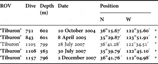
Other material: the station data for the other material examined are shown emboldened in Table 2. These remain in the collections of P.R.P. One further specimen was identified from in situ photographs but was not collected.
List of specimens of Resomia persica sp. nov. examined (emboldened) and/or identified from in situ frame grabs (*).

DIAGNOSIS
Nectophores with complete upper lateral ridges. Lower lateral ridges weak and relatively short, ending on a level with the top of the nectosac. Two pairs of lateral ridges, one pair of which can connect with upper lateral ridge and runs to the ends of the lateral ectodermal tracts extending from the ostium. The second pair is short, not connecting with any other ridge. Nectophoral buds with digitate processes arising at mid-length of upper lateral ridges.
Bracts kite-shaped with complete transverse ridge and distal group of nematocysts. Cnidoband of zigzagged tentilla with short fourth zag (cf. Resomia convoluta) and entirely enclosed by involucrum; the latter not forming a narrow tube around the proximal end of the terminal filament as in R. convoluta.
DESCRIPTION
Photographs of specimens from ‘Tiburon’ Dives 843 and 1108 are shown in Figure 12. The Dive 843 specimen was very young and retained only a single fully developed nectophore, although the in situ frame grabs showed before capture in possessed five. Unfortunately there are no photographs for the complete type specimen. Like Resomia ornicephala in situ specimens of this species (Figure 12) were extremely transparent, particularly in the nectosomal region where no pigmentation was observed. The siphosomal zooids, apart from the bracts and mature gastrozooids, had a milky white appearance, and the cnidobands of the tentilla were white or pale yellow. The only other pigmentation seen, particularly on the small specimens, was the peach coloration of the stomach region of the more mature gastrozooids. Nonetheless, the species was easily distinguished from R. ornicephala by the large white patches, containing nematocysts, at the distal ends of the bracts as opposed to the arcs of nematocysts seen on those of R. ornicephala.
Resomia persica sp. nov. (A & B) In situ pictures of specimen from ‘Tiburon’ Dive 1108. Specimen ~10 cm in length; (C) small specimen from ‘Tiburon’ Dive 843. Scale bar: 5 mm.

Pneumatophore: the pneumatophore of the type specimen measured 2.4 mm in length and 0.9 mm in diameter. It showed no characteristic features and was without any obvious pigmentation. The gas within the pneumatosaccus had expanded and ruptured the base of the gas-gland (pneumadenia) so that gas passed into the gastrovascular cavity of the nectosome, causing the gas-gland to become detached from the base of the pneumatophore.
Nectosome: the nectophores were clearly budded off on the ventral side of the nectosome. No pigmentation could be seen in either the stem or the nectophores, which were completely transparent.
Nectophore: fourteen nectophores at various stages of development were found with the type specimen, and some small buds were still attached to the nectosome. The young specimen from ‘Tiburon’ Dive 843 retained only a single nectophore and a young bud (Figure 13).
Resomia persica sp. nov. Photographs of nectophore bud from ‘Tiburon’ Dive 843 specimen. (A) Ostial view; (B) axial view; (C) lateral view; (D) upper, slightly lateral, view. Scale bar: 0.5 mm.

The young nectophoral bud (Figure 13) showed characteristic digitate processes; reminiscent of those described for Resomia similis (see Pugh, Reference Pugh2006). However, whereas in R. similis the digitate processes were attached toward the proximal end of the axial wings, in R. persica they were attached approximately at the mid-length of the upper lateral ridges. As the nectophore buds enlarged so did the bases of these digitate processes (Figure 14, ldp), thus forming triangular flaps. The upper lateral ridges (rul) could be seen to cross these processes before continuing distally to reach the ostium. The lower lateral ridges (rll) united with the upper laterals toward their proximal ends. They were relatively short and in the young buds extended to only about one-third the length of the nectophore and with nectophoral growth became very indistinct. Just distal to where the digitate processes were inserted each upper ridge divided off a lateral ridge (rl), although the connection was weak and in more developed nectophores it was difficult to tell whether such a connection still existed. These clearly visible lateral ridges continued obliquely across the lateral sides of the nectophore to reach the ends of the lateral ectodermal tracts extending from the ostium, which were marked by clumps of up to twelve nematocysts (ne). These nematocysts measured 140 × 21 µm and were probably microbasic mastigophores; although no devaginated ones were found. In addition to these ridges there was another pair of weak ridges (ral) that ran obliquely across the lateral surfaces of the nectophores, from upper proximal to lower distal and paralleled the lateral ridges but did connect with any of the other ridges. As the nectophores grew these ridges became less and less distinct.
Resomia persica sp. nov. Ostial (left) and adostial (right) views of very young nectophore from type specimen. ldp, lateral digitate process; ne, nematocysts; pc, pallial canal; pec, patches of ectodermal cells; ral, additional lateral ridge; rl, lateral ridge; rll, lower lateral ridge; rul, upper lateral ridge. Scale bar: 1 mm.

The nectosac occupied the majority of the nectophore and bore straight radial canals. Both ascending and descending pallial canals (pc), at this stage approximately equal in length, were present. Patches of large ectodermal cells, probably photocytes (pec), were found on the velum close to the ostial ring canal. These were situated on either side of the point of insertion of the upper canal and on the lateral sides, mainly below the insertion of the lateral canals.
Most of the mature nectophores of the type specimen were flaccid and very flimsy, several having almost disintegrated, and their characters were difficult to distinguish. They measured up to 10 mm in length and 11 mm in width. A younger, next up in size from that shown in Figure 14, and a medium-sized nectophore have been drawn in upper view (Figure 15). The nectophores somewhat differed from the heart-shape of the previously described Resomia species (see Pugh, Reference Pugh2006), in being narrower and having extensive axial wings (Figure 15, aw). The thrust block (tb) was very weakly demarcated as were most of the ridges. The upper lateral ridges (rul), as they approached the ostium, diverged laterally from each other. The lateral ridges (rl) appeared to connect with the upper laterals, although the connection was often weak. The lower laterals (rul) arose from the junction with the upper laterals on the proximal tips of the axial wings, but were very weak and terminated on a level with the top of the nectosac. The additional pair of lateral, or vertical lateral, ridges (ral) became difficult to distinguish in the larger nectophores.
Resomia persica sp. nov. Upper views of a younger (left) and medium-sized (right) nectophores of the type specimen. aw. axial wing; et, ectodermal tract; tlp, terminal patch of nematocyst at end of lateral ectodermal tracts extending from the ostium. For other annotations see legends to Figures 3 & 4. Scale bar: 1 mm.

The lateral ectodermal tracts extending from the ostium were now less distinct, and comprised four rows of ovoid cells arranged regularly with their long axes in line with the axis of the tract. The tract expanded at its end into a relatively small ovoid patch of cells (tlp) that were not nematocysts. Its central region contained large circular cells, with large nuclei, surrounded by a series of smaller ovoid cells. Presumably the nematocysts present on the younger nectophores had been lost, by abrasion or devagination and subsequent disposal, and not replaced.
Ascending and descending pallial canals (Figure 15, pc) were present, with the upper canal, which ran up onto the upper surface, being longer then the lower one. The pedicular canal ran straight to the nectosac and was inserted close to the proximal (axial) face of the nectosac on its lower side. The canal gave rise to all four radial canals that ran straight to the ostium. The nectosac occupied the distal half of the nectophore and did not have a muscle-free zone. The ostium was widely open with few signs, in larger nectophores, of any large ectodermal cells in the vicinity of the ring canal (see Figure 14, pec). This is probably the result of abrasion as these nectophores were in poor condition. When present, the cells on either side of the upper canal insertion onto the ring canal were large and spherical, and probably functioned as sources of bioluminescence. Because of the rarity of specimens, however, we were unable to confirm the luminescence for this species. The lateral groups of cells, when present, were usually more obvious than upper ones, and often formed swollen clumps that usually were inserted below the level of the insertion points of the lateral canals. The single nectophore from ‘Tiburon’ Dive 843 had a broad, but short strip of cells on either side of the junction of the upper canal with the ring canal. There was an additional pair of short ectodermal tracts (et) from the ostium, lying midway between the points of insertion of the lateral and lower canals onto the ostial ring canal. These had a short narrow stalk and a small, expanded terminal region. Such structures were not found on the younger nectophores.
Siphosome: in mature specimens (Figure 12) the siphosomal stem was colourless and its component zooids, apart from the bracts, had a milky white appearance. From the in situ photographs of specimens it appeared that the siphosome was permanently contracted. In the youngest specimen, from ‘Tiburon’ Dive 843 (Figure 12), there was no sign of any pigmentation apart from some peach coloration in the stomach region of the only well-developed gastrozooid. In this specimen the distinctly whitish distal tips to the bracts formed by clusters of nematocysts were very evident, as they were relatively large in comparison to the size of the bract itself. Nonetheless they could still be seen clearly on the older specimens, whose bracts were much larger while the size of the cluster of nematocysts remained the same or decreased.
Again the clustered nature of the siphosome made it impossible to discern the exact arrangement of the zooids in each cormidium. However, there were on average 6 palpons and ~30 bracts to each gastrozooid.
Bract: the type specimen included over 160 bracts of which the great majority were elongate and narrow (Figure 16A), with the distal region narrower than the proximal one. They measured up to 15 mm in length and 5.5 mm in width. A few of the bracts were somewhat shorter and wider than the others (Figure 16B, C), measuring up to 10 mm in length and 7.2 mm in width. The 40+ bracts of the youngest specimen (‘Tiburon’ Dive 843) were all of the narrow, elongate type and measured up to 7.5 mm in length and 3.5 mm in width.
Resomia persica sp. nov. Upper views of bracts. Proximal end at bottom, distal at top. (A) Small; (B) medium; (C) large bracts from the type specimen. nec, cluster of nematocysts at distal end of bracteal canal. For other annotations see legend to Figure 6. Scale bar: (A) 1 mm; (B) 2 mm; (C) 3 mm.

Both types of bract had a complete transverse ridge on the upper side (Figure 16, tr) separating the thinner distal facet (df) from the thicker proximal part. For the elongate bracts this ridge was usually positioned at its mid-length, although this was somewhat variable. Distal to this ridge the bract was narrower than proximal to it. For the squatter bracts the position of the transverse ridge varied considerably, unrelated to their size, generally lying closer to the distal end of the bract, and distal to it there was a broad triangular facet. In both types, distal to the transverse ridge, the bract rapidly lost depth on its upper side so that it was only half as thick as it was just proximal to the ridge. For the squatter bracts it then tapered down both laterally and distally, so that the distal end was pointed. For the elongate bract, the facet tapered down only gradually in the distal direction so that the distal end of the bract was blunt, but laterally it tapered down rapidly so that in cross-section the distal part of the bract was approximately triangular.
Some of the largest of the elongate type of bract bore a single small lateral cusp on the more proximal part of the distal facet. In general this was positioned on the outer side of the bract (Figure 16, lc), but occasionally there was a cusp on both sides. A few bracts from the ‘Tiburon’ Dive 1157 specimen showed a further enlargement of this pair of cusps to form relatively large flaps on the lower lateral sides of the bract just distal to the transverse ridge, but such were not seen with the type specimen.
Small patches of ectodermal cells were found on the upper side of the bract just proximal to the transverse ridge. These have not been illustrated as they were very indistinct, without staining, and were variable in their disposition and shape. The cells themselves were small with darkly staining nuclei. The number of these patches varied from zero to five, and the number of cells in each from four to ~50.
Proximally, the bracteal canal extended on to the upper side of the bract, then curved over onto the lower side and ran distally, remaining in contact with the lower wall of the bract for about one-quarter of its length (Figure 16, bc). In the youngest bract, the canal in this region was surrounded by relatively thick endodermal walls, with a reticulated appearance, but this was not apparent in the older ones. The canal then penetrated into the mesogloea and ran obliquely to the distal tip of the bract, where it terminated below a cluster of nematocysts (nec). There were ~150 nematocysts in the distal cluster on the youngest bracts, which were almost certainly microbasic mastigophores and which were relatively large, measuring 160 × 26.5 µm. The shaft length of the devaginated nematocysts was ~220 µm. In the larger bracts not only did the number of nematocysts decrease, possibly by abrasion, to ~50, but they were also slightly smaller, measuring ~130 × 21 µm.
Gastrozooid and tentacle: only three mature gastrozooids, with tentacles bearing tentilla, were present with the type specimen. The largest measured ~12 mm in length. The proximal basigaster was greatly expanded (Figure 17A, bs), up to 1.2 mm in diameter, while the stomach (st) and proboscis (pr) regions formed a narrow tube, with hepatic stripes visible in the latter region. The stomach region contained peach-coloured pigmentation, the density of which depended on the degree of contraction of the gastrozooid.
Resomia persica sp. nov. (A) Gastrozooid and tentacle showing both forms of tentilla; (B) fully formed spirally-coiled tentillum. For (B–D) proximal end at top, distal at bottom; (C) tentillum in the process of reconfiguration; (D) fully formed zigzagged tentillum. bs, basigaster; eb, elastic band; in, involucrum; pr, proboscis; st, stomach; tf, terminal filament. Scale bar: (A) 1 mm; (B–D) 0.5 mm.

The course of development of the tentilla was the same as has been described for other Resomia species (Pugh, Reference Pugh2006). The youngest were simply undifferentiated straight tubes that, as they increased in length, differentiated the three main zones: pedicle, cnidoband and terminal filament (cf. Figure 7B). The terminal filament then began to coil up, the cnidoband began to form loose spirals, and the pedicle began to lengthen (Figure 17A). At this stage no mastigophores could be seen at the proximal end of the cnidoband. Only when the cnidoband had formed ~6 tight spirals did these nematocysts begin to appear. The pedicle continued to elongate and the first signs of the involucrum appeared. The involucrum then increased in length until it covered almost the entire cnidoband (Figure 17B), being widely open at its distal end. The tentilla then began to change their configuration. This transformation consisted of an unwinding and straightening out of the cnidoband, which then extended beyond the involucrum (Figure 17C). Following this it began to form into zigzags within the involucrum, culminating in a cnidoband with 3.25 zags. At this stage the involucrum did not entirely cover the cnidoband, but at a later stage it extended further to cup not only the cnidoband but the entire contracted terminal filament (Figure 17A, in; Figure 17 D). It did not form a narrow tube around the terminal filament as in R. convoluta (see Pugh, Reference Pugh2006), but remained widely open. The elastic band (Figure 17A, eb) could clearly be seen to connect the pedicle to the distal end of the cnidoband. The means of activation of the cnidoband is presumably the same as for R. ornicephala, as is discussed below, but would also involve the rupture of the flimsy involucrum.
As previously described for resomiid species (Pugh, Reference Pugh2006), the cnidoband consists of two types of nematocysts, with rows of microbasic mastigophores (74–80 × 18.5–21 µm) lying laterally and a large number of anisorhizas (~55 × 8 µm) lying between them. On the form of tentillum with a zigzagged cnidoband, microbasic mastigophores were only present on the proximal zag. At the proximal end of the cnidoband there were two of these nematocysts on either side in each row. By the seventh row their number had increased to three but at about half the length of the proximal zag decreased again to two for the next five rows, and then to one per row. Distally only five pairs of these nematocysts were counted and they became increasingly spaced apart with the most distal pair being situated at about 3/4 the length of the proximal zag. The anisorhizas filled the space between the lateral rows of mastigophores. Proximally, there was only a single anisorhiza between the mastigophores, but this gradually increased to 5–6 per row at the point where the mastigophores reduced from three to two pairs in each row, and at the point where the they finally disappeared there were about 16 per row. The middle zag of the cnidoband was wider than the other two and comprised approximately 24–30 rows of anisorhizas. Although the third zag appeared narrower, it too had ~24 rows of anisorhizas, with the number reducing to 18 on the short fourth zag. The terminal filament bore what were presumed to be acrophores (32–34.5 × 8–9 µm) and relatively large desmonemes (30 × 10 µm) in an apparent ratio of about 4:1.
The arrangement of the nematocysts on the form of tentillum with a spirally coiled cnidoband was basically the same as for the zigzagged form. Microbasic mastigophores were only present laterally on the proximal 2–3 spirals. Most proximally there were two of these nematocysts on either side, which then increased to three in the eighth row, decreasing again to two and then one by the fifteenth row. There were then about 9 individual mastigophores on each side, which gradually became spaced further and further apart before disappearing altogether. Only a few anisorhizas were found in the proximal two spirals.
Palpon: in the preserved state the palpons had a milky white appearance and measured up to 7 mm in length, but were variously laterally expanded, measuring up to 2 mm in diameter (Figure 18). The surface of the palpon was covered in irregular groupings of 6–12 rounded ectodermal cells, which in the living specimens was suffused with peach-coloured pigmentation. There usually was a distinctive distal proboscis region, and a small basal disc from which the palpacle arose. No annulations were discerned on the palpacle. No nematocysts were found on any part of the palpon itself, but a row of irregularly spaced small desmonemes (6.5 × 5.5 µm) was present along one side of the palpacle.
Resomia persica sp. nov. Palpon. Proximal end at bottom, distal at top. Scale bar: 1 mm.

Gonophores: only two mature male gonophores (Figure 19A) were found with the type specimen, and these measured 7.9 mm in length and 0.9 mm in diameter.
Resomia persica sp. nov. (A) Mature and (B) developing male gonophore; (C) mature female gonophore. Proximal end at bottom, distal at top. Scale bar: (A & C) 1 mm; (B) 0.5 mm.

However, a large number of developing ones (Figure 19B) was also found. There was a ring of nematocysts, probably microbasic mastigophores and measuring 32 × 13 µm, around the distal opening of each gonophore. The mature female gonophores (Figure 19C) measured up to 2.6 mm in length and 1.2 mm in diameter, with their proximal halves comprising a conical mesogloeal process through which the canal from the gastrovascular cavity penetrated. The single large egg filled most of the distal half and, again there was a ring of nematocysts, up to 80 in number, surrounding the distal mouth opening. Because of the extreme contraction of the stem it was not possible to discern the arrangement of the gonophores in each cormidium. However, on one part of the stem a ventral swelling was found to have individual female gonophores budding from most of its surface, while a cluster of male gonophores all arose from a single stalk.
DISTRIBUTION
Only five specimens of Resomia persica have been positively identified, four of which were collected. They all came from a very limited area of the north-eastern Pacific Ocean (see Table 2) within Monterey Bay. The specimens occurred over a relatively narrow depth-range (563–799 m).
REMARKS
The fragility of the mature nectophores would probably mean that they would be difficult to recognize if the specimen was in poor condition. Otherwise the characteristic ridge pattern, particularly the very short lower laterals, and the terminal swelling at the end of the fairly long lateral ectodermal tracts extending from the ostium are characteristic. In addition, if nectophoral buds are present, the position of the digitate processes from the upper lateral ridge again is characteristic. The elongate kite-shaped bracts, with a transverse ridge on the upper surface and a distal cluster of nematocysts over the end of the bracteal canal, would also help in identification. The tentilla are very similar to those of Resomia convoluta (see Pugh, Reference Pugh2006), in particular the zigzagged form where there is a short fourth zag with the terminal filament arising from its end. However, there is a slight difference in that the involucrum, although enclosing the whole cnidoband, does not form a narrow tube around the proximal part of the terminal filament as it does in R. convoluta. Because of the rarity of the specimens and their fragility they were not subjected to tests to discover whether any parts of them were bioluminescent.
ETYMOLOGY
The species name persica is from the Latin for peach, and refers to the pale orange coloration of the stomach region of the gastrozooids, together with the fuzzy look of the young bracts, and the overall fragility of the organism.
TYPE MATERIAL
All collected specimens were fixed in 5% borax-buffered formalin and later transferred to Steedman's preserving fluid.
Holotype: specimen collected during the ROV ‘Ventana’ Dive 2974 from a depth of 979 m (21 February 2007; 36°41.74′N 122°03.55′W). The specimen has been deposited at the United States National Museum (Smithsonian Institution), Washington, DC (USNM 1121485).
Paratype: specimen collected during the ROV ‘Tiburon’ Dive 856 from a depth of 462 m (9 January 2005; 36°36.22′N 122°22.55′W). The specimen has been deposited at the United States National Museum (Smithsonian Institution), Washington, DC (USNM 1121486).
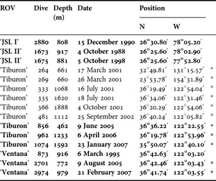
Other material: the station data for the other material examined are shown emboldened in Table 3. The three specimens collected by the ‘Johnson Sea Link’ submersibles also have been donated to the USNM (USNM 1121487-9), while the remainder are housed in the collections of P.R.P. The remaining data refer to specimens identified from in situ photographs but not collected.
List of specimens of Resomia dunni sp. nov. examined (emboldened) and/or identified from in situ frame grabs (*).

DIAGNOSIS
Nectophores with upper lateral ridges that demarcate the edges of the axial wings. Just proximal to the ostium they diverge laterally and continue outwards, terminating close to the short lateral ectodermal tracts extending from the ostium. Lower lateral ridges absent. Pair of indistinct but quite separate lateral ridges. Nectophoral buds without digitate processes.
Bracts kite-shaped with complete transverse ridge and distal group of nematocysts. Patch of distinct ectodermal cells on upper side proximal to transverse ridge. Proximal tentilla with coiled cnidoband including one reverse turn. Involucrum covers distal end of pedicle. After reconfiguration distal tentilla with involucrum covering cnidoband, which consists of individual coils and zags.
DESCRIPTION
Photographs of three complete specimens, including the type specimen, are shown in Figure 20. Like the other two Resomia species described herein, R. dunni appears, from the in situ photographs, to have a permanently contracted siphosome. Similarly the nectophores are extremely transparent and without any pigmentation, although the nectosomal stem and the pneumatophore can be suffused with orange-red coloration, particularly in the smaller specimens (Figure 20B). However, the deep orange-red pigmentation of the gastrozooids and palpons on the siphosome clearly distinguishes this species from the other Resomia species. Although, like R. persica, the white spots at the distal ends of the bracts represent an accumulation of nematocysts, in R. dunni they are better defined as cupulate processes rather than their more fuzzy appearance in R. persica. One other distinguishing feature is that the specimens often appear to bear only one well-developed gastrozooid and tentacle.
Photographs of Resomia dunni sp. nov. from: (A) ‘Tiburon’ Dive 366 (taken in situ, estimated length 15–20 cm); (B) ‘Tiburon’ Dive 962 (stem length approximately 4 cm); (C) ‘Ventana’ Dive 2974 (5 cm in length).

Pneumatophore: the pneumatophore of the type specimen measured 1.7 mm in length and 0.85 mm in diameter. To a greater or lesser extent, perhaps dependent on the size of the specimen, it was suffused with a light orange to deep red coloration, but otherwise showed no characteristic features.
Nectosome: like the pneumatophore, the nectosomal stem was suffused, to a greater or lesser extent, with a light orange to deep red coloration (Figure 20). The nectophores, which were attached to the ventral side of the nectosome, were mainly colourless, although occasionally the radial canals had some light orange pigmentation in their walls.
Nectophore: eleven detached nectophores remain with the type specimen, together with some young buds still attached to the nectosome immediately below the pneumatophore. This specimen was not one of the largest known as, for instance, the ‘Tiburon’ Dive 366 (Figure 20A) specimen, which was observed but not collected, possessed thirty-five, fully developed nectophores. However, most of the specimens collected had about the same or fewer nectophores as the type specimen.
The young nectophoral buds showed no signs of any digitate lateral processes, as were noted on those of both Resomia similis and R. persica. They, and a slightly larger young nectophore (Figure 22) showed no signs of a thrust block or axial wings. The ridge system was, however, very distinct and consisted of a pair of upper lateral ridges (Figure 22, rul), which demarcated a median groove on the upper side of the nectophore, and a pair of entirely separate laterals (rl). No lower lateral ridges were present. Proximally, the upper lateral ridges extended (Figure 22C) to the approximate level of where the pedicular canal (ped) was inserted onto the nectosac. Distally, they diverged as they approached the ostium (ost) and continued out laterally, ending just short of the lateral ectodermal tracts (let) extending from the ostium, without reaching the ostium itself. The upper median groove was quite shallow on the youngest buds, but deepened considerably as the nectophore enlarged (Figure 22C).
Photographs of tentilla from type specimen of Resomia dunni sp. nov. before preservation. (A–D) Proximal tentilla; (E–H) distal tentilla. eb, elastic band; in, involucrum; scn, distal straight part of cnidoband. Scale bar: 1 mm.

Young nectophore of Resomia dunni sp. nov. (A) Upper, (B) lower and (C) lateral views. let, lateral ectodermal tract; ost, ostium; pec, patch of ectodermal cells; uet, upper ectodermal tract. For other annotations see legend to Figure 3. Scale bar: 1 mm.

On the young nectophores the ascending pallial canal (Figure 22, pc) was almost twice as long as the descending one. The pedicular canal ran directly to the nectosac, which occupied the majority of the nectophore, where it gave rise directly to the four radial canals. Because of the deep median groove on the upper side of the nectophore, the nectosac possessed large lateral wings over which the lateral radial canals looped first upwards and then downwards toward the ostial opening, which lay obliquely on the lower side of the nectophore at its distal end. Distinct upper (uet) and lateral (let) ectodermal tracts arose from the ostium, but no nematocysts were found in association with them. The lateral tracts tended to increase in width away from the ostium. In addition, groups of large ectodermal cells, probably photocytes were found close to the ostial ring canal in the vicinity of the insertion of the upper radial canal, and extending down the ostium from the insertion points of the lateral radial canals. There was also a small patch of ectodermal cells (pec), also probably photocytes, on each side of the nectophore, lying over the lateral radial canals toward the apex of the nectosac. This is the only Resomia species found to have these lateral patches on the nectophores, although they are known to occur on several other physonect species. Because of the fragility and rarity of the specimens their potential bioluminescence was not investigated.
As the nectophores grew, axial wings were developed and increased considerably in size such that on an intermediate-sized nectophore (Figure 23, aw) they occupied about one-third of its length. The thrust block (tb) formed a small swelling between these wings. The upper lateral ridges (rul) demarcated the edges of these wings and continued distally toward the ostium (ost) but, as in the young nectophores, they diverge laterally before reaching it and terminated close to the lateral ectodermal tracts (let) extending from the ostium. Proximally the median gutter between the ridges was wide and deep, while distally it was narrower and shallower. The lateral ridges (rl) became very indistinct, particularly in their upper regions.

On more developed nectophores, the ostium opened obliquely onto the distal end of the nectophore but otherwise the arrangement of the radial canals and lateral ectodermal tracts from the ostium remained the same. The photocytes around the ostium became less obvious. The ascending pallial canal was still twice the length of the descending canal. The lateral patches of ectodermal cells (Figure 23, pec) remained small and thereby less conspicuous.
The largest nectophores (Figure 24) differed little from those described above, except that the upper lateral ridges were further apart from each other and that the thrust block increased considerably in size. In preserved specimens the latter often became folded distally such that it was not as conspicuous as on the living nectophores. The lateral ridges were indistinct and their course difficult to follow. Similarly the lateral patches of ectodermal cells remained small and inconspicuous. The ostial opening was now at the distal end of the nectophore.
Resomia dunni sp. nov. Larger nectophores. Upper (left) and lower (right) views. Scale bar: 2 mm.

Siphosome: the in situ photographs showed the siphosome to be permanently contracted and rigid (Figure 20). In the larger specimens (Figure 20A) the siphosomal stem and its component zooids, apart from the bracts, had orange-red pigmentation, with the often single mature gastrozooids being a deeper red. In the smaller specimens (Figure 20C) the coloration was much paler, but again only one gastrozooid seemed to be well developed. The clusters of nematocysts at the distal tips of the otherwise transparent bracts were visible as white dots.
Many of the gastrozooids on the siphosome were so slightly developed that they were indistinguishable from other developing zooids and, therefore, could not be enumerated. In general the ratio of the number of bracts to that of palpons was approximately the same as that found for the previous two species, i.e. approximately 5:1, although again there was much variation between the individual specimens.
Bract: the bracts measured up to ~9 mm in length and 7 mm in width. They were kite-shaped and bore a complete transverse ridge (Figure 25, tr) on the upper surface that divided off the larger proximal facet from the smaller distal one. On the preserved bracts this transverse ridge usually slightly overhung the distal facet (df), and at its inner end it could form a slight cusp (lc), particularly in the larger bracts. In the youngest the whole surface of the bract was mottled with small protuberances, particularly on the distal facet, but these were less obvious or absent on the larger ones, and may have been a preservation artefact. However, most bracts had a distinct patch of small ectodermal cells (pec), with darkly staining nuclei, on the upper side and a short distance proximal to the transverse ridge. This patch was asymmetrically placed, a short distance from the mid-line on the outer side of the bract, although for the ‘Tiburon’ Dive 962 specimen these patches were often further displaced laterally. Very occasionally the patch was absent and possibly had been abraded, although no evidence for this could be found. It is presumed that the cells were photocytes and produced bioluminescence but this was not investigated further.

At the proximal end of the bract the bracteal canal extended a slight distance on to the upper side. The canal was thickened where it remained in contact with the lower wall of the bract (bc), for about one-third to one-half its total length, but once it passed into the mesogloea (pbc) it became considerably thinner. The canal continued to the small swollen process at the distal tip of the bract and ended below a distinctive cupulate cavity that was filled with nematocysts (nec). There were about 100 nematocysts housed in these processes, although for the larger bracts their number was often greatly reduced or the cavity was completely empty. The nematocysts were microbasic mastigophores. For the type specimen they measured up to 380 µm in length and 55 µm in diameter, but for the ‘Tiburon’ Dive 962 specimen they were somewhat smaller—275 × 50 µm. The shaft length was also about 275–300 µm in length and the total length of the thread reached at least 3.4 mm.
Gastrozooid and tentacle: all the specimens examined had only one well-developed gastrozooid (Figure 20A). This consisted of a large cupulate basigaster attached directly to the stem and to which the tentacle was attached. The stomach region often was narrow proximally, clearly distinguishing it from the basigaster, but greatly swollen distally. The proboscis region was often widely open and distinct hepatic stripes could be seen.
Quite why each specimen only has one fully developed gastrozooid and tentacle is unknown, but it made the study of the development of the tentilla difficult as so few were present and often they became detached and lost during collection. In actuality only 10 well-developed tentilla were found with the type specimen, while the other specimens examined had less. No tentilla of the transformed type were found with the ‘Johnson Sea Link’ specimens.
The tentilla arose in the usual way as simple undifferentiated tubes on which, subsequently, the three main regions of the tentillum were differentiated. The cnidoband then began to become loosely coiled and, at the same time, large microbasic mastigophores began to accumulate in the pedicle causing a part of it to become swollen. These mastigophores measured 190 µm in length and 80 µm in diameter, but they were not as yet fully formed. With regard to the other types of nematocyst, it was not clear when the anisorhizas appeared within the cnidoband or the desmonemes and acrophores within the terminal filament, but at the start of the accumulation of mastigophores no other types of nematocyst were noted within the tentillum. The mastigophores then began to move into the cnidoband, forming a single line on each side and extending along the more proximal three to four coils, but becoming more and more widely spaced distally. After this stage the pedicle was depleted of nematocysts, but its distal end remained swollen.
The development of the involucrum (Figure 21A–D) only started once the cnidoband was fully formed. Because of the dearth of material it was difficult to obtain an exact picture of its development, but it appeared to start to develop from close to the base of the distal swelling on the pedicle. At first it did not form a complete ring around the pedicle, being only present over about two-thirds of its circumference. As it developed further it became deeply emarginate in the centre of its distal margin, but at a later stage this emargination disappeared, and it began to grow entirely around the circumference of the pedicle. The involucrum initially extended in a distal direction, but as it reached its maximum size, on the spirally-coiled form of tentillum, it folded back to form an inflated covering to the distal end of the pedicle (Figure 21D, in). The pedicle itself lost its distal swelling and the involucrum now appeared to be attached at its distal end. Within the cnidoband there was always a reversal in the direction of coiling (Figure 21A arrowed), which occurred only once but at any point along its length.
At some stage it is presumed that the involucrum increased in size and came to enclose the cnidoband as the latter began its transformation, although no tentilla were observed in this state. Unlike in the Resomia species described above this transformation did not result in a regular zigzagged arrangement of the cnidoband but in a mixture of coils and zags, although the distal third of the cnidoband was usually quite straight (Figure 21E, scn). The involucrum (Figure 21E, in) came to enclose the entire cnidoband and the proximal part of the terminal filament (Figure 21F–H), although occasionally the distal end of the cnidoband projected out from a relatively small distal opening (Figure 21E). Although the arrangement of the cnidoband appeared haphazard it actually was quite consistent for all the tentilla examined. The double elastic band (Figure 21E, eb) was the only part of the cnidoband that connected, proximally, with the pedicle. The proximal end of the cnidoband was loosely attached to this band just distal to that point, and quite often the connection between them was broken. The double elastic band then passed through the involucral capsule quite separate from the cnidoband itself before uniting with the distal end of the cnidoband. In the preserved specimens the involucra covering the cnidobands of the distal tentilla (Figure 21E-H) usually became detached and their flimsy nature meant that they were easily torn.
As mentioned above, microbasic mastigophores formed a single row on either side of the cnidoband. There were approximately 100 on each side and, in the mature cnidoband, they measured up to 280 × 45 µm. Many devaginated ones were found and these had a shaft length of ~300 µm, with numerous long spines being present on the distal half. Between the mastigophores were innumerable anisorhizas of variable size ranging from 75 × 10 µm to 120 × 22 µm. At the proximal end of the cnidoband there were only about 4–6 rows of these anisorhizas, but the number of rows gradually increased until there were at least 24 on the more distal parts of the cnidoband. Desmonemes (up to 50 × 23 µm) and acrophores (~30 × 13 µm) were present in the terminal filament, but their exact arrangement could not be assessed.
Palpon: the palpons of the type specimen measured up to 5 mm in length and 0.7 mm in diameter, appearing as long, narrow cylindrical structures (Figure 26, centre). The proximal half of the palpon was covered in rounded vesicles of various sizes, while the distal half appeared more granular. However, in life, at least for the ‘Tiburon’ Dive 856 specimen, the surface of the palpon was covered in irregular shaped and dispersed patches of cells containing orange-red pigmentation, and there was a concentration of red pigment in its gastrovascular cavity just proximal to the mouth opening. A long filiform palpacle arose from its base and appeared to have annulations only in the part closest to where it was attached. The most characteristic feature of the palpon was that, at its base and on the opposite side to the attachment point of the palpacle, there was a protuberance consisting of large ovoid structureless vesicles.
Resomia dunni sp. nov. Palpons of specimen from ‘Tiburon’ Dive 962 (left and right) and ‘Ventana’ Dive 2974 (centre). Proximal end at bottom, distal at top. Scale bar: 1 mm.

Some of the palpons of the ‘Tiburon’ Dive 962 specimen (Figure 26 left, right) were different in that they were globular structures up to 6 mm in length and 4 mm in diameter. Most of their surface, apart from a distinctive granular ring around the base of the proboscis, was covered in rounded vesicles of various sizes, but these were denser in the proximal half, while in the distal half they were more sparsely distributed with clear patches between them. The distal proboscis region usually had a distinctive cone-shape structure. The basal protuberance, on the opposite side to the attachment point of the palpacle, was prominent and constructed in the same way as for the type specimen. On one of these palpons (Figure 26, left) the large ovoid cells only formed an annulus around a central denuded, sunken region. It is most likely that this region had been damaged in some way.
On one side of the palpacle there was an irregular series of desmoneme nematocysts (Figure 27) measuring 18 µm in length and 10 µm in width. Similarly sized desmonemes were also found in the region of the proboscis of the palpon, but there they were quite sparsely distributed.
Resomia dunni sp. nov. Desmonemes (18 µm long) on one side of a palpacle of type specimen.

Gonophore: the type specimen only bore immature gonophores, while the ‘Tiburon’ Dive 962 specimen bore larger ones of both sexes (Figure 28). The male gonophore may still have been immature, but measured up to 1 mm in length and 0.6 mm in diameter. The female ones were somewhat larger—up to 1.5 mm in length and 1.2 mm in diameter. Neither showed any characteristic features, nor were any nematocysts found in the region of the subumbrella opening. The gonophores of each sex formed clusters originating from a single gonostyle. However, the contracted condition of the preserved specimen prevented their arrangement within a cormidium to be assessed.
Resomia dunni sp. nov. Male (left) and female (right) gonophores from ‘Tiburon’ Dive 962 specimen. Proximal end at bottom, distal at top. Scale bar: 0.5 mm.

DISTRIBUTION
Three specimens of Resomia dunni have been collected by ‘Johnson Sea Link’ submersibles in the ‘Tongue of the Ocean’, The Bahamas, and six others by the ROVs ‘Tiburon’ and ‘Ventana’, with a further six having been identified from in situ low definition frame grabs, from the north-eastern Pacific (Table 3). All but one of the north-eastern Pacific specimens have been found in an extremely restricted area, of less than 100 km × 100 km, west of Monterey Bay, California, the exception coming from the Gulf of California. They were found over a fairly extensive depth-range, from 462 to 1888 m, although most of them occurred at depths >900 m. The Bahamian material came from depths between 808 and 917 m.
Remarks: although the Bahamian material lacked any tentilla of the second form, and indeed very few tentilla at all, all the other characters were in accord with the Pacific material, especially the patch of ectodermal cells on the bracts and the basal swelling on the palpons. Similarly only one large gastrozooid was found with the siphosomal fragments. There is no doubt in our minds that all the material belongs to the same species.
As with the other two Resomia species described herein, the mature nectophores of R. dunni are very flimsy and may be difficult to recognize. However, the presence of large axial wings, a relatively large thrust block, the absence of lower lateral ridges, and the small lateral patch of ectodermal cells may serve to identify them. The presence of a similar patch of cells on the upper surface of the bracts, proximal to the transverse ridge, is also a distinguishing feature, as is the distinct swelling at the base of the palpon, on the opposite side to where the palpacle is attached. However, by far the most characteristic feature of R. dunni are the tentilla. The proximal, spirally coiled ones can be distinguished on the basis of the shape of the involucrum and the reversal in spiralling at some point along the cnidoband. The distal tentilla are quite unlike any of Resomia species in being only partially zigzagged and partially coiled, and entirely enclosed in a bag-like involucrum.
The method of activation of the cnidobands of these distal tentilla most probably will be along the same lines as the other Resomia species, as is discussed below, despite the difference in its arrangement, but the dearth of available material does not shed any light on this. As mentioned above, it is entirely unclear why, for the specimens observed, there appeared to be only one well-developed gastrozooid and tentacle. It would seem unlikely that the abundance of prey over the depth-range that R. dunni has been found would be sufficient to allow so little fishing effort.
ETYMOLOGY
Resomia dunni is named after Casey W. Dunn, for his important contributions to siphonophore phylogeny, taxonomy, and biology.
DISCUSSION
Pugh (Reference Pugh2006) diagnosed the genus Resomia as ‘Physonect siphonophores with simple heart-shaped mature nectophores, bearing only upper and lower lateral ridges. Ascending and descending pallial canal present; straight radial canals on nectosac. Tentacles bearing two forms of tentilla; the more proximal ones, with spirally coiled cnidobands, transforming into the more distal ones, with a zigzagged cnidoband. Monoecious.’ At that time only two species, R. convoluta (Moser, 1925) and R. similis (Margulis, 1977), had been described.
Although neither of these species was included in the molecular phylogenetic study by Dunn et al. (Reference Dunn, Pugh and Haddock2005a), Pugh (Reference Pugh2006) surmised that the genus belonged to the ‘Monoecious’ group of Physonectae not only because the species were monoecious, but also because a descending pallial canal was present on the nectophores. As the nectophores were budded on the ventral side of the nectosome, the genus did not belong to the family Agalmatidae sensu stricto and like several other genera that had once been included in that family (see Totton, Reference Totton1965) its exact taxonomic status became uncertain. Since the presence of two forms of tentilla on the same tentacle, the proximal form with a spirally coiled cnidoband that then reconfigured itself into the more distal zigzagged cnidoband, was a unique character Pugh (Reference Pugh2006) decided that there was sufficient reason to establish the family Resomiidae to encompass the genus Resomia.
Molecular sequences of 18S ribosomal RNA genes were obtained from each of the new Resomia species herein described. Based on these preliminary results, the three new resomiid species fall, as expected, among the monoecious physonects and closest to the family Forskaliidae, with R. persica and R. ornicephala as sister taxa to each other. The position of R. dunni, however, remains unclear as it falls on a node. Sequences for the three species have been deposited in Genbank under accession numbers EU880275 (R. ornicephala), EU880274 (R. persica; T751–SS9), and EU880273 (R. dunni; V2701–D8). Using only 18S data, the phylogenetic position of many siphonophore species remains unresolved. Even with the addition of data for another gene (mitochondrial 16S) the positions of several species, for example Cordagalma ordinata (Haeckel, 1888) remain unresolved (Dunn et al., Reference Dunn, Pugh and Haddock2005a). Thus, without further phylogenetic analysis, including sequences from additional genes, the exact relationships of the resomiids within the monoecious physonects remain unclear.
The new species of Resomia described herein show a number of morphological characters quite different from those described for the original two species. In particular, the shape of the nectophores is not a simple heart shape, and the arrangement of the ridges on them is specifically variable particularly with regard to the lower lateral ridge, which is entirely missing in R. dunni. Although the bracts of all the species are somewhat similar in shape, the distal division of the bracteal canal in R. ornicephala is very unusual although, as noted above, two Marrus species also possess this character. Nonetheless the presence of two forms of tentilla on the same tentacle, the more proximal of which transforms into the more distal form, is a character shared by all the Resomia species and, although there is much variation in the shape of the nectophores in particular, there presently appears no reason to separate into separate genera.
The morphological characters of the five Resomia species are compared in Tables 4 and 5. Through a combination of the characters of the nectophores, bracts and tentilla it should be easy to distinguish them. The structure of the more distal form of tentillum should be sufficient to distinguish between all the species, but for the new species described herein at least, these tentilla are often few in number and easily lost during collection.
Comparisons of the morphological characters of the nectophores of the five Resomia species.

1Maximum Length × Width.
2Data from Margulis (1977).
Comparisons of the morphological characters of the siphosomal structures of the five Resomia species.

In the light of the present descriptions of new species, the diagnosis of the family Resomiidae, together with its monotypic genus Resomia, needs to be modified as: monoecious physonect siphonophores the tentacles of which bear two forms of tentilla. The more proximal form has a spirally coiled cnidoband (with a reverse coil in R. dunni) that undergoes a reconfiguration into the more distal form with zigzags, or a combination of single coils and zags. The involucrum usually, but not in R. ornicephala, covers or encloses the cnidoband. Ascending and descending pallial canal present on the nectophores; radial canals on nectosac straight.
The method by which the cnidoband of many physonect and all calycophoran siphonophores is brought into action seems never to have been explained adequately. Chun (Reference Chun1891) summarized previous observations and added his own interpretation with regard to the calycophoran Stephanophyes superba Chun, Reference Chun1891, but little consideration has been given to this subject since. Chun considered that the first point of contact of any potential prey with the tentillum would likely be by way of its greatly extended terminal filament. Not only would the latter become entangled with the prey but the discharge of its nematocysts would further aid the ensnarement. Any escape response by the prey would likely lead to the stretching of the cnidoband resulting in the rupture of the tissues connecting it with the so-called elastic bands. In this way only the distal end of the cnidoband would remain in connection with the pedicle of the tentillum, by way of the elastic bands. This rupture was easy to observe when the zigzagged tentilla of Resomia ornicephala were mechanically stretched.
Chun (Reference Chun1891, p. 598) appeared to think that the nematocysts on the terminal filament were penetrant as he said ‘Ist es durch die Projektile des letzteren bereits betäubt’. However, the filaments of the two types of nematocysts usually found on the terminal filament, acrophores (physonects) or anacrophores (calycophorans) and desmonemes, have closed tips, and their action is to adhere to or entangle the prey. In order to paralyse the prey it would be necessary to bring into effect the penetrant nematocysts on the cnidoband itself. It is this process that remains little understood. Chun's interpretation appears to be wrong in that he believed it was necessary for the basal tissue supporting the nematocysts to be stripped off, in order that the latter could then discharge their filaments from their, previously embedded, basal ends. However, evidence suggests that it is from the opposite, unembedded end of the nematocyst that the filament is discharged.
For Resomia ornicephala the whole process is made more complicated by the presence of the involucral lure. As noted above, this lure is positively buoyant and floats above the cnidoband and, by the slow jigging movements of the whole tentacle, the lure is animated. However, what function this lure has in attracting the principal prey item of R. ornicephala, which appear to be krill, remains a mystery. Nonetheless, whether the prey is attracted to the lure or merely becomes entangled in the terminal filament, its escape responses will result in the freeing of the proximal end of the cnidoband such that the whole array of its penetrant nematocysts can be brought to bear on the animal. Occasionally, by presenting specimens of R. ornicephala with freshly dead krill, we were able to observe how quickly the animal became entangled in the terminal filament and how the freeing of the cnidoband enabled the discharge of its nematocysts. However, the details of exactly how all the processes involved in prey capture are brought into play remain to be fully resolved.
Mechanisms of speciation in the midwater are sometimes difficult to identify. For physonect siphonophores, and in particular with members of the genus Resomia discussed herein, it appears that niches are largely defined by depth and diet. This differentiation is most visible in the morphological diversity of the specialized tentilla that define the genus. As also seen in the genus Erenna (Pugh, Reference Pugh2001), species with otherwise similar structures may have easily distinguishable tentilla. These distinctions, in combination with the evidence for the use of lure-like structures, support the idea of diversification through prey-specific morphological adaptations.
ACKNOWLEDGEMENTS
We thank Casey Dunn, Dhugal Lindsay and Gill Mapstone for their help in improving the manuscript, Bruce Robison for the opportunity to collect some specimens, and Lynne Christianson for her work on DNA sequencing. The crews of the ROVs ‘Tiburon’ and ‘Ventana’ were essential for collection and observation of the present specimens, and the ‘Johnson Sea Link’ submersible was used to obtain earlier samples, for which one of us (P.R.P.) thanks Richard Harbison for the opportunity to participate in his cruises. Research was supported by the David and Lucile Packard Foundation.