Case report
A 6-year-old girl (weight: 25.2 kg) with a history of hypoxic–ischemic encephalopathy, spastic cerebral palsy and global developmental delay presented acutely with jerking movements, increased work of breathing and hyperpyrexia. She was discharged from our hospital only 1 day prior after receiving a month of inpatient treatment for respiratory failure secondary to multi-drug resistant organisms. The patient had a normal echocardiogram and electrocardiogram, and no ventricular ectopy was noted on telemetry during the month-long hospital stay.
Within 24 hours of discharge, she developed a low-grade fever which rapidly progressed to hyperpyrexia (105 °F). She was hypoxic (SpO2 of 88%) in room air during transport and received oxygen therapy via a non-rebreather mask. In the emergency department, she was noted to have increased work of breathing. Her vitals were salient for hyperpyrexia (temperature: 108.3 °F), tachypnoea (respiratory rate: 34 breaths/minute) and tachycardia (heart rate: 239–250 bpm). The blood pressure measured 124/67 mmHg. Her chest radiograph revealed no significant change in lung aeration compared to prior.
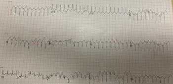
A 12-lead electrocardiogram revealed a monomorphic-wide QRS tachycardia (rate ∼250 bpm) with a right bundle branch block pattern, abrupt QRS transition in lead V2 with deep S waves in lateral precordial leads and a positive QRS polarity in lead aVR. These findings were consistent with a left ventricular origin tachycardia (Fig 1). Synchronised cardioversion (30 J) was unsuccessful. Intravenous lidocaine bolus (1 mg/kg) decreased the ventricular tachycardia rate to 210 bpm. An intravenous magnesium bolus was also administered but did not terminate the ventricular tachycardia. It rather led to severe hypotension (blood pressure 49/27 mmHg). She was administered low volume intravenous fluid boluses (5 m/kg each), three intravenous epinephrine boluses (0.1 mg each) and then started on intravenous epinephrine infusion at 0.2 µg/kg/minute. After initial stabilisation, hyperpyrexia was treated with ice packs, intravenous acetaminophen and ketorolac. A focused echocardiogram showed rapid ventricular motion with severely reduced ventricular function. Serum electrolytes were normal (sodium: 145 meq/L, potassium: 3.7 meq/L, calcium: 8.7 mg/dL [ionised calcium: 1.17] and magnesium: 2.7 mmol/L).
A 12-lead electrocardiogram at presentation shows a monomorphic-wide QRS tachycardia (rate ∼250 bpm) with right bundle branch block pattern, abrupt QRS transition in V2 with deep S waves in lateral precordial leads and a positive QRS polarity in lead aVR which is diagnostic of ventricular tachycardia.

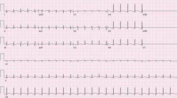
The patient developed circulatory arrest during transport to the paediatric intensive care unit due to profound hypotension. Cardiopulmonary resuscitation was initiated and continued for 2 minutes followed by the successful return of spontaneous circulation. Norepinephrine (0.15 µg/kg/minute) infusion was also initiated, and a slow bolus of intravenous calcium chloride (20 mg/kg) was administered. With a decrease in body temperature (104 °F), the patient reverted to normal sinus rhythm (Fig 2). Laboratory workup revealed a white cell count of 11,200/µL, haemoglobin of 10 g/dL, a platelet count of 59,000/µL, creatine kinase of 216 U/L, N-terminal pro-brain-type natriuretic peptide concentration of 2140 pg/mL and serum troponin concentration of 2137 ng/L (mildly elevated). The patient has weaned-off inotropes over the next 48 hours. An infectious workup did not reveal any abnormality (negative blood and urine cultures and COVID-19 RT-PCR). Empiric antibiotics (vancomycin and piperacillin–tazobactam) were discontinued on day 3 of admission. Echocardiogram obtained 48 hours after the admission revealed normal biventricular systolic function. There was no recurrence of ventricular tachycardia. Her respiratory status gradually improved over the next 2 weeks, following which the patient was discharged to home. Since then, the patient has done well since then without any recurrence of arrhythmia.
The 12-lead electrocardiogram after termination of tachycardia does not reveal any abnormality.

Discussion
Hyperthermia is defined as an elevated body temperature above the normal range due to a failure of heat regulatory mechanisms. Its direct effects on cardiac rhythm include sinus tachycardia, ventricular extrasystoles, junctional rhythm, sustained supraventricular (atrial flutter, atrial fibrillation, ectopic atrial tachycardia) and ventricular tachyarrhythmias. Reference Paul, Alex, Jacob and Yadav1 Indirectly, hyperthermia can precipitate or potentiate arrhythmogenesis by affecting other organ systems. Acute kidney injury, skin burns, gastrointestinal symptoms (nausea, vomiting, diarrhoea), disseminated intravascular coagulation, hyperglycaemia and electrolyte abnormalities (hypocalcaemia, hypomagnesemia, hypophosphataemia and hypokalaemia) have been reported in patients with hyperthermia. Reference Barlogie, Corry and Yip2
Our patient presented with extreme hyperthermia (108.3 °F) and symptomatic sustained ventricular tachycardia. The latter was refractory to cardioversion and anti-arrhythmic drugs but responded to cooling. This life-threatening presentation is rare in patients with a structurally normal heart and is hitherto unreported in a child. In fact, only one adult patient has been reported to present in a similar manner to date. Shimada et al. reported a 38-year-old male with extreme hyperthermia (108 °F) who presented with ventricular tachycardia which was unresponsive to repeated cardioversions. He converted to sinus rhythm after the temperature decreased to 106 °F with aggressive cooling. Reference Shimada, Tsai and Marshall3
Autonomic modulation of cardiovascular response to heat stress is important to understand the aetiology of hyperthermia-related arrhythmogenesis. The arterial blood pressure is regulated by a tight negative feedback loop mechanism predominantly mediated peripherally via carotid sinus and aortic arch baroreceptors. A decrease in mean arterial pressure or pulse pressure decreases the vascular wall stretch, resulting in the decreased firing of these receptors. This in turn triggers the medullary neurons to increase sympathetic outflow resulting in vasoconstriction and tachycardia. These physiological responses lead to an increase in cardiac output and blood pressure. This pathway is likely dysregulated in pathophysiological states such as hyperthermia. Yamazaki et al studied the cardiovascular effects of hyperthermia in healthy humans and found no change in the carotid baroreflex sensitivity during heat stress. However, the sympathetic response to hypotension was diminished and bradycardia was exaggerated. Reference Yamazaki, Sagawa, Torii and EndoY4 These changes can lead to orthostatic hypotension, decreased end-organ perfusion and oxygen demand–supply imbalance in patients with hyperthermia. Reference Yamazaki, Sagawa, Torii and EndoY4 Furthermore, during hyperthermia, there is redistribution of blood flow to cutaneous vasculature due to associated vasodilation. This causes a reduction in circulating blood volume and central venous pressure, further contributing to orthostasis. Reference Crandall, Wilson and Marving5 Inotropic effects of heat stress have also been evaluated. Hyperthermia increases LV systolic function by increasing peak septal and lateral mitral annular systolic velocities leading to an increase in stroke volume. Reference Brothers, Bhella and Shibata6
Given the profound autonomic and inotropic effects of hyperthermia, it is not surprising that under pathological conditions, heat stress can lead to cardiac arrhythmias. In patients with malignant hyperthermia, a disease which is characterised by extreme hyperthermia, ventricular tachycardia and cardiac arrhythmias have been reported in 36% and 72% of patients, respectively. Reference Fenoglio and Irey7
The mechanisms that underlie arrhythmogenesis in patients with hyperthermia are not well understood. However, several hypotheses have been postulated. Ultrastructural changes have been reported in both atrial and ventricular myocardium during heat stress. These include overstretching of sarcomeres with marked contraction bands, prominent I-bands and H-zones, foci of loss of actin and myosin filaments, persistent anaerobic glycolysis leading to intracellular acidosis and disrupted sarcolemma with consequent potassium and calcium leak. Reference Fenoglio and Irey7,Reference Overgaard and Overgaard8 A number of detrimental electrocardiographic changes have been reported in patients with hyperthermia including both marked sinus tachycardia and bradycardia, atrial and ventricular ectopy, intraventricular conduction delays, complete or incomplete right bundle branch block (but not isolated left bundle branch block), corrected QT and PR interval prolongation, and diffuse ST elevation, ST depression and T wave inversion. A low p wave voltage (≤0.01 mV) has also been reported and is associated with poor clinical outcomes in patients with hyperthermia. Reference Paul, Alex, Jacob and Yadav1,Reference Akhtar, Al-Nozha, Al-Harthi and Nouh9 Additional electrophysiological mechanisms reported in hyperthermia include a faster activation and inactivation kinetics of cardiac sodium channels, which could enhance early influx and attenuate late influx of sodium, thus predisposing to cardiac arrhythmias. Reference Nagatomo, Fan and Ye10 The effects of temperature on human sodium channels have been well characterised in patients with Brugada syndrome and can perhaps provide insights into mechanisms that underlie hyperthermia-induced arrhythmogenesis. Dumaine et al showed that mutant cardiac sodium channels in patients with Brugada syndrome demonstrated worsening of the biophysical properties at higher temperatures, leading to further loss of function of sodium channel current. Reference Adler, Topaz and Heller11,Reference Dumaine, Towbin and Brugada12 These findings are in contrast to findings reported by Keller et al who studied mutations found in patients with fever-induced type I Brugada electrocardiogram and fever-induced arrhythmias. Reference Keller, Rougier and Kucera13 In these patients, the mutated channels exhibited severe to total loss of sodium current at physiological temperature. Thus, further loss of function during fever was not expected and indeed not found when expression studies of these channels were performed at higher temperatures. The authors, therefore, hypothesised that in heterozygous patients, the effect of temperature on the wild-type channels is responsible for fever-induced arrhythmias. As per this model, wild-type (ie, normal) channels are less efficient at high temperatures, but this slight loss of function becomes clinically significant only in the presence of other factors that reduce the repolarization or depolarization reserve. Reference Adler, Topaz and Heller11,Reference Keller, Rougier and Kucera13
Heat stress-induced acute heart failure, myocardial infarction and stress cardiomyopathy have also been reported. Reference Chen, Lin, Hsieh, Huang and Yeh14,Reference García-Rubira, Aguilar and Romero15
Lack of response to cardioversion in this patient would argue against re-entry as the mechanism of ventricular tachycardia in this patient. Since the patient did not respond to magnesium infusion, it is likely that the ventricular tachycardia was not mediated by triggered activity. Given the variability in ventricular tachycardia rate and gradual decrease in rate prior to termination, enhanced automaticity is the likely mechanism of arrhythmogenesis in this patient.
It is important to note that any febrile illness can trigger ventricular tachyarrhythmia (usually polymorphic ventricular tachycardia) in patients with Brugada syndrome. A 12-lead electrocardiogram with standard and high (V1 and V2 second intercostal space) lead position should therefore be obtained after the restoration of sinus rhythm in all patients with fever-induced arrhythmias. Reference Antzelevitch and Brugada16 The presence of any maladaptive response to hyperthermia and potential for an exaggerated effect on cardiac arrhythmogenesis are not well studied in patients with brain injury or neurological sequelae. We, therefore, cannot confidently postulate any mechanistic link between the presence of cerebral palsy in our patient and her clinical presentation.
In conclusion, as seen in our patient, heat stress-induced ventricular tachyarrhythmia can be life-threatening and remain refractory to conventional therapies including cardioversion. Therefore, immediate lowering of the core body temperature via aggressive cooling should be initiated to facilitate reversion to sinus rhythm.
Acknowledgements
None.
Financial support
This research received no specific grant from any funding agency, commercial or not-for-profit sectors.
Conflicts of interest
None.
Ethical standards
This report does not include any human or animal experimentation.