Introduction
Ultrasound is a valuable diagnostic tool used in many areas of medicine. It has been described as quick, portable, non-invasive and cost effective, and does not involve ionising radiation.Reference Rozycki, Ochsner, Jaffin and Champion1–3 In mainland Europe, it is almost the exception for the radiologist rather than the clinician to perform ultrasound in some specialties. However, in the UK, with the exception of obstetric ultrasound, radiologists and radiographically trained sonographers have traditionally provided a service from centralised departments of radiology, where equipment and manpower can be concentrated cost-effectively.
There are increasing demands for other medical specialists to utilise ultrasound as a direct adjunct to clinical examination, and in some specialties it is becoming an integral part of the physician's diagnostic armamentarium and training. This trend is likely to be exacerbated by the increase in referrals and shortage of radiologists.4 A recent survey distributed by ENT UK discussed the prospects and usefulness of British otolaryngologists learning this skill. Additionally, there is a demand by some European training boards to incorporate ultrasound into clinical training and accreditation. The Royal College of Radiologists recognises that it is appropriate for medical practitioners other than clinical radiologists to develop skills in ultrasound.5
The role of head and neck ultrasound performed by the ENT clinician, and the ability of the clinician to carry out the ultrasound and accurately interpret the findings, have not been investigated. This prospective study essentially describes the learning process of an ENT trainee with no previous specialist imaging experience, in acquiring neck ultrasound skills.
Materials and methods
Training
An ENT trainee attended head and neck ultrasound sessions in the radiological ultrasound department of a large teaching hospital for 12 months. A well-established 2-day practical ultrasound course (The Head and Neck Ultrasound Workshop, Morriston Hospital, Swansea) provided a basic introduction. Thereafter, the trainee attended several sessions with one of the course faculty members, observing neck ultrasound examinations. Informal tutorials covered physics and instrumentation, and ultrasound anatomy of the neck. Early practical experience was gained by practising on normal volunteer colleagues.
Following this induction, the trainee worked alongside a consultant radiologist with over two decades of experience in head and neck ultrasound (LB). This consultant radiologist works closely with all clinical departments at our centre, including surgery, endocrinology and oncology, helping with the management of patients. Ultrasound sessions included a weekly dedicated ‘head and neck lump’ clinic. These sessions include patients with no palpable mass, which typically involves a search for an undiagnosed parathyroid lesion in a patient with hypercalcaemia. This arrangement afforded the trainee one-to-one mentorship.
Following the studies of normal volunteers, the second stage of the learning process involved 50 ultrasound examinations of clinical referrals observed by the radiologist. All examinations were repeated by the radiologist who provided immediate feedback to the trainee. These 50 examinations were excluded from the final analysis of the 250 cases that comprise the current study. If any aspect of the trainee's examination was considered technically suboptimal, and where time constraints permitted, the scan was repeated by the ENT trainee following the radiologist's study.
Learning objectives included the identification of variations in normal neck structures and anatomical relationships, the recognition of any deviation from normal, and correct interpretation of an abnormality. A systematic approach to examination was emphasised. This included comprehensive scanning of neck anatomical triangles, comparing both sides of the neck, and use of Doppler ultrasound where appropriate. Teaching included advanced use of the machine controls, to a much higher level than usually achieved by practitioners other than radiologists or sonographers.
Main study
After the induction and training period described above, the trainee undertook examinations on patients referred to the neck ultrasound clinic. The trainee's study and conclusion was compared with the examination and conclusion of the radiologist. The ‘gold standard’ was taken to be the radiologist's report rather than eventual surgical or histological diagnosis if biopsy or surgery was undertaken.
Examinations were performed with Toshiba Aplio XG ultrasound apparatus (Toshiba Medical Systems, Crawley, UK) using appropriate high-frequency linear array transducers. All patients referred with palpable neck masses were included. Scans were undertaken with the patient in a semi-recumbent position with neck extension.
Following the scan, the trainee completed a proforma, on which the trainee indicated the presence or absence of a lesion, and commented on its nature and significance. If the lesion was considered indeterminate, the most likely diagnosis was described. Minor findings (e.g. reactive lymph nodes) were considered as lesions and were included in our analysis. The radiologist repeated the study and completed a similar proforma. It was not possible to blind the radiologist to the ultrasound findings described by the trainee because of time constraints and the evaluation process: as part of the evaluation, the radiologist scrutinised, and, if necessary, criticised and corrected the trainee's scanning technique.
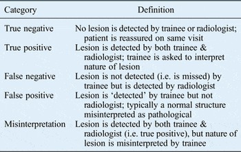
Anonymised data were entered into a database. Results were placed in one of five categories (Table I): true negative (normal study), true positive (abnormal study), false negative (missed abnormality), false positive (normal study misinterpreted as abnormal), and misinterpretation (abnormality detected, but the nature or significance misinterpreted). There were therefore two aspects to the trainee's assessment. Firstly, identifying whether an abnormality was present, and secondly correctly interpreting any abnormal findings.
Outcome categories and definitions

Results
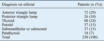
A total of 250 consecutive patients with suspected head and neck masses who attended over a 12-month period were included in the study. The median patient age was 50 years, with a male to female ratio of 1:1.7. The range of clinically suspected pathologies at the time of referral is shown in Table II.
Suspected pathology

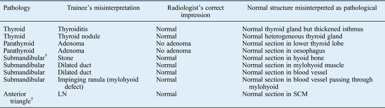
Scans performed by the trainee indicated a positive finding in 155 patients. The findings of radiological repeat examinations concurred with the trainee's study in 144 examinations (true positives). Eleven of the trainee's 155 ‘positive’ findings were considered normal by the radiologist and were therefore deemed to be false positives (Table III).
Trainee false positive results*
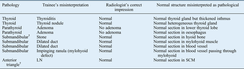
* 11 patients. †n = 2. LN = lymph node; SCM = sternocleidomastoid muscle
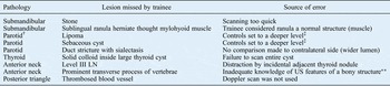
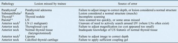
The trainee examination indicated a negative finding in 95 patients. The radiologist's repeat examination indicated normal findings in 63 patients (true negatives). Therefore, according to the radiologist gold standard, the trainee missed abnormalities in 32 (34 per cent) of the abnormal scans (false negatives). These abnormalities included palpable and impalpable neck masses (Tables IV and V).
Trainee false negatives: palpable lumps*

* 10 patients. †n = 2. ‡Lesion was in superficial skin layers. **Appears as white line as it reflects sound. LN = lymph node; US = ultrasound
Trainee false negatives: impalpable lumps*

* 23 patients. †n = 8; ‡n = 2; **n = 3. LN = lymph node; IJV = internal jugular vein; US = ultrasound
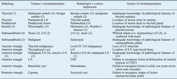
Of the trainee's 144 true positives, the trainee's interpretation of the lesion was concordant with that of the radiologist in 117 (81 per cent) of the abnormal scans. The trainee's interpretation of detected pathology was considered a misinterpretation in 28 cases (19 per cent of all abnormal scans) (Table VI).
Trainee misinterpretations*

Numbers in parentheses represent number of lesions.
* 28 patients. LN = lymph node; CBT = carotid body tumour
Using the radiological opinion as a gold standard, the overall figures for sensitivity, specificity, positive predictive value, negative predictive value and accuracy of the trainee examinations were: 82, 85, 93, 67 and 83 per cent, respectively.
Of all the 250 examinations, we were able to reassure 127 patients by excluding serious pathology (50 patients) or excluding any lesion (77 patients). Only 16 patients required biopsies, of which 10 proved to be malignant. Of the 16 patients that underwent biopsy, the trainee failed to detect 1 malignant lesion (false negative) and misinterpreted 4 malignant lesions as benign (interpretive error).
Although it was not the purpose of this study to evaluate the use of ultrasound in expert hands, with a minimum follow-up period of two years, none of the patients have re-attended with a significant lesion.
Discussion
This is the first study to describe the process of an ENT trainee undertaking structured training in neck ultrasound. Head and neck ultrasound is difficult, and fraught with pitfalls. Nevertheless, the radiologist in this study (LB) has trained numerous radiologists to a level consistent with non-specialist general radiology practice. The experience required to define or interpret some lesions may be measured in years rather than months, and this would apply equally to a radiologist or sonographer learning head and neck ultrasound.
Surgeon-performed neck ultrasound is infrequently discussed in the literature, with most reports describing the value of peri-operative localisation of parathyroid lesions in shortening operation time.Reference Jabiev, Lew and Solorzano6–Reference Solorzano, Carneiro-Pla and Irvin8 Other studies focused on the advantage of clinic-based ultrasound in changing decisions about operative management of thyroid disease when compared to scans performed by a conventional ultrasound practitioner before the clinic visit.Reference Mazzaglia9 Spurious lesions are frequent in head and neck ultrasound (Table III), commonly the result of a misinterpretation of a normal neck structure. This more likely occurs at an early stage, before the trainee becomes familiar with the radiological anatomy of the neck. Bony structures such as the hyoid or prominent transverse processes of vertebrae can simulate macrocalcification in a lesion or a calculus in Wharton's duct. A blood vessel can be confused with a duct, but this distinction can usually be made by skilled Doppler ultrasound technique.
The process of palpation before the scan does not necessarily facilitate the ultrasound study. Table IV comprises 10 cases where the ENT trainee suspected a definite palpable abnormality prior to performing the ultrasound study, yet nevertheless went on to miss the abnormality on the scan. The ultrasound study may need to go beyond confirming the organ of origin of a positive palpation finding. An example of this is the quest for a calculus following the identification of a sialectatic salivary gland or duct. It may be important to further characterise a lesion; for example, defining a solid component that may require a biopsy within an otherwise cystic lesion. Extremely superficial lesions such as lipomas or sebaceous cysts may easily be overlooked if the focus of the ultrasound apparatus is suboptimal or too much pressure is applied to the ultrasound transducer.
It is notable that false negative results and misinterpretations on the part of the trainee were the most frequent types of errors (Tables V and VI). We regard this as a constructive rather than a discouraging learning outcome, as we will continue to develop this skill. It is likely that many of these errors would have been made by radiologically qualified practitioners less experienced than the gold standard radiologist of the current study. We analysed the trend of our false negative results by equally dividing the total number of examinations into five consecutive blocks. Interestingly, most errors occurred at the initial stages; the learning curve showed subsequent improvement (10 of the 32 missed lesions occurred in the first 50 examinations, and this figure was reduced to 8, 6, 6 and 2 in subsequent blocks). Individual readers of this study will decide whether this is acceptable following a regime of training that is unlikely to be equalled or surpassed in other centres. The subjective impression of the radiologist participating in this study is that the level of the ENT trainee's ability surpasses that of general radiology trainees.
The use of ultrasound is expanding rapidly in the emergency room, surgical ward and critical care unit, and more recently in office practice.Reference Jabiev, Lew and Solorzano6, Reference Iannotti, Ciccone, Buss, Visotsky, Mascha and Cotman10–Reference Buzzas, Kern, Smith, Harrison, Helmer and Reed15 The impetus driving this trend may sometimes be suspect, and will vary between differing medical cultures such as private fee-for-item practice as opposed to a British model of salaried public health provision. A catalogue of objections to clinician-based ultrasound frequently raised by radiologists has included: access to an ultrasound machine, medicolegal liability, lack of specific training and fear of lost revenue.Reference Yiengpruksawan, Ganepola and Freeman11, Reference Filly16, Reference Staren, Knudson, Rozycki, Harness, Wherry and Shackford17 The policy adopted by the Royal College of Radiologists is that it is appropriate for practitioners other than clinical radiologists to seek to develop skills in the performance of ultrasound.5, 18
There is growing literature to suggest that clinicians with limited experience in radiology can perform niche ultrasound examinations at a level comparable to radiologists. Specific studies have included the gall bladder,Reference Fang, Pilcher, Putnam, Smith and Smith19 breast,Reference Whitehouse, Baber, Brown, Moskovic, King and Gui13 parathyroid gland,Reference Jabiev, Lew and Solorzano6 joints,Reference Iannotti, Ciccone, Buss, Visotsky, Mascha and Cotman10 emergency hepatobiliary pathology,Reference Kell, Aherne, Coffey, Power, Kirwan and Redmond14 general trauma,Reference Buzzas, Kern, Smith, Harrison, Helmer and Reed15 and chest in both critical care and trauma settings.Reference Rozycki, Pennington and Feliciano12, Reference Knudtson, Dort, Helmer and Smith20 Similarly, radiographers performed well when they were adequately trained.Reference Leslie, Lockyer and Virjee21 Ultrasound has been shown to be a more sensitive technique than clinical evaluation in certain conditions and has been recommended as an extension to physical examination.Reference Mazzaglia9, Reference Rozycki, Pennington and Feliciano12, Reference Chen, Lin, Hsieh and Chen22 A further advantage of office-based ultrasound is that it allows clinical and imaging assessment at a single visit.Reference Rahman, Crawford, Hall, Bavosiet and Quinlan23
Ultrasound teaching programmes for surgeons have been established for decades in mainland Europe, as pioneered at the University of Göttingen in 1982. Subsequently, the German Association of Surgery began requiring experience and competence in ultrasound for certification in general surgery, orthopaedics and urology.Reference Yiengpruksawan, Ganepola and Freeman11 In 1996, the American College of Surgeons launched an educational programme to train surgeons on the use of this technology, supported by interested surgical societies and professional bodies.Reference Staren, Knudson, Rozycki, Harness, Wherry and Shackford17, 24–26 The American Board of Surgery advocates that surgeons ‘have the opportunity to gain a working knowledge of ultrasonography of the head and neck, breast, abdomen, and endorectal ultrasound’.Reference Grosfield27 Residents in the US are expected to complete a basic ultrasound course.Reference Rozycki, Feliciano, Schmidt, Cushman, Sisley and Ingram2, Reference Buzzas, Kern, Smith, Harrison, Helmer and Reed15, Reference Rozychi28
There are many specialties (obstetrics and gynaecology, cardiology, emergency medicine, urology, and family practice) where ultrasound skills are included in the training, and model curricula have been developed.Reference Mateer, Plummer and Heller29 Similarly, a robust training model exists for radiographically qualified ultrasonographers, which is delivered in a relatively short timescale.4 The Royal College of Radiologists stated that radiologists have the background to provide guidelines for the training of medical non-radiologists, which should be to the same standard as those for radiologists, albeit restricted to the relevant area of their clinical expertise.5 They proposed three levels of minimum training requirement, ranging from the ability to recognise normal anatomy, to performing specialised examinations and interventions. This is consistent with the minimum requirements of the European Federation of Societies for Ultrasound in Medicine and Biology.5
Many criteria would need to be met before the experience of the current authors could be extrapolated. Ultrasound training requires a motivated ENT trainee, and a dedicated head and neck radiologist with relevant ultrasound expertise. Short courses are adequate as an introduction, but adequate one-to-one training more than doubles the time taken for each patient ultrasound study. Additionally, there may be competing radiology trainees in a teaching radiology department, and it would be impractical to train more than one person on each patient.
• Ultrasound is a valuable diagnostic tool used in many areas of medicine including ENT
• Provision of ultrasound service by clinicians other than radiologists has gained wide acceptance in USA and Europe, but less in UK
• A recent survey published by ENT UK discussed the prospects of otolaryngologist-performed neck ultrasound as a diagnostic tool
• This study reports the unique experience, outcomes and lessons of an ENT trainee learning this technique
• Although trainee results were less favourable compared with an experienced head and neck radiologist, improvements were steady
• We regard this as a constructive learning outcome and will continue to develop this skill
Specific training and a range of supervised examinations have been suggested before a non-radiologist can be considered competent and credentialled to perform ultrasound. The number of necessary examinations before applying for certification can be between 50 and 400. This wide range probably reflects the individual variation in aptitude and the varying complexity of different organ systems.5, 24, Reference Mateer, Plummer and Heller29–31 Some studies have been hyperbolically optimistic about the length of training. In a study evaluating surgeon-performed ultrasound in trauma patients, it was demonstrated that with only 8 hours of didactic and hands-on training, surgeons could acquire the necessary skills to obtain and interpret ultrasound images to accurately detect haemoperitoneum.Reference Staren, Knudson, Rozycki, Harness, Wherry and Shackford17 The radiologist author of the current study (LB) is sceptical about much of this literature and would disregard studies where there has not been participation of a skilled radiologist.
The National Ultrasound Steering Group (a subgroup of the National Imaging Board in the UK) recommends the establishment of a Clinical Governance Board for all providers of ultrasound imaging services that includes a clinical lead for each department using ultrasound.3 Quality assurance is emphasised with regard to maintaining professional standards equivalent to those issued by the General Medical Council, the latter of which recommends that doctors recognise and work within the limits of their competence. The Royal College of Radiologists states that National Health Service trusts in the UK are unlikely to be able to mount any defence to an action brought against an untrained practitioner.5
In this series, we describe a unique one-to-one training process in neck ultrasound. We consider this model the gold standard for any ENT trainee attempting to learn this technique, as it allows close supervision and input by the radiologist. Although it might look labour intensive to some readers, the process becomes less demanding as skills are learned. Following our study period, the department acquired an ultrasound machine and the radiologist joined our one-stop neck lump clinic, which improved our partnership and made the training more streamlined.
Conclusion
This study evaluated a one-to-one training model of neck ultrasound for an ENT trainee. We identified important learning outcomes and explored potential errors during the initial stages of training that we significantly improved. Neck ultrasound performed by an otolaryngologist, while less accurate than an experienced radiologist, is a useful adjunct to clinical assessment, and can facilitate assessment in a one-stop clinical setting. A close collaboration with the radiology department is a key element in learning this technique. This study can become a platform for the incorporation of ultrasound training in future ENT curricula. The authors consider that the overriding consideration for extending head and neck ultrasound skills beyond the radiology department should be the welfare and management of the patient, rather than the academic or financial competing interests of other professional groups.