Implications
This review article aims to provide an overview of the pig as a biomedical model for humans and specific categories of diseases are considered, metabolic, cardiovascular, digestive and bone in particular. Previous reviews have considered different disease categories and the relevant references are included here. The main limitation is the depth to which each disease can be considered, particularly with respect to space and the availability of information in the primary references.
Introduction
Animals allow a multi-disciplinary approach to be taken aimed at improving our understanding of the pathophysiology of diseases in humans. The underlying physiological, biochemical and molecular biological mechanisms can be examined in animal models in a more invasive manner that would be unethical in humans, under controlled conditions and without the confounding genetic/social factors. It is acknowledged that there is generally not a direct numerical equivalence between animal and human experiments but responses to treatments are, on the whole, qualitatively similar allowing responses of animals to more extreme treatments to be used to predict the anticipated response in humans.
Various species have been used as biomedical models to study human medicine (e.g. rat, mouse, guinea pig, dog, pig and sheep). Although rodents are small, inexpensive and are ideal in multivariate experiments, there are numerous differences between rodent models and humans. For example, such differences in adipose tissue characteristics (i.e. adipsin, leptin, resistin, tumor necrosis factor-α and other adipokines) have prevented the translation of data obtained from rodents into preventative/treatment strategies aimed at improving human health (Spurlock and Gabler, Reference Spurlock and Gabler2008). It is becoming increasingly apparent that pigs are a valuable translational model to bridge the gap between classical rodent models and humans in developing new therapies to aid human health (Schnieke and Wolf, Reference Schnieke and Wolf2008).
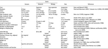
Pigs are an appropriate biomedical model for humans due to many genetic (Lunney, Reference Lunney2007), anatomical (Pracy et al., Reference Pracy, White, Mustafa, Smith and Perry1998; Prestige World Genetics Korea, 2006) and physiological similarities to humans (Book and Bustad, Reference Book and Bustad1974; Miller and Ullrey, Reference Miller and Ullrey1987; Mei and Xu, Reference Mei and Xu2003) (Table 1). However, it should be noted that swine also have unique behavioural characteristic and husbandry requirements, which must be taken into consideration when used as a laboratory animal (Swindle et al., Reference Swindle, Nolan, Jacobson, Wolf, Dalton and Smith2005). Other advantages of using the pig are that it is possible to use standard medical technologies to image internal organs and vessels, repeatedly collect blood samples and obtain an adequate quantity of tissue sample at slaughter without the need to pool samples. Pigs also share a high sequence and chromosome structure homology with humans, and genomic and proteomic tools are improving (Lunney, Reference Lunney2007). Furthermore, by using pigs from the same litter, or cloned or transgenic pigs, genetic variation is reduced making genetic mapping easier.
A comparison of reproductive and general anatomical/physiological factors between human, swine and rats

1Similarity compared to humans.
The two main categories of pigs used as biomedical models are the domestic pig (e.g. modern commercial breeds such as the Large White and Pietrain or ancient breeds such as the Meishan) and the minipig (e.g. Göttingen, Yucatan), both of which can differ in their suitability as a model for specific disorders. Some of the advantages of using the minipig compared with the domestic pig are its smaller and similar in size to humans, even at full maturity, slower growth during studies, ease of handling, and controlled genotype as well as its microbiological characteristics (Nunoya et al., Reference Nunoya, Shibuya, Saitoh, Yazawa, Nakamura, Baba and Hirai2007). However, one drawback of using minipigs is that they are in short supply and considerably more expensive to purchase. In contrast, domestic pigs are more costly to house, feed and medicate. Minipigs also have an advantage over traditional non-rodent animals, such as the dog, because of increasing ethical concerns about their use in experiments.
Apart from using whole animals, porcine cell lines have been established for studying metabolic function (e.g. Nishitsuka et al., Reference Nishitsuka, Kashiwagi, Tojo, Kanno, Takahashi, Yamamoto, Heldin and Yamashita2007) in a wide range of tissue and these well-defined cell lines are readily available making it easier to examine gene expression and drug susceptibility for example. It should, however, be noted that some characteristics of porcine embryonic stem cells differ from murine and ovine embryos (Piedrahita et al., Reference Piedrahita, Anderson and Bondurant1990). Cell lines can be used for some diseases, for example post-weaning diarrhoea (Pavlova et al., Reference Pavlova, Volf, Alexa, Rychlik, Matiasovic and Faldyna2008) and the influenza virus (Seo et al., Reference Seo, Goloubeva, Webby and Webster2001), but this aspect will not be discussed further in this review.
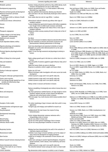
Pigs have been used for research in a number of diseases (Table 2) including metabolic disorders (e.g. obesity and diabetes), cancer, chronic alcoholism, psychiatric disorders and regenerative therapies (e.g. use of stem cells). Recently, the transgenic pig has been developed for many uses within the areas of agriculture and biomedicine. For example, pigs have been modified to produce recombinant proteins in the mammary gland (Niemann and Kues, Reference Niemann and Kues2003), and anti-rejection work in xenotransplantation (Fodor et al., Reference Fodor, Williams, Matis, Madrit, Rollins, Knights, Velander and Squinto1994). Genomic studies of melanoma and of infectious disease resistance, and issues for consideration in designing such genomic studies, have been recently reviewed by Lunney (Reference Lunney2007) and will not be discussed further in the present review. The aim of this review article is to give an overview of the use of the pig as a biomedical model for humans, and in particular focuses on the Metabolic Syndrome, digestive tract-related dysfunction and bone disorders.
An overview of the use of the pig as a biomedical model

NIDDM = Non-insulin dependent diabetes mellitus; GI = gastrointestinal.
The metabolic syndrome
The metabolic syndrome consists of a cluster of risk factors for non-insulin dependent diabetes mellitus (NIDDM; Type II diabetes) and cardiovascular disease, which are generally classified as a combination of insulin resistance (DeFronzo and Ferrannini, Reference De Fronzo and Ferrannini1991), central obesity, raised plasma triacylglycerol concentrations, reduced high-density lipoprotein cholesterol, increased low-density lipoprotein cholesterol and hypertension (Eckel et al., Reference Eckel, Grundy and Zimmet2005; Pi-Sunyer, Reference Pi-Sunyer2007). It is estimated that the current global prevalence of the metabolic syndrome is approximately 16% (95% confidence intervals 10 to 23; Wild and Byrne, Reference Wild and Byrne2005) and that this figure is growing at an alarming rate. A multitude of research has been undertaken to improve our knowledge of underlying causes and subsequent prevention and treatment of the metabolic syndrome, but much of this research has been conducted in rat models. One of the problems associated with rat models is that they rarely exhibit three of the clinical signs of this disorder, whereas in humans three or more of these symptoms are observed simultaneously; this is due to fundamental differences in metabolism and physiology between these two species. As a consequence, the porcine model, which exhibits three or more of the clinical signs, is now generally considered the optimum non-primate model for investigating the metabolic syndrome (Spurlock and Gabler, Reference Spurlock and Gabler2008). These authors have recently reviewed the development of porcine models of obesity and the metabolic syndrome in juveniles and adults, and so this aspect will not be covered in depth herein. The components contributing to the onset of the Metabolic Syndrome are discussed below.
Developmental programming of the metabolic syndrome
Evidence from numerous epidemiological studies has suggested that there is a relationship between intrauterine growth restriction (IUGR) and the subsequent development of the metabolic syndrome (Hansen, Reference Hansen1999; Horvath and Bruning, Reference Horvath and Bruning2006; Palinski, Reference Palinski2007; Taylor and Poston, Reference Taylor and Poston2007). Sheep, rats, guinea pigs and more recently pigs have extensively been used to investigate the mechanisms which link prenatal programming to adult diseases (Hoet and Hanson, Reference Hoet and Hanson1999; Clarke et al., Reference Clarke, Kirth, Heasman, Juniper, Budge, Stephenson and Symonds2000; Poore and Fowden, Reference Poore and Fowden2004a).
Placental and Foetal growth and development
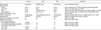
It is well established that in mammals the principal determinant of IUGR is placental nutrient supply, which in turn depends on the size, morphology, blood supply and transporter abundance of the placenta, and on synthesis and metabolism of nutrients and hormones by the uteroplacental tissues (Fowden et al., Reference Fowden, Ward, Wooding, Forhead and Constancia2006). Both human and rat placentas belong to the general category of haemochorial placentas; however, there are fundamental differences (Table 3). For example, adiponectin and adiponectin receptor 2 (Adipo-R2) are present in cytotrophoblast and syncytiotrophoblast cells in humans, whereas adiponectin expression occurs in rat trophoblast cells and Adipo-R2 in giant cells and viteline membranes (Caminos et al., Reference Caminos, Nogueiras, Gallego, Bravo, Tovar, García-Caballero, Casanueva and Diéguez2005). Pigs, on the other hand, have epithelial chorial (or diffuse) placentas making them a good model for studies during pregnancy as the maternal and foetal components of the placenta can be cleanly dissected without cross contamination (Tayade et al., Reference Tayade, Black, Fang and Croy2005).
Placental and metabolic comparisons between foetal and neonatal humans, swine and rats

NEFA = non-esterified fatty acids.
*Values in bracket are for premature human infants.
Foetal growth is a combination of numerous interrelated mechanisms that include cell replication and tissue differentiation, matrix formation and cell death, all of which need to be regulated (Gluckman, Reference Gluckman1986). There is a well-defined, programmed, sequence of developmental changes that occurs at specific times in the growth of the embryo and foetus (Figure 1). The underlying principles of foetal maturation appear to be comparable between mammals, but it should be noted that quantitative differences occur between species. For example, stage 3 foetal development occurs in utero in humans and swine but postnatally in rats (Figure 1). In addition, rat pups are much smaller than either babies or piglets (Figure 1), which means that it is often necessary to pool tissue samples, thereby reducing indications of natural variation. When compared with the young of other commonly used research animals such as the dog, cat, goat and sheep, the piglet appears to show the greatest similarity to the human infant in terms of anatomy, physiology, nutrition and metabolism (Book and Bustad, Reference Book and Bustad1974; Miller and Ullrey, Reference Miller and Ullrey1987; Mei and Xu, Reference Mei and Xu2003).
Comparison of prenatal development and crown-to-rump length of human (—), pig (– -) and rat (- - - -) foetuses throughout gestation. (a) Adapted from Book and Bustad (Reference Book and Bustad1974) and Hill (Reference Hill2008). (b) Adapted from Book and Bustad (Reference Book and Bustad1974), and Evans and Sack (Reference Evans and Sack1973).

Measurements of embryos and foetuses are generally cited as crown-to-rump length (Figure 1), but while this parameter is quite reliable for man, it must be used with caution for domestic mammalian foetuses because of the longer neck and the increased chance in error due to changes in posture (Evans and Sack, Reference Evans and Sack1973). In humans, the rate of foetal growth is relatively slow up to the 20th week of pregnancy and, as with the pig and rat; the majority of growth occurs during the second half of pregnancy (Manners and McCrea, Reference Manners and McCrea1963; Ashworth, Reference Ashworth2006). Human foetal growth accelerates to reach a maximum around 30 to 36 weeks, declining thereafter until birth as the mother fails to meet the increasing energy demand of her growing foetus. On the contrary, the pig and rat have not reached their peak growth rate at birth and so the decline in growth rate observed during late gestation in humans does not occur (Figure 1). In litter-bearing species the number of developing foetuses is inversely related to the ultimate size of each individual at full term (Pond and Houpt, Reference Pond and Houpt1978). The pattern of foetal growth in pigs is determined by the genome of the foetus, maternal nutrition and health, uterine capacity and foetal position in the uterine horn (McCance and Widdowson, Reference McCance and Widdowson1974; Antipatis et al., Reference Antipatis, Finch and Ashworth2008).
Protein accumulation tends to occur early in human foetal development to reach its maximum (∼300 g) around week 35, and precedes fat accumulation, most of which is subcutaneous, and only exceeds the weight of protein deposition by week 38 (Prentice et al., Reference Prentice, Spaaij, Goldberg, Poppitt, van Raaij, Totton, Swann and Black1996). By term, there is approximately three-times more energy stored as fat than as protein in human infants. Unlike humans and sheep, both pigs and rats are born with little adipose tissue, but in this sense their early post partum development can be viewed as equivalent to the late development of human babies in utero (Table 3). It should be noted that the amino acid compositions differ appreciably among species that have relatively short periods of gestation such as the rat and species that have relatively long periods of gestation such as the pig and human for the amino acids: histidine, glycine, lysine, proline and hydroxyproline (Wu et al., Reference Wu, Ott, Knabe and Bazer1999).
The newborn pig’s body fat is minimal and usually constitutes no more than 2% of total body weight (BW). However, this situation is reversed postnatally; piglets can double their birth weight due to high rates of protein deposition and lean tissue growth (Wood and Groves, Reference Wood and Groves1965; Whittemore and Kyriazakis, Reference Whittemore and Kyriazakis2006), and there can be a 10- to 20-fold increase in body fat in 7 to 10 days, whereas the human infant doubles its birth weight by about 5 months of age (Shulman, Reference Shulman1993; Pond and Mersmann, Reference Pond and Mersmann2001). This means that piglets can provide an excellent model of accelerated growth and development (Odle, Reference Odle1997), which is advantageous when studying the effects of postnatal nutrition on health in later life (Taylor and Poston, Reference Taylor and Poston2007). Moreover, the newborn piglet is more akin to the premature than to the full-term human infant in several aspects, such as body composition (i.e. low fat and hepatic glycogen content) and a reduced thermoregulatory ability (Widdowson, Reference Widdowson1971; Book and Bustad, Reference Book and Bustad1974; Herpin et al., Reference Herpin, Damon and Le Dividich2002; Mostyn et al., Reference Mostyn, Litten, Perkins, Euden, Corson, Symonds and Clarke2005).
Low birth weight (LBW) modern domestic pigs exhibit impaired mitochondrial metabolism in adipose tissue, which may in part explain their reduced body temperature (Mostyn et al., Reference Mostyn, Litten, Perkins, Euden, Corson, Symonds and Clarke2005). However, physiological studies have shown that porcine genotype must be taken into consideration when selecting an appropriate model because despite the minipig being smaller, it possesses more fat (0.117 to 0.158 kg fat for a 1.25 kg 5-day old neonatal piglet, (Sheng et al., Reference Sheng, Adolph, O’Brian Smith and Garza1988)) compared with modern domestic breeds (0.03 kg fat for a 1.5 kg 1 day old neonatal piglet, Shields et al., Reference Shields, Mahan and Graham1983)) and exhibits altered uncoupling protein (UCP) expression and endocrine profile at birth compared with modern domestic breeds of pigs (Knol et al., Reference Knol, Leenhouwers and van der Lende2002; Mostyn et al., Reference Mostyn, Litten, Perkins, Alves-Guerra, Pecqueur, Miroux, Symonds and Clarke2004, Reference Mostyn, Sebert, Litten, Perkins, Laws, Symonds and Clarke2006). Compromised UCP abundance in adipose tissue may contribute to excess fat deposition in later life resulting in obesity, hence providing a natural model for obesity (Dyson et al., Reference Dyson, Alloosh, Vuchetich, Mokelke and Sturek2006).
Maternal environment and foetal development
Foetal metabolism and neonatal outcome are altered by factors such as maternal body composition, nutrition, endocrine environment (Gluckman and Pinal, Reference Gluckman and Pinal2002) and complications like gestational diabetes (Ray et al., Reference Ray, Vermeulen, Shapiro and Kenshole2001). Ethical restraints forbid the use of studies investigating human foetal metabolism. Size and length of gestation in rodents makes it difficult to study metabolic activity in the developing foetus. In contrast, piglets are large enough to catheterize in utero (Silver et al., Reference Silver, Fowden, Comline and Bloom1986), and one further advantage is that since the pig is a litter-bearing species several different metabolic studies can be performed using replicates within a litter.
Protein intake determines placento-foetal growth (Godfrey et al., Reference Godfrey, Robinson, Barker, Osmond and Cox1996). Rats are currently the best-characterized animal model with many studies showing maternal low protein diet (LPD) consumption throughout (Langley-Evans, Reference Langley-Evans2001), or at specific gestational periods leading to hypertensive offspring (Kwong et al., Reference Kwong, Wild, Roberts, Willis and Fleming2000). The magnitude of effect is partly dependent on timing of exposure and in some (Kwong et al., Reference Kwong, Wild, Roberts, Willis and Fleming2000), but not all studies, is gender specific. The rat, however, may be particularly vulnerable to any nutritional imbalance during gestation given the exceptional rate of foetal protein accretion compared with the human foetus and the far larger total weight of the products of conception relative to maternal weight. For example, the rate of protein accretion (Table 1) is approximately 25 g/kg BW/day in the rat (Goldspink and Kelly, Reference Goldspink and Kelly1984) and 13 g/kg BW/day in the human foetus near-to-term (Chien et al., Reference Chien, Smith, Watt, Scrimgeour, Taylor and Rennie1993). In the pig, the rate of foetal protein accretion is approximately 19 g/kg BW/day after day 69 of gestation (McPherson et al., Reference McPherson, Ji, Wu, Blanton and Kim2004).
Maternal protein restriction during early pregnancy influences foetal and placental growth throughout pregnancy, whereas protein restriction throughout the whole of gestation stunts postnatal growth of swine progeny (Schoknecht et al., Reference Schoknecht, Pond, Mersmann and Mauer1993 and Reference Schoknecht, Newton, Weise and Pond1994). Protein restriction during the first or last trimester, however, does not have a permanent effect upon postnatal growth of pigs once balanced nutrition is restored (Schoknecht et al., Reference Schoknecht, Pond, Mersmann and Mauer1993). It has also been demonstrated that piglet size at birth is correlated to changes in the transport of leucine across the porcine placenta (Finch et al., Reference Finch, Yang, Nwagwu, Page, McArdle and Ashworth2004), which is in accordance with the findings that a low protein maternal diet induces foetal growth retardation in rats and humans (Metges, Reference Metges2005). Although there is a somewhat limited amount of information on the effect of a LPD on the development of adulthood diseases in pigs, given the amino acid composition of the foetal pig is also similar to that of the human foetus (Wu et al., Reference Wu, Ott, Knabe and Bazer1999), it seems feasible to suggest that results from porcine studies would be comparable to that observed in human epidemiological studies. Further work is required to determine the suitability of the pig as a model for studying the effects of a maternal LPD and the ontogeny of the metabolic syndrome in their offspring.
Essential fatty acids are known to be important for the maintenance of normal growth and development, as they form precursors for the components of the endocrine and immune systems (Milner and Allison, Reference Milner and Allison1999; Wainwright, Reference Wainwrigh2000). Supplementing the sow diet with lipids during pregnancy not only alters the lipid composition of the developing pig foetuses (Rooke et al., Reference Rooke, Bland and Edwards1998), but also subsequent milk composition (Laws et al., Reference Laws, Amusquivar, Laws, Herrera, Lean, Dodds and Clarke2008 and Reference Laws, Litten, Laws, Lean, Dodds and Clarke2009). As observed in humans (Innis, Reference Innis2004), the fatty acid composition of the milk mirrors that of the type of oil that is added to the sow diet, which can subsequently influence neonatal outcome in modern domestic breeds (Laws et al., Reference Laws, Amusquivar, Laws, Herrera, Lean, Dodds and Clarke2008 and Reference Laws, Litten, Laws, Lean, Dodds and Clarke2009). Supplementing the maternal diet with sunflower oil during the first half of gestation resulted in a greater proportion of LBW piglets (20 ± 3%), whereas supplementing with olive oil reduced the incidence of LBW offspring (5 ± 3%), and hence growth rate was higher in these piglets. Irrespective of the type of oil added to the maternal diet, LBW offspring of supplemented mothers possessed more fat compared with the offspring born to unsupplemented sows at birth. Follow-up studies on these LBW were not undertaken with respect to their glucose metabolism in later life. However, these results further support a role for the modern domestic pig as a biomedical model for understanding the relationship between maternal nutrition, birth weight and the development of the metabolic syndrome.
It is worth mentioning here the differences in milk composition between humans and pigs. There is no passive transfer of immunoglobulin across the porcine placenta as seen in humans (Table 3), and as such the sow-colostrum is rich in immunoglobulins (Laws et al., Reference Laws, Amusquivar, Laws, Herrera, Lean, Dodds and Clarke2008). Modern domestic sow milk contains significantly higher amounts of fat and protein (∼8% and 6%, respectively; Laws et al., Reference Laws, Amusquivar, Laws, Herrera, Lean, Dodds and Clarke2008) than human milk (4% and 1%, respectively; Mitoulas et al., Reference Mitoulas, Kent, Cox, Owens, Sherriff and Hartmann2002). This, in part, explains the higher rate of fat deposition observed during the neonatal period in pigs. In contrast, lactose is reduced in sows’ milk (∼4%) compared to human milk (∼6%) (Mitoulas et al., Reference Mitoulas, Kent, Cox, Owens, Sherriff and Hartmann2002; Laws et al., Reference Laws, Amusquivar, Laws, Herrera, Lean, Dodds and Clarke2008).
Size and shape at birth
More recently, human studies (Jarvelin et al., Reference Jarvelin, Elliott, Kleinschmidt, Martuzzi, Grundy, Hartikainen and Rantakallio1997; Laitinen et al., Reference Laitinen, Taponen, Martikainen, Pouta, Millwood, Hartikainen, Ruokonen, Sovio, McCarthy, Franks and Jarvelin2003 and Reference Laitinen, Pietilainen, Wadsorth, Sovio and Jarvelin2004) have demonstrated that body shape at birth, rather than weight per se, may be a better indicator of future health and development of the human infant. The modern domestic pig, rather than the ancient domestic pig or minipig, is an excellent model for developmental programming due to the 2- to 3-fold variation in BW among littermates, which provides a naturally occurring form of foetal growth restriction with less genetic variation than seen in man or other monotocous species (Bauer et al., Reference Bauer, Walter, Hoppe, Gaser, Lampe, Kauf and Zwiener1998; Corson et al., Reference Corson, Laws, Laws, Litten, Lean and Clarke2008a). In addition, it is usually possible to identify asymmetrical and symmetrical body shapes within a domestic pig litter. Piglets that are long and thin at birth grow more slowly compared with those that are short for weight (i.e. fatter) over the first month of life and have a lower developmental score (Litten et al., Reference Litten, Drury, Corson, Lean and Clarke2003). Moreover, LBW piglets that exhibited ‘catch-up’ growth had a higher neonatal developmental score than those animals that remained on their prenatal growth trajectory.
There is a growing body of evidence to suggest that postnatal ‘catch-up’ or ‘catch-down’ growth may also be a contributory factor to predisposition to adulthood diseases (Ong et al., Reference Ong, Ahmed, Emmett, Preece and Dunger2000 and Reference Ong, Kratzsch, Kiess and Dunger2002 and Ong and Dunger, Reference Ong and Dunger2004). Practically it is difficult to follow the growth of individual rat pups from birth and the importance of size at birth remains controversial. In addition, the programming of higher blood pressure appears to be greatly amplified in rats compared with human epidemiological and large animal studies (Langley-Evans, Reference Langley-Evans2001). The pig provides an excellent model for longitudinal studies as individuals are easily identifiable, large volumes of blood can be taken and exogenous substances can be infused in both chronic and acute studies (Poore et al., Reference Poore, Forhead, Gardner, Giussani and Fowden2002; Poore and Fowden, Reference Poore and Fowden2003, Reference Poore and Fowden2004a and Reference Poore and Fowden2004b; Litten et al., Reference Litten, Mostyn, Perkins, Corson, Symonds and Clarke2005 and Reference Litten, Mostyn, Laws, Corson, Symonds and Clarke2008; Corson et al., Reference Corson, Laws, Laws, Litten, Lean and Clarke2008a and Reference Corson, Laws, Litten, Lean and Clarke2009). Furthermore, the pig can be trained to remain conscious but relatively unstressed while procedures are carried out (Larsen and Rolin, Reference Larsen and Rolin2004). Postnatal dietary studies can continue as individual feed intake can be recorded using feed intake recording equipment feeders without removing the pig from its social group (Litten et al., Reference Litten, Corson, Hall and Clarke2004). The similarity of pathological response to high caloric intakes with humans supports the use of pig models for identifying genes and their variants associated with energy storage defects through the activation of both hormonal and biochemical pathways (Brambilla and Cantafora, Reference Brambilla and Cantafora2004).
Centile charts for BW and height are routinely used to track the normal growth and development of children alongside their birth percentile, allowing their health and progress to be monitored. Many children deviate from their early percentile, but it is still not clear as to when the point at which they deviate becomes of pathological importance (Wright et al., Reference Wright, Matthews, Waterston and Aynsley-Green1994). Percentile curves for pigs, similar to those used for human studies, have recently been developed, and allow an efficient method of following an individual pig’s growth, particularly during experimental manipulations (Corson et al., Reference Corson, Laws, Laws, Litten, Lean and Clarke2008a). Such curves could be used to determine if a pig’s growth deviates from the expected normal growth curve, and identify when, or if, the point of deviation is of pathological importance. However, an air of caution must be taken if using these porcine growth curves as data were collected only for a specific domestic breed combination (i.e. Large White X Landrace), reared under standard commercial conditions, not experimental conditions; it is known that growth and development varies between porcine genotypes (Mostyn et al., Reference Mostyn, Litten, Perkins, Alves-Guerra, Pecqueur, Miroux, Symonds and Clarke2004) and also under different management regimes (Kusec et al., Reference Kusec, Baulain, Kallweit and Glodek2007).
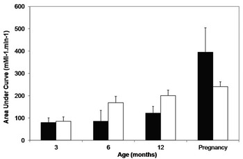
Studies in older pigs have demonstrated that LBW is associated with glucose intolerance (Poore and Fowden, Reference Poore and Fowden2004a; Corson et al., Reference Corson, Laws, Litten, Lean and Clarke2009), reduced insulin sensitivity and increased mean arterial blood pressure (Poore et al., Reference Poore, Forhead, Gardner, Giussani and Fowden2002) and altered Hypothalamo–pituitary–adrenal axis function (Poore and Fowden, Reference Poore and Fowden2003) in conjunction with changes in body composition and endocrine profile (e.g. plasma leptin) (Poore and Fowden, Reference Poore and Fowden2004b; Morise et al., Reference Morise, Louveau and Le Huërou-Luron2008). LBW pigs exhibit glucose intolerance (Figure 2), irrespective of whether they are reared under standard commercial (Corson et al., Reference Corson, Laws, Litten, Lean and Clarke2009) or experimental conditions (Poore and Fowden, Reference Poore and Fowden2004b), and the methodology used for a glucose tolerance test (GTT). This suggests that their postnatal environment had little effect on their risk of developing adulthood diseases, and that the LBW pig provides a robust model for studying the metabolic syndrome.
The effect of low (L) or normal (N) birth weight on the area under the curve following a glucose tolerance test (GTT) at 3, 6 and 12 months of age and during pregnancy. Values are presented as Mean ± s.e.m., L pigs are represented by black bars, N pigs by white bars. GTT: intravenous administration of 0.5 g/kg BW dextrose 40%. Data taken from Poore and Fowden, Reference Poore and Fowden2004a (3 and 12 months) and Corson et al., Reference Corson, Laws, Litten, Dodds, Lean and Clarke2008b (6 months and pregnancy).

Studies have shown that the birth weight of a human baby is related to both their mother’s and maternal grandmother’s birth weight (Hackman et al., Reference Hackman, Emanuel, Belle and Daling1983; Klebanoff et al., Reference Klebanoff, Graubard, Kessel and Berendes1984). Women who were themselves small for gestational age (SGA) are twice at risk of having a SGA baby (Klebanoff et al., Reference Klebanoff, Meirik and Berendes1989; Skjærven et al., Reference Skjærven, Wilcox, Øyen and Magnus1997) as well as being at a greater risk of developing adulthood diseases. Moreover, women who had parents with NIDDM had children with higher birth weights than women with non-diabetic parents. Grandchildren of grandparents with NIDDM are usually in the top percentiles in growth charts (i.e. 90th centile and above) compared with non-diabetic grandparents (McCarron et al., Reference McCarron, Smith and Hattersley2004). In the past intergeneration studies have been somewhat limited to rodent models (Harrison and Langley-Evans, Reference Harrison and Langley-Evans2009), but more recently it has been demonstrated that an intergeneration relationship between maternal size at birth and altered glucose tolerance in later life exists in the domestic pig (Corson et al., Reference Corson, Laws, Litten, Lean and Clarke2009). The added advantage of using a porcine model for intergeneration investigations is that it allows both LBW and high birth weight piglets within a litter to be studied, further reducing the number of animals required as well as decreasing genetic variation.
Cardiovascular disease and the metabolic syndrome
Pigs are frequently used as models for the study of cardiovascular disease and atherosclerosis as the morphology and physical function of the cardiovascular system in swine are similar to humans’ (Smith and Swindle, Reference Smith and Swindle2006; Tables 1 and 2). Exceptions are that the swine heart has a left azygous vein that contains systemic blood from the inter-costal vessels; the conduction system is more neurogenic than myogenic; and the nerve cells within the atrioventricular node also differ. Having said that, the blood supply to the myocardium and the conduction system of the swine is similar to 90% of the human population (Smith and Swindle, Reference Smith and Swindle2006). Atherosclerosis in the pig develops both spontaneously and when induced by an experimental atherogenic, high-cholesterol diet in pigs (Gerrity et al., Reference Gerrity, Natarajan, Nadler and Kimsey2001). Plaque histology and pathogenesis in the atherogenic swine appears to be similar to that of humans (Gal and Isner, Reference Gal and Isner1992; Rand, Reference Rand2009). Atherogenic diets offered to rodents result in some of characteristics of atherosclerosis and the metabolic syndrome observed in humans (Buettner et al., Reference Buettner, Schölmerich and Cornelius Bollheimer2007; Oron-Herman et al., Reference Oron-Herman, Kamari, Grossman, Yeger, Peleg, Shabtay, Shamiss and Sharabi2008), however they generally provide poor models of the changes in plasma lipids associated with coronary heart disease.
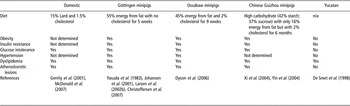
Some metabolic and cardiovascular disorders occur naturally in domestic pigs as a consequence of intense selective breeding and selection for increased growth rate and leanness (Brambilla and Cantafora, Reference Brambilla and Cantafora2004). Feeding the modern domestic pig excess amounts of lard and cholesterol can induce atherosclerosis (Gerrity et al., Reference Gerrity, Natarajan, Nadler and Kimsey2001; McDonald et al., Reference McDonald, Gerrity, Jen, Chen, Wark, Wight, Chait and O’Brien2007; Table 4) without some of the other clinical signs of the metabolic syndrome. The minipig, particularly the ossabaw swine, has proved to be an excellent model because an atherogenic diet induces three or more of the clinical signs of the metabolic syndrome (Johansen et al., Reference Johansen, Hansen, Richelsen and Malmlöf2001; Spurlock and Gabler, Reference Spurlock and Gabler2008).
A comparison of diet-induced metabolic syndrome in different breeds of pig

Yes indicates whether the animal expresses diseases.
The natural history of restenosis (i.e. the reoccurrence of stenosis) in pigs, both with respect to neointimal formation and remodelling, resemble the human disease closely. A combination of denudation and atherogenic diet can be used to induce atherosclerosis in pigs. Although the Yucatan breed of minipig is not a successful model for metabolic-syndrome-induced atherosclerosis, as it fails to exhibit insulin resistance and obesity, it does provide a model for studying the arterial response after balloon angioplasty and natural remodelling (De Smet et al., Reference De Smet, Van Der Zande, Van Der Helm, Kuntz, Borst and Post1998).
Over the past decade, pigs have been increasingly used to study changes in the collagen of the extra cellular matrix of the heart during hypertrophic cardiomyopathy (Liu et al., Reference Liu, Chiu, Shyu, Factor, Chu, Lin, Hsou, Fox and Yang1994; Chiu et al., Reference Chiu, Liu, Liu, Chen, Lin, Mao and Chu1999); abdominal aortic aneurysm (Ruiz et al., Reference Ruiz, Zhang, Butt and Whittaker1997) and chronic ischaemia because their coronary anatomy, with minimal preexisting coronary collateral vessels (Maxwell et al., Reference Maxwell, Hearse and Yellon1987), cardiac physiology, and cardiac conduction systems are very similar to humans (Swindle et al., Reference Swindle, Horneffer, Gardner, Gott, Hall, Stuart, Baumgartner, Borkon, Galloway and Reitz1986). Likewise, the heart weight to BW ratio for the typical 30 kg minipig used in most laboratory studies is identical to that of humans at around 0.005:1 or 5 g heart tissue per kilo of BW (Joseph, Reference Joseph1908; Hughes, Reference Hughes1986; Kist et al., Reference Kist, Thomas, Horner and Laughlin1999) and from a metabolic standpoint non-esterified fatty acids (NEFA) are the preferred substrate of myocardial energy production in both species (Abdel-Aleem et al., Reference Abdel-Aleem, St Louis, Hughes and Lowe1999). The development of a porcine model of myocardial ischaemia to evaluate the efficacy of therapeutic angiogenesis in the preclinical setting is ongoing (Sodha et al., Reference Sodha, Boodhwani, Ramlawi, Clements, Mieno, Feng, Xu, Bianchi and Sellke2008). Despite the success in some of these models (Hughes, 2003), randomized placebo controlled clinical trials failed to demonstrate a similar degree of success (Kastrup et al., Reference Kastrup, Jørgensen, Rück, Tägil, Glogar, Ruzyllo, Bøtker, Dudek, Drvota and Hesse2005). There are multiple reasons for the disparate results seen between animal models and human patients, including the presence of comorbidities such as hypercholesterolemia and diabetes, and medication usage, which may alter the milieu of signalling molecules involved in the angiogenic cascade (Simons et al., Reference Simons, Bonow, Chronos, Cohen, Giordao, Hammond, Laham, Li, Pike, Sellke, Stegmann, Udelson and Rosengart2000).
Diabetes and the metabolic syndrome
NIDDM
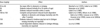
Atherosclerosis is accelerated in diabetic patients and contributes to the majority of deaths of diabetic patients (Ruderman et al., Reference Ruderman, Williamson and Brownlee1992). Pigs represent a valuable model for diabetes-induced accelerated atherosclerosis. Administration of streptozotocin (STZ) induces DNA strands to break within β-cells. The activation of repair mechanisms leads to a reduction in cellular nicotinamide adenine dinucleotide and adenosine triphosphate levels to below physiological levels, resulting in cell death and ultimately diabetes (Yamamoto et al., Reference Yamamoto, Uchigata and Okamoto1981). Various studies have investigated the effectiveness of different dose rates of STZ to induce diabetes (Table 5) and the differences in response have been attributed to age and gender variations (Larsen and Rolin, Reference Larsen and Rolin2004).
Response to increasing dose rates of streptozotocin in juvenile pigs

Insulin dependent diabetes mellitus
Pigs provide a good model for Type I or insulin-dependent diabetes as there are many similarities between pigs and humans in terms of the structure and function of the pancreas (Larsen and Rolin, Reference Larsen and Rolin2004) and pharmacokinetics following administration of trial drugs. However, the spontaneous development of Type-I diabetes is very rare in pigs and so this condition must be induced experimentally either via surgery, chemical administration, such as STZ (Larsen and Rolin, Reference Larsen and Rolin2004) or diet as described above (Xi et al., Reference Xi, Win, Wang, Kusunoki, Lian, Koike, Fan and Zhang2004).
The severity of Type I diabetes after surgical-induction by pancreatectomy is dependent on the degree of excision/ablation. For example, a 40% pancreatectomy results in mild changes, 80% in significant alterations (Lohr et al., Reference Lohr, Lubbersmeyer, Otremba, Klapdor, Grossner and Kloppel1989) and 100% in severe hyperglycemia (Wilson et al., Reference Wilson, Dhall, Simeonovic and Lafferty1986; Stump et al., Reference Stump, Swindle, Saudek and Strandberg1988; Mellert et al., Reference Mellert, Hopt, Hering, Bretzel and Federlin1991 and Reference Mellert, Hering, Liu, Brandhorst, Brandhorst, Brendel, Ernst, Gramberg, Bretzel and Hopt1998). The problems associated with using this method are that surgery involves the removal of exocrine and endocrine tissue, which is not characteristic of the disease in humans (Wilson et al., Reference Wilson, Dhall, Simeonovic and Lafferty1986), and that it is an extremely invasive technique with huge welfare implications.
Digestive physiology and metabolism
Total parenteral nutrition
When an infant is born prematurely, although its gastrointestinal (GI) tract is fully formed at about 20 weeks of pregnancy, it is not fully functional at that time; for example peristalsis does not commence until around 29 weeks of gestation and it may not be able to synthesize the enzymes required to digest milk and formula (Sangild, Reference Sangild2006). As a consequence, it can often not tolerate enteral feeding due to its immature GI tract and so must be fed intravenously (known as total parenteral nutrition (TPN)). As nutrition provided by TPN bypasses the gut and absorption at the intestinal mucosa, it results in varying degrees of gut atrophy, depending on the period of treatment and whether the individual is receiving any nutrient enterally. Most infants on TPN are subject to numerous clinical problems, which complicate the nutritional and metabolic requirements and make interpretation of any experimental results difficult (Jadhav et al., Reference Jadhav, Parimi and Kalhan2007). The presence of central venous catheters and mechanical ventilation associated with TPN may increase the incidence of late-onset sepsis in these very LBW (<1500 g) babies (Kansagra et al., Reference Kansagra, Stoll, Rognerud, Niinikoski, Ou, Harvey and Burrin2003). TPN results in deprivation of luminal nutrition that also adversely affects the mucosal integrity and may compromise the barrier function of the neonatal gut, resulting in ‘gut-derived’ sepsis, and the subsequent translocation of luminal bacteria and toxins into the blood (Berg, Reference Berg1995).
Rat pups are not a suitable model for studying human premature infants receiving TPN due to their immaturity. When rats have reached a BW of around 180 g (∼60% mature BW), they can be used as a model to study gut function and epithelial regeneration following TPN after GI trauma, surgery or cancer (Jordinson et al., Reference Jordinson, Goodlad, Brynes, Bliss, Ghatei, Bloom, Fitzgerald, Grant, Bardocz, Pusztai, Pignatelli and Calam1999). As mentioned above, piglets possess poor thermoregulatory mechanisms, have high metabolic rates and are prone to hypoglycemia, thus providing an excellent model for premature human neonates receiving TPN (Mei and Xu, Reference Mei and Xu2003). Although immature at parturition, piglets have the ability to survive when delivered preterm allowing modelling of different levels of prematurity visible in human infants. Piglets can also be used with or without being given colostrum to provide immunoglobulins (Shulman, Reference Shulman1993). Work with colostrum-deprived (i.e. receiving no colostrum and so immumocompromised) minipiglets was first carried out by Mehrazar and Kim (Reference Mehrazar and Kim1988) who used germfree colostrum-deprived piglets delivered 3 to 5 days preterm to study the ontogeny of the immune system. Borum (Reference Borum1993) developed their work by using non-germfree (i.e. born and housed in non-sterile conditions), colostrum-deprived pigs as a model for studies into human TPN. This model has been further developed by Sangild et al. (Reference Sangild, Petersen, Schmidt, Elnif, Petersen and Buddington2002), with the use of colostrum-deprived piglets with limited immunity gained via an infusion of maternal serum; they also delivered the piglets at 93% of gestation (i.e. 8 days preterm) with a good degree of success. Pigs in a germfree (i.e. sterile) environment can be administered TPN successfully for up to 21 days (Mehrazar and Kim, Reference Mehrazar and Kim1988; Borum, Reference Borum1993).
Apart from anything else, piglets have a large enough body size to allow the same TPN methods and procedures to be used as in humans and for regular blood sampling without significant diminution of the total blood volume. At the same time, their body size is small enough to limit experimental costs (Burrin, Reference Burrin2001). Pigs have the advantage of a large litter size and 2 to 3 litters per sow per year (Pond and Mersmann, Reference Pond and Mersmann2001) making repeat experiments easily achievable, and reducing, to some extent, genetic influences. A typical TPN solution (energy content 1.67 kJ/ml) is usually administered with 20% lipid emulsion (8.36 MJ/l) (Hyde et al., Reference Hyde, Amusquivar, Laws, Corson, Geering, Lean, Putet, Dodds, Herrera and Clarke2008). One factor important to emphasize is that premature piglets, as with premature human babies, are prone to fluid retention and so infusion rates must be altered accordingly (Hyde et al., Reference Hyde, Amusquivar, Laws, Corson, Geering, Lean, Putet, Dodds, Herrera and Clarke2008) by gradually stepping up the infusion rates over the first 24 h of life. Infusion rates also differ between piglets delivered by Caesarean section (112 days of gestation) and vaginally at term (115 days) (Hyde et al., Reference Hyde, Amusquivar, Laws, Corson, Geering, Lean, Putet, Dodds, Herrera and Clarke2008 and Reference Hyde, Griffin, Herrera, Byrne, Clarke and Kemp2010).
Over the last two decades the newborn piglet has proved to be a good model for studying: (i) calcium metabolism (Draper et al., Reference Draper, Yuen and Whyte1991); (ii) growth and development of the gut and gut barrier function (Burrin et al., Reference Burrin, Shulman, Storm and Reeds1991; Van Aerde et al., Reference Van Aerde, Keelan, Clandinin and Thomson1997; Kansagra et al., Reference Kansagra, Stoll, Rognerud, Niinikoski, Ou, Harvey and Burrin2003); (ii) the immune system (Mehrazar and Kim, Reference Mehrazar and Kim1988); (iv) amino acid metabolism (House et al., Reference House, Pencharz and Ball1997 and Reference House, Pencharz and Ball1998); and (v) non-alcoholic fatty liver disease (Hyde et al., Reference Hyde, Perkins, Laws, Dodds, Symmons, Geering, Litten, Corson, Lean and Clark2005) in TPN-fed human infants. More recently, the effects of the fatty acid composition of the lipid emulsion on fatty acid profiles of tissues from Caesarean-sectioned delivered piglets has shown that the fatty acid composition of key tissues mirrors the fatty acid profile of the lipid emulsion supplied (Amusquivar et al., Reference Amusquivar, Sánchez, Hyde, Laws, Clarke and Herrera2008); this could have important consequences, particularly for neurodevelopment, in premature human babies.
Postnatal digestive physiology
The piglets’ GI tract has a high degree of anatomical and physiological similarity to that of the human infant (Siggers et al., Reference Siggers, Siggers, Boye, Thymann, Molbak, Leser, Jensen and Sangild2008), and their protein and lipid metabolism are comparable with humans (Canavan et al., Reference Canavan, Flecknell, New, Alberti and Home1997; Davis et al., Reference Davis, Suryawan, Orellana, Nguyen and Fiorotto2008). Therefore, pigs are an accepted and extensively used model for specific types of nutritional studies (Moughan and Rowan, Reference Moughan and Rowan1989; Spurlock and Gabler, Reference Spurlock and Gabler2008). For example, pigs have been used to study protein digestion in human infants (Darragh and Moughan, Reference Darragh and Moughan1995; Lin et al., Reference Lin, Mahan, Wu and Kim2009); digestibility of dietary amino acids (Rowan et al., Reference Rowan, Moughan, Wilson, Maher and Tasman1994; Wu, Reference Wu1998; Reeds and Burrin, Reference Reeds and Burrin2000) and the effect of infant milk formulas on organ development and digestive enzyme activities (Moughan et al., Reference Moughan, Pedraza, Smith, Williams and Wilson1990 and Reference Moughan, Birtles, Cranwell, Smith and Pedraza1992). A review of the pig as an experimental model for elucidating the mechanisms governing dietary influence on mineral absorption has recently been published by Patterson et al. (Reference Patterson, Lei and Miller2008), and so this aspect of digestion will not be considered herein.
Another area of interest is the effect of dietary fats on the growth and development of the human infant, particularly the brain (Innis, Reference Innis2004). Piglets undergo a growth spurt in the brain at a similar time as that observed in humans; the porcine brain is relatively well developed at birth, having achieved 38% of its adult brain weight (Pond et al., Reference Pond, Boleman, Fiorotto, Ho, Knabe, Mersmann, Savell and Su2002). This growth spurt is accompanied by subcutaneous lipid accumulation and research shows that the fatty acid profile of the nutrients supplied to the developing piglet postnatally can influence the fatty acid profile of the brain and other tissues (Amusquivar et al., Reference Amusquivar, Sánchez, Hyde, Laws, Clarke and Herrera2008; Hyde et al., Reference Hyde, Amusquivar, Laws, Corson, Geering, Lean, Putet, Dodds, Herrera and Clarke2008). Hence, the piglet is an excellent model for the studies on fatty acid metabolism during the perinatal period (Purvis et al., Reference Purvis, Clandinin and Hacker1982; Laws et al., Reference Laws, Litten, Laws, Lean, Dodds and Clarke2009).
In addition to the physiological changes associated with the transition from the intrauterine to extrauterine environment, pig and human infants both are also prone to diarrhoea. Diarrhoea during this phase of development is usually associated with rotavirus and Escherichia coli, although Coronavirus (Transmissible Gastroenteritis) and Clostridium perfringens type C also cause enteritis in pigs. The beneficial effect of the probiotic lactic acid bacteria in human or animal health has interested scientists over the last century. Probiotics are essentially ‘living microorganisms which favourably influence the health of the host by improving the indigenous microflora’ (Fuller, Reference Fuller1989). The World Health Organization (WHO, 2002) defines them as ‘live microorganisms which, when administered in adequate quantities, confer a health benefit to the host’. Positive health effects may also be obtained by ‘prebiotics’ – non-digestible substances that increase the growth or the activity of specific microorganisms in the gastro-intestinal tract. Potential effects of prebiotics and probiotics include increased nutritional value of food due to improved digestibility and absorption, improvement of the immune system, and prevention of intestinal tract infections, cancer, atherosclerosis and osteoporosis (Ziemer and Gibson, Reference Ziemer and Gibson1998; Gill and Guarner, Reference Gill and Guarner2004).
Necrotizing enterocolitis (NEC) is a medical condition primarily seen in premature infants in which portions of the bowel undergo necrosis, and this condition has been investigated using Caesarean-delivered preterm pigs as a model for premature human infants (Sangild, Reference Sangild2006). A probiotic-containing formula can reduce the severity of NEC in this model. Probiotic administration immediately after birth appears to promote the colonization of a beneficial commensal microbiota capable of limiting the formula-induced mucosal atrophy, dysfunction, and pathogen load in preterm neonates, thereby reducing the incidence and severity of NEC (Siggers et al., Reference Siggers, Siggers, Boye, Thymann, Molbak, Leser, Jensen and Sangild2008).
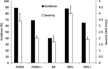
Weaning piglets at an age of 3 to 5 weeks changes the flora, morphology and function of the porcine intestine (Nabuurs, Reference Nabuurs1998). The effectiveness of feeding probiotic (Bifidobacterium lactis HN019 concentration 108 cfu/mL) against naturally acquired enteric infectious diseases caused by rotavirus and E. coli has been studied using a piglet model (Shu et al., Reference Shu, Qu and Gill2001). Results suggest that diarrhoea was significantly reduced in probiotic-administered weanlings. It is of interest to note that the reduction in the incidence and severity of diarrhoea in neonatal and weanling pigs receiving probiotics is comparable, further supporting the pig as a good model for neonatal GI infection in human infants (Figure 3).
The incidence and severity of diarrhoea as influenced by breast (colostrum) milk (BR), milk formula (FORM) and milk formula with probiotics (FORM+) in caesarean-delivered preterm pigs and weaned pigs orally administered either a placebo (PRO−) or probiotic (PRO+). Values are presented as means for incidence & mean ± s.e.m. for severity. FORM+ = Bifidobacterium animalis and Lactobacillus: L. acidophilus, L. casei, L. pentosus, L. plantarum. PRO+ = Bifidobacterium lactis HN019 (109 colony-forming units (cfu)/piglet per day). Caesarean-delivered preterm pigs data adapted from Siggers et al. (Reference Siggers, Siggers, Boye, Thymann, Molbak, Leser, Jensen and Sangild2008). Weaned pigs data adapted from Shu et al. (Reference Shu, Qu and Gill2001).

Alternative animal models have been used, for example probiotics have been shown to reduce bacterial infection in the acute pancreatitis rat model, the major advantage of this model is that it resembles the human to such an extent that bacteriologic results, reaction to treatment and disease course can all be predicted (Van Minnen et al., Reference Van Minnen, Timmerman, Lutgendorff, Verheem, Harmsen, Konstantinov, Smidt, Visser, Rijkers, Gooszen and Akkermans2007). Supplementation with Bifidobacterium infantis in the rat model resulted in intestinal colonization and a significant reduction in intestinal colonization and a significant reduction in the incidence of necrotising enterocolitis in comparison to the controls (Caplan et al., Reference Caplan, Miller-Catchpole, Kaup, Russell, Lickerman, Amer, Xiao and Thomson1999). However, due to the small size of the rat, the tissues samples obtained are also small and often require pooling.
It is well documented that diet modulates immune functions in different ways and affects host resistance to infections. In addition to the essential nutrients, non-digestible carbohydrates such as inulin (IN) and oligofructose (OF) modulate the systemic immune system (Seifert and Watzl, Reference Seifert and Watzl2007). IN and OF are classified as prebiotics, which occur naturally as plant storage carbohydrates in vegetables, cereals and fruits. Results from human intervention studies suggest that the intake of IN and OF has beneficial effects on the gut-associated lymphoid tissue. Both porcine and rat models have demonstrated similar anti-inflammatory changes in the gut-associated lymphoid tissue as those observed in humans and give more insight into the immune tissue- specific effects of IN and OF (Roller et al., Reference Roller, Rechkemmer and Watzl2004; Seifert and Watzl, Reference Seifert and Watzl2007). More recently an in vitro dynamic model to simulate porcine ileal digestion (Meunier et al., Reference Meunier, Manzanilla, Anguita, Denis, Pérez, Gasa, Cardot, Garcia, Moll and Alric2008) has been developed, but the potential benefits of using a similar system to investigate the effects of probiotics/prebiotics remains to be established.
Ulcers
Damage to the human GI tract can be caused by a number of different conditions such as repeated use of non-steroidal anti-inflammatory drugs (NSAID) or surgery due to cancer and other GI tract-related diseases. Bleeding peptic ulcers remain a major cause of upper-GI bleeding (Laine and Peterson, Reference Laine and Peterson1994) and although endoscopic haemostasis is now the primary treatment for bleeding peptic ulcers (Sacks et al., Reference Sacks, Chalmers, Blum, Berrier and Pagano1990; Cook et al., Reference Cook, Guyatt, Salena and Laine1992) there remains a subgroup of patients with bleeding who are not amenable to endoscopic control, and in this cohort salvage surgery carries a mortality of ∼25% (Rockall, Reference Rockall1998).
The pig provides a good model for testing the efficacy and safety of new endoscopic equipment before clinical trials (Hu et al., Reference Hu, Sun and Chung2004). A feasibility study using a porcine model for endoscopic plication of massively bleeding peptic ulcer by using the Eagle Claw VII device has recently been conducted (Chiu et al., Reference Chiu, Hu, Lau, Sun, Sung and Chung2006). Although this model simulated bleeding ulcers with a large vessel at the base, the chronicity of such an ulcer could not be reproduced primarily because it is often matted with hard and fibrotic tissue (Hu et al., Reference Hu, Chung, Sun, Lau, Kawashimak, Yamamoto, Cotton, Gostout, Hawes, Kalloo, Kantsevoy and Pasricha2005). Consequently, although the authors suggest that the Eagle Claw VII provides a feasible and reliable technique in achieving endoscopic plication on bleeding peptic ulcers, it remains to be established whether a needle of the Eagle Claw VII device would be able to penetrate such ulcer (Chiu et al., Reference Chiu, Hu, Lau, Sun, Sung and Chung2006).
A non-survival porcine model that simulates acute peptic ulcer bleeding has recently been established that can be used to develop future endoscopic therapies and for training purposes (Chen et al., Reference Chen, Marks, Wong, McGee, Faulx, Isenberg, Schomisc, Deng, Ponsky and Chak2008). Similarly, a porcine model is currently being used to investigate the possible role of high-intensity focused ultrasound in the treatment of acute peptic ulcer haemorrhage (Marks et al., Reference Marks, Chen, Rosen, Mcgee, Chak, Onders, Faulx, Schomisch and Ponsky2006).
Many unresectable primary (gastric, duodenal, pancreatic) or metastatic (colorectal, renal, etc.) malignancies can cause gastric outlet and duodenal obstruction in humans (Alam et al., Reference Alam, Baines and Parker2003; Cogliandolo et al., Reference Cogliandolo, Scarmozzino, Pidoto, Pollicino and Florio2004; Mittal et al., Reference Mittal, Windsor, Woodfield, Casey and Lane2004). Moreover, open surgery for palliation of this obstruction is associated with high morbidity and mortality (DeMaria et al., Reference De Maria, Schweitzer, Kellum, Meador, Wolfe and Sugerman2002; Reed et al., Reference Reed, Cacchione, Allen, Arlauskas, Casey, Larson and Vitale2003). Although the laparoscopic approach is less traumatic than open surgery, the laparoscopic creation of a gastrojejunostomy is technically difficult, requires extensive surgical and laparoscopic skills, and is associated with numerous complications, primarily anastomotic stricture (3.1% to 8.8%) and leak (1.2% to 3.0%) (Hamad et al., Reference Hamad, Mentges and Buess2003; Sundbom and Gustavsson, Reference Sundbom and Gustavsson2004). The safety and feasibility of transgastric endoscopic gastrojejunostomy, with survival, has recently been studied in a porcine model, and has demonstrated the potential advantages of this procedure in comparison with surgical or laparoscopic gastroenteric anastomosis. Benefits include minimal invasiveness, no need for an anterior abdominal wall and skin incisions, thereby eliminating the risk of skin wound infection and post-operative hernias (Kantsevoy et al., Reference Kantsevoy, Jagannath, Niiyama, Chung, Cotton, Gostout, Hawes, Pasricha, Magee, Vaughn, Barlow, Shimonaka and Kalloo2005). A common treatment for gastric cancer is a resection; the pig has been effectively used to study an endoscopic full-thickness resection with sutured closure before clinical trials (Ikeda et al., Reference Ikeda, Fritscher-Ravens, Mosse, Mills, Tajiri and Swain2005).
As mentioned earlier, humans often suffer GI diseases following repeated daily enteral administration of NSAID, but it is often difficult to study certain regions of the GI tract for example, the proximal regions of the large intestine using routine endoscopic investigations. A pig model has successfully been developed to assess the resultant damage to the GI tract from long-term use of NSAID (Rainsford et al., Reference Rainsford, Stetsko, Sirko and Debski2003).
Renal physiology
In the past canines have been successfully used as a large animal model for kidney investigative work. The larger animals are often selected as their size makes arterial catheter navigation easier; pigs have the additional benefit over dogs of possessing kidneys which are similar in anatomy and physiology to the human kidney (Yokota et al., Reference Yokota, Benyajati and Dantzler1985; Tumbleson and Schook, Reference Tumbleson and Schook1996). Similar to humans, the porcine kidney is multi-pyramidal with an undivided cortex and has several different medullary structures. Each medullary pyramid forms a separate papilla and fusion results in the formation of some compound papillae. The rat kidney has a single papilla, and the medulla and cortex are undivided. Pig and Man have similar maximal urine concentration (pig 1080 and man 1160 mOsmol/l), maximal urine-to-plasma osmolal ratio (pig 3.7 and man 4.0), glomerular filtration rate (pig 130 and man 126 to 175 ml/min per 70 kg) and total renal blood flow (pig 4 and man 3.0 to 4.4 ml/min per g; Sachs, Reference Sachs1994).
When atherosclerosis causes stenosis or an abnormal narrowing of blood vessels, the pig has been successfully used to study not only atherosclerosis but also renal artery stenosis. Angioplasty (widening of the blood vessel) work has been completed using magnetic resonance to guide the procedure and while the authors state that the work is preliminary, these authors believe that future hardware, and software advances will improve the performance of the procedures further (Omary et al., Reference Omary, Gehl, Schirf, Green, Lu, Pereles, Huang, Larson and Li2006; Park et al., Reference Park, Rhee, Cashen, Shin, Schirf, Gehl, Larson, Prasad, Li, Carroll and Omary2007a and Reference Park, Rhee, Cashen, Shin, Resnick, Gehl, Schirf, Wang, Larson and Carroll2007b).
Swine have also been shown to be an excellent model for urological studies, including the formation of renal calculi (Borden and Vermeulen, Reference Borden and Vermeulen1966; Assimos et al., Reference Assimos, Boyce, Lively, Weidner, Lewis and Howard1986; Terris, Reference Terris1986). Papillary calcifications have been induced in pigs fed oxamide (Borden and Vermeulen, Reference Borden and Vermeulen1966) and the implantation of kidney stones into pig kidneys has been used as a model for lithotripsy research (Paterson et al., Reference Paterson, Lingeman, Evan, Connors, Williams and McAteer2002). To-date the rat has been used as the main reproducible animal model of calcium oxalate crystalluria and urolithiasis. However it is desirable to have an animal model, such as the pig, of oxaluria and urolithiasis with physiological, anatomical and nutritional characteristics that more closely resemble man. It has been shown that feeding pigs trans-4-hydroxy-l-proline causes hyperoxaluria and calcium oxalate crystalluria in which calcium oxalate papillary deposits form that may be precursors of kidney stones (Mandel et al., Reference Mandel, Henderson, Hung, Wille and Wiessner2004). Consequently, further development of the pig as a model of human hyperoxaluria and stone formation should be successful for studying these human diseases.
Bones
Bones and osteoporosis
Osteoporosis is a multigenic complex disorder. Though the mouse and rat are used as experimental models for human osteoporosis, the pig bone remodelling cycle is histologically more similar to human than the rat or mouse. Moreover, livestock genomics have many advantages over model organisms and human studies for complex trait dissection (Onteru et al., Reference Onteru, Fan and Rothschild2008). In addition to other features mentioned elsewhere in this review, the reproductive cycle of the pig is similar in duration to the human (18 to 21 days) and is continuous, also like the human (Turner, Reference Turner2001).
A syndrome of spontaneous vertebral fracture has been reported in the pig, a rarity in the animal world (Spencer, Reference Spencer1979), pigs are large enough to receive prosthetic implants, withstand serial bone biopsies and large volumes of blood sampling (Turner, Reference Turner2001). The rate of bone removal and deposition of the trabecular and cortical bone is also similar to that observed in humans (Mosekilde et al., Reference Mosekilde, Weisbrode, Safron, Stills, Jankowsky, Ebert, Danielsen, Søgaard, Franks, Stevens, Paddock and Boyce1993b). A porcine model has also been used to evaluate the initial fixation strength of a biodegradable interference screw in anterior cruciate ligament reconstruction using a bone-patellar tendon-bone graft (Seil et al., Reference Seil, Rupp, Krauss, Benz and Kohn1998; Walsh et al., Reference Walsh, Wijdicks, Parker, Hapa and LaPrade2009).
Findings suggest that the Sinclair Si minipig fed a 0.75% calcium restricted diet may be a good model to study the bone remodelling and perimenopausal bone loss in women when an ovariectomy is performed (Mosekilde et al., Reference Mosekilde, Weisbrode, Safron, Stills, Jankowsky, Ebert, Danielsen, Søgaard, Franks, Stevens, Paddock and Boyce1993a and Reference Mosekilde, Weisbrode, Safron, Stills, Jankowsky, Ebert, Danielsen, Søgaard, Franks, Stevens, Paddock and Boyce1993b; Boyce et al., Reference Boyce, Ebert, Youngs, Paddock, Mosekilde, Stevens and Gunderson1995). Lafage et al. (Reference Lafage, Balena, Battle, Shea, Seedor, Klein, Hayes and Rodan1995) have also used minipigs to investigate bone active agents such as sodium fluoride, assessed on bone quality and remodelling. Dynamic 18F-fluoride ion positron-emission tomography (PET) has demonstrated that porcine bone loss after total gastrectomy is related to a high-turnover bone disease without associated changes in bone blood flow. It is thought that the increased bone metabolism observed in minipigs is probably related to an elevated parathyroid hormone secretion, thus maintaining serum calcium homeostasis at the expense of the bone mineral content. Normalizing bone metabolic activity by the specific bone mass increases the sensitivity in the detection of osteopenic high turnover bone diseases. Therefore, the combination of quantitative computed tomography and 18F-fluoride ion PET seems to be the method of choice for the classification of metabolic bone diseases and for monitoring treatment effects quantitatively (Piert et al., Reference Piert, Zittel, Jahn, Stahlschmidt, Becker and Machulla2003).
Dental research
Since the tooth structure of the Prestige World Genetics (PWG) Micro-pig® closely resembles that of human more than any other species, the pigs are valuable in dental research. Adult stem cells originated from pulp show that new dental material can be created and implanted. As the size of the Micro-Pig® jawbone is as thick as humans, these pigs are frequently used in dental surgery, especially in the testing of dental implants and the use of dental stem cells to grow new teeth (PWG Genetics Korea, 2006; Mantesso, Reference Mantesso2008).
By examining the ontogeny of porcine enamel our understanding of human enamel deposition and maturation has been significantly improved (Robinson et al., Reference Robinson, Kirkham, Weatherell, Richards, Josephsen and Fejerskov1987 and Reference Robinson, Kirkham, Weatherell, Richards, Josephsen and Fejerskov1988; Kirkham et al., Reference Kirkham, Robinson, Weatherell, Richards, Fejerskov and Josephsen1988). Work has been completed on implants in pigs to consider loading, however caution needs to be used here as porcine bone remodels slightly faster than human bone. That noted this work provides a valuable insight into micro damage strains following implants (Ko et al., Reference Ko, Swift, DeLong, Douglas, Kim, An, Chang and Huang2002 and Reference Ko, Douglas, DeLong, Rohrer, Swift, Hodges, An and Ritman2003). A novel device for a bite force measurement system in a porcine model has recently been described (Bousdras et al., Reference Bousdras, Cunningham, Ferguson-Pell, Bamber, Sindet-Pedersen, Blunn and Goodship2006) which enables the biomechanical adaptation of the bone-implant interface to masticatory loads to be assessed with either natural dentition or single implant crowns.
Conclusions
In conclusion, the pig is an extremely useful biomedical model for improving our understanding of the pathophysiology and for investigating potential treatment/prevention strategies under rigorously controlled conditions for many human diseases and ailments. The optimum porcine model to use is often dependent on the specific disease being studied. For example, the domestic pig can be used to study developmental programming due to the natural variation in BW within a litter, while the minipig is considered the best breed for investigation on the metabolic syndrome. Pigs provide a valuable translational model to bridge the gap between classical rodent models and humans in developing new therapies to aid human health.
Acknowledgements
The authors would like to thank Dr Colin Litten-Brown for his assistance in the preparation of this manuscript.