

Introduction
Humans and animals perceive inspiratory hypoxia and the ensuing hypoxaemia as a major threat to mortality and physiological homeostasis and are genetically designed to incur acute (e.g. hyperventilation via carotid bodies) and chronic (e.g. polycythaemia, angiogenesis, up-regulation of hypoxia-inducible factor) adaptations to defend oxygen transportReference Hochachka, Gunga and Kirsch1, Reference Taylor and McElwain2. Central to these adaptations is the increased haemoglobin concentration ([Hb]) that is driven by renal hypoxic sensitivity, which stimulates increased production and circulating plasma concentrations of the peptide hormone erythropoietin ([EPO])Reference Abbrecht and Littell3–Reference McKeever, Agans, Geiser, Lorimer and Maylin11.
With respect to arterial hypoxaemia at sea level, humans may experience exercise-induced arterial hypoxaemia, but it is neither as common nor as severe as that experienced during heavy- and severe-intensity exercise in the horseReference Bayly, Schultz, Hodgson and Gollnick12–Reference Poole and Erickson16. Thus, one would not expect to find elevated [EPO] following exercise in humans and, with one exception following marathon runningReference Schwandt, Heyduck, Gunga and Rocker17, this is trueReference Ekhardt, Kurtz and Bauer18–Reference Bodary, Pate, Wu and McMillan21. Notwithstanding that the lactic acidosis of heavy/severe-intensity exercise is expected to oppose [EPO] productionReference Ekhardt, Kurtz and Bauer18, whether short-term heavy or severe exercise increases plasma [EPO] in the horse at sea level is unknown. It is pertinent that this mechanism has been proposed to account, in part, for overtraining-induced ‘red cell hypervolaemia’ diagnosed in Swedish Standardbred race horsesReference Persson22, Reference Persson and Osterberg23. Moreover, a further interesting question is whether the plasma [EPO] response to exercise might be different against the background of hyperventilation-induced alkalaemia consequent to hypoxaemic stimulation of the peripheral chemoreceptors at altitude.
Unlike humans, the horse has a large (up to 14 kg) spleen that acts as a reservoir for red blood cells (RBCs) such that, during severe-intensity exercise, the circulating haematocrit can be almost doubled (i.e. from ~30 to 60–70%, in the extreme)Reference Poole and Erickson16. Hence, with regard to O2 transport, the horse has a degree of freedom for increasing tissue(s) O2 supply without the necessity for making new RBCs per se. Thus, it might be argued that the presence of this mechanism may reduce the necessity or inclination for the horse to elevate its whole-body RBC mass via [EPO]-induced stimulation of RBC production. Counter to this argument, we found that total RBC volume increased by ~26% over a ~10 day sojourn at high altitude (i.e. 3800 m)Reference Wickler and Anderson24. Whether this was the result of increased plasma [EPO] was not determined.
The purpose of this investigation was to explore the effects of altitude and exercise in separatum and combined on plasma [EPO] in the equid. Specifically, we tested the following hypotheses: (1) acute exercise would increase plasma [EPO]; (2) exposure to 3800 m altitude, which is accompanied by a substantial fall in the partial pressure of O2 (PO2) in arterial blood from 94.9 ± 6.9 to 54.9 ± 4.2 mmHgReference Green, Wickler, Anderson, Cogger, Lewis and Wyle25, would increase plasma [EPO]; and (3) combined exercise and altitude would potentiate the increase in plasma [EPO] compared with either perturbation on its own.
Materials and methods
Animals
Six healthy horses (four Arabians, one Quarter Horse and one Shetland Pony), four females and two males, with an average age of 9.0 ± 4.5 years (mean ± SD) were used in the study. Body masses averaged 467 ± 15 kg for the horses and 257 kg for the pony. Each animal was accustomed to running on the treadmill and to all the test protocols used in this investigation. The animals were physically conditioned for at least 4 months, after which they began a monitored training program, where they were conditioned 3 days per week on a treadmill (SÄTO I; Equine Dynamics Inc., Lexington, KY, USA) at walk, trot and canter for a total of 30 min each session. Training also included an exercise test performed on a dirt track, 2 days a week. Heart rates (HRs) were monitored during all exercise periods using a HR meter (Polar Vantage XL; Polar CIC, Inc., Port Washington, NY, USA). Prior to transport to high altitude (2.5 weeks), horses were exercised on the treadmill to determine their individual maximal HRs and maximal exercise-induced haematocrit. This exercise regime involved an 8 min warm-up at 3.5 m s− 1 and increases in treadmill speed by 1 m s− 1 at 1 min intervals until the horse would no longer maintain its position on the treadmill despite humane encouragement. This test was repeated 1 week later.
General protocol
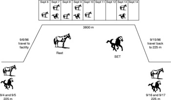
The experimental paradigm and general testing protocol used for this experiment are detailed in Fig. 1. Blood gases, haematological changes and performance effects of this protocol have been publishedReference Wickler and Anderson24, Reference Green, Wickler, Anderson, Cogger, Lewis and Wyle25. [EPO] was measured in venous blood samples obtained at rest and following exercise on a dirt track both at sea level (~225 m) and high altitude, and on a treadmill at sea level in horses. Resting [EPO] was measured initially on 2 days at sea level (mean day T a, 21°C and average P b, 743 mm Hg), over the course of a 10 day exposure to 3800 m altitude (mean day T a, 15°C and P b, 487 mm Hg), and at rest for 2 days after return to sea level. The effects of exercise were examined by measuring [EPO] in samples obtained before and after the submaximal test (speed set-to-HR), which was performed on either of the 2 days pre-high-altitude exposure and on 3 days (days 2, 4 and 8) at 3800 m. Additional samples were obtained during a maximal incremental treadmill exercise test performed on either of the 2 days prior to high-altitude exposure and on one of the 2 days following return to sea level.
The experimental paradigm and general testing protocol. The study consisted of 2 pre-altitude days at sea level (P 1 = 9/4 and P 2 = 9/5), 10 days at altitude (A 1–A 8 = 9/6–9/14) and 2 days after return to sea level (R 1 = 9/16 and R 2 = 9/17)

Jugular catheters (volume ~1.5 ml− 1, ®Check-Flo Blue Introducer Set, 8F; Cook Inc., Bloomington, IN, USA) were used for all sampling. Catheters were inserted using aseptic procedures and the site protected with bandaging material. Catheters were flushed twice daily with heparinized saline and were replaced approximately every 72 h (or as needed). Twenty millilitres of blood were withdrawn prior to actual blood sampling to ensure that residual blood/saline was cleared from the catheter.
Blood samples were collected in pre-chilled heparinized syringes from resting horses on each of the 2 days prior to transport. On the third day of the study (i.e. day 1 at altitude), the animals were transported 500 km to the Barcroft Facility of the University of California, White Mountain Research Station in the White Mountain range (3800 m), an approximately 13 h journey. On the day of transport to altitude, the blood sample was collected 3 h after reaching the research station (at ~17.00 hours). Subsequently, blood samples were collected 90 min postprandial (08.15 hours) on days 2, 4 and 8 of exposure to high altitude. The animals were transported back to sea level on study day 10, with resting blood samples collected 2 days, following return from high altitude.
The submaximal (track) test was performed on either of the 2 days, pre-altitude transport (i.e. at sea level), and on days 2, 4 and 8 of exposure to high altitude (3800 m). The maximal incremental treadmill test was performed at sea level once (within 2 days) prior to altitude exposure (on the alternate day from the submaximal track test) and on each of the 2 days upon return to sea level (no track test was run upon return to sea level because of potential interference with the treadmill test).
Submaximal exercise test on the track
The track test was performed on a 200 m long, compacted dirt, oval track. The test was preceded by a 15 min walking warm-up period. Subsequently, the submaximal test was standardized using HR, and consisted of 5 min walking, a 4 min trot (HR 110 bpm), a 4 min canter (HR 150 bpm) and a 2 min gallop (HR 180 bpm). At the completion of the gallop phase, the animals were allowed a 15 min recovery period consisting of 5 min walking and 10 min standing. An oval track was also prepared at the high-altitude site, and the submaximal test at altitude used the same HRs to standardize exercise ‘intensity’.
Maximal incremental exercise test on the treadmill
The incremental test consisted of 5 min at 1.6 m s− 1, 4 min at 3.5 m s− 1 (HR 110 bpm) and 4 min at 5.0 m s− 1 at a 10% grade (HR 150 bpm). The animals then exercised for 1 min at maximal speed (10% grade). This speed (8.23 ± 0.40 m s− 1, n = 5) was individually determined to produce a maximal HR response (214 ± 3 bpm). This test concluded with a 15 min recovery. For the pony, treadmill running speeds were reduced, as necessary, for this animal's capabilities. Treadmill tests were not done at high altitude.
Blood samples and assays
An aliquot of blood (10 ml− 1) was collected into pre-chilled tubes containing sodium heparin (Vacutainer; Becton-Dickinson, Inc., Parsippany, NJ, USA). Blood was immediately centrifuged, and the plasma was separated and frozen in liquid nitrogen. Upon returning to sea level, the samples were transferred and stored at − 70°C until analysis. [EPO] was measured using a commercially available radioimmunoassay kit (Incstar, Inc., Stillwater, MN, USA). Previous studies have demonstrated that equine [EPO] is highly homologous with other mammalian speciesReference Wen, Boissel, Tracy, Gruniger, Mulcahy and Czelusniak26–Reference Breyman28, and this particular radioimmunoassay has a reported 100% specificity for α-erythropoietin. Serial dilutions of equine plasma demonstrated that the radioimmunoassay and laboratory procedures utilized were reproducible and accurate. Samples were run in one batch against the manufacturer's standards with a within-assay coefficient of variation (CV) of 7%.
Statistical analysis
Data were analysed (Sigmastat 2.0; Jandel Scientific, Chicago, IL, USA) using ANOVA for repeated measures. Post hoc separation of means utilized the Student–Newman–Keuls method. The null hypothesis was rejected when P < 0.05.
Results
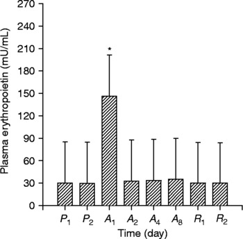
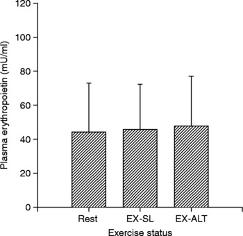
High-altitude exposure increased [EPO] by 387% (P < 0.05) within the first day (Fig. 2). However, 12 h later on the second day at high altitude, [EPO] had returned to pre-altitude levels (P>0.05) (Fig. 2). [EPO] did not change significantly over the remaining 8 days at altitude nor during the 2 days following return to sea level. Moreover, [EPO] did not change significantly in response to acute exercise performed either at sea level or at 3800 m (Fig. 3).
Mean ± SD plasma [EPO] from samples collected at rest on 2 days at sea level (P 1 and P 2); on four occasions at altitude (i.e. days A 1, A 2, A 4 and A 8); and at rest on 2 days after return to sea level (R 1 and R 2). Means marked with an asterisk (*) are (P < 0.05) different from those measured during the pre-altitude period

Mean ± SD plasma [EPO] from samples collected at rest (Rest), following exercise at sea level (EX-SL) or at altitude (EX-ALT) (see Materials and methods section for exercise protocols). There were no differences (P>0.05) in plasma [EPO] measured in the resting samples collected before exercise at sea level or at altitude; therefore, the mean indicated as ‘Rest’ in the graph represents a pooled value based on all three resting sample periods

Discussion
The principal original findings from this investigation are that: (1) acute exercise does not increase plasma [EPO]; (2) exposure to 3800 m altitude does increase plasma [EPO] substantially (~4-fold) within 24 h, but this effect is only transient and resolves within the second day at high altitude; and (3) combined exercise and high-altitude exposure do not potentiate the increase in plasma [EPO] observed at high altitude.
Comparison with previous findings
Whereas the baseline [EPO] levels found for the equine in this study were well within the range reported for other speciesReference Wen, Boissel, Tracy, Gruniger, Mulcahy and Czelusniak26, Reference Breyman28, they were substantially higher than those found previously for horsesReference Jaussaud, Audran, Gareau, Souillard and Chavanet29, Reference Souillard, Audran, Bressolle, Jaussaud and Gareau30. This investigation used standard radioimmunoassay techniques and a kit validated for use in the measurement of [EPO] in several mammalian species, including horsesReference Wen, Boissel, Tracy, Gruniger, Mulcahy and Czelusniak26–Reference Breyman28. The difference in basal [EPO] found in equids between this investigation and previous studies may be due to contrasting methodologies. For instance, Jaussaud et al. Reference Jaussaud, Audran, Gareau, Souillard and Chavanet29 and Souillard et al. Reference Souillard, Audran, Bressolle, Jaussaud and Gareau30 used different storage times and temperatures, a different anticoagulant (EDTA versus heparin) and a different immunoassay technique (immunoradiometric sandwich technique versus standard RIA kit) to measure [EPO]. Nevertheless, values reported in this investigation are in substantial agreement with those reported for other species and are internally consistent with acceptably low CV. Confidence in the experimental data and the effects of altitude and lack thereof for acute exercise are enhanced by each horse serving as its own control and the very robust altitude response evident. Because the only other studies reporting equine [EPO] focused solely on the effects of exogenous recombinant human [EPO] administration on the [EPO] kinetics, this experiment presents original and valuable data on baseline [EPO] and its plasticity in response to high-altitude exposure but not acute exercise.
Experimental considerations
The molecular structure and molecular weight of equine [EPO] are highly homologous with those characterized for other mammalian speciesReference Wen, Boissel, Tracy, Gruniger, Mulcahy and Czelusniak26–Reference Breyman28. However, this experiment is one of very few to use commercially available radioimmunoassay kits to measure [EPO] in equidsReference Jaussaud, Audran, Gareau, Souillard and Chavanet29, Reference Souillard, Audran, Bressolle, Jaussaud and Gareau30. The Incstar, Inc. assay kit used in this study employs human recombinant [EPO] antibodies and, thus, to provide additional confidence that the [EPO] measurements were valid, tests of parallelism were performed using serially diluted plasma. The resulting measured concentrations demonstrated linear parallelism over a broad range of concentrations. A concentration-dependent decrease in optical density has also been demonstrated using serially diluted horse plasma and Western blot techniquesReference Kearns, Lenhart and McKeever27. Collectively, these results provide confidence that the Incstar, Inc. [EPO] radioimmunoassay kit used in this study measured [EPO] with high fidelity.
A second concern was that the appreciable stresses involved in transporting the animals several hundred miles to the high-altitude facility, rather than the hypobaric hypoxia itself, might have contributed to, or indeed caused, the significant [EPO] elevation reported. This concern was addressed by examination of effects of the return trip to sea level, which was of similar duration and therefore potential to induce stress, on [EPO]. As evident in Fig. 3, there was absolutely no increase in [EPO] following the return journey, effectively alleviating this concern.
Effects of high altitude on plasma [EPO]
It is pertinent that [EPO] was increased after only 3 h at 3800 m, which would appear to be too rapid to be explained by de novo synthesis of [EPO] after a hypoxic stimulation, which reportedly takes ~6 h, at least in humansReference Johnson5. However, the total time taken to ascend to the Barcroft Research Station was ~13 h; thus, ample time was available for stimulation of [EPO] synthesis. Interestingly, the rapid [EPO] increase was temporary, with a return to baseline evident by the second day at altitude. The temporary increase in equid [EPO] in this investigation is similar to observations made in some (4900 and 7600 m)Reference Boning, Maassen, Jochum, Steinacker, Halder and Thomas31 but certainly not all (2315 m)Reference Abbrecht and Littell3, Reference Milledge and Cotes4, Reference Gunga, Kirsch, Rocker and Schoberberger8, Reference Gunga, Rocker, Behn, Hidebrandt, Koralewski and Rich9 altitude studies of humans. As mentioned earlier, one substantial structural and functional attribute of the equine (but not human) O2 transport system is the large, contractile spleen, which, under conditions of exerciseReference Lekeux, Art, Hodgson and Rose14, Reference Poole and Erickson16, Reference Persson22, Reference McKeever and Hinchcliff32 or altitudeReference Wickler and Anderson24 stress, can augment systemic haematocrit and ameliorate or counter the O2 transport deficits resulting from arterial hypoxaemia. Indeed, it is likely that this effect, in addition to a reduced plasma volume (revealed by increased plasma [protein]Reference Wickler and Anderson24, Reference Green, Wickler, Anderson, Cogger, Lewis and Wyle25, was principally responsible for the elevated resting haematocrit (from ~35 to 45%) found for these horses after just 2 days at high altitudeReference Wickler and Anderson24; this response occurred too rapidly for [EPO]-induced erythropoiesis and thus that increase in haematocrit must have been due to mobilization of the splenic red cell reserve. Notwithstanding the above, there is some suggestion that body RBC mass may have increased for the animals in this investigationReference Wickler and Anderson24. This includes the following: (1) resting packed RBC volume measured at sea level increased by ~6% from pre- to post-high (i.e. after 10 days) altitude in the face of altitude-induced elevation of plasma and total blood volume; and (2) during the track exercise test, which was equated to cardiovascular stress (HR), there was a modest, though significant, increase in packed RBC volume from that observed pre-altitude and at day 2 at altitude compared with that present at day 8 of altitude exposure. This would represent evidence of altitude-induced erythropoiesis only if normalizing for HR did indeed mean that sympathetic splenic contraction was not greater at day 8 than previously – which could not be determined. One consideration, which mitigates against an increased rate of erythropoiesis, is that an [EPO]-induced red cell increase in RBC production in the horse is thought to take multiple and/or sustained elevations in [EPO]Reference McKeever and Wade10, Reference McKeever and Hinchcliff32, Reference McKeever, McNally, Kirby, Farris and Hinchcliff33 beyond the exposure observed in this study. Such is also the case in humans and other speciesReference Johnson5, Reference Giger6, Reference Geor, Weiss, Hinchcliff and Sams7, Reference MacDougall, Roberts, Coles and Williams34, and it is pertinent that high-altitude exposure does cause a sustained elevation of [EPO] in humans and miceReference Abbrecht and Littell3, Reference Gunga, Kirsch, Rocker and Schoberberger8, Reference Gunga, Rocker, Behn, Hidebrandt, Koralewski and Rich9. Studies of the effects of rhuEPO administration on the horse have shown that increases in RBC volume are not apparent until after 2–3 weeks of repeated rhuEPO administrationReference McKeever and Wade10, Reference McKeever and Hinchcliff32, Reference MacDougall, Roberts, Coles and Williams34. Finally, while purely speculative, the observed lack of a sustained plasma [EPO] response in the horses of this study may also suggest that any drop in the level of arterial oxygen tension in renal blood was not low enough to stimulate sustained elevations of [EPO] release despite the combined effects of exercise and altitude. In contrast to humans and other species, it may be that, with regard to hypoxaemic stimulation of sustained renal [EPO] release, the horse has developed a greater tolerance than humans and other species, possibly due to its history of exercise-induced arterial hypoxaemia.
Effects of exercise on plasma [EPO]
Elevated RBC mass, circulating haematocrit and arterial O2 concentration elevates maximal O2 uptake (i.e.  O2 max) and enhances exercise performanceReference Poole and Erickson16, Reference Gledhill35–Reference Poole, Crystal, West, Weibel and Barnes37. Contrary to our first hypothesis, acute short-duration exercise, on its own or in combination with high altitude-induced arterial hypoxaemia, does not appear to stimulate increased [EPO] in equids (Fig. 3). This corresponds with most of the evidence in humans that, across multiple intensities and durations, acute exercise does not elevate [EPO]Reference Ekhardt, Kurtz and Bauer18–Reference Bodary, Pate, Wu and McMillan21. The single report that a transient increase in [EPO] occurred 31 h following marathon running may have been the result of that particular event causing dehydration and an extended period of exercise-induced decreases in renal blood flowReference Schwandt, Heyduck, Gunga and Rocker17. In contrast, a compelling weight of evidence indicates that exercise training does not increase RBC massReference Shoemaker, Green, Coates, Ali and Grant38, and further that highly trained humans might actually evidence a mild reduction in haematocritReference Shaskey and Green39. However, beyond an initial erythropoiesis stimulation to counter the rapid plasma volume increase that typically attends the first week of exercise trainingReference Convertino40, teleologically, if repeated exercise did cause a sustained [EPO] elevation, this might lead eventually to RBC polycythaemia and potentially pathological increases in blood viscosity such as observed in the high altitude-derived Monge's diseaseReference Richalet, Rivera-Ch, Maignan, Privat, Pham and Macarlupu41. In marked contrast to humans, horses evince a marked exercise-induced arterial hypoxaemiaReference Poole and Erickson16, and this supported our hypothesis that acute exercise might elevate [EPO]. However, this investigation suggests that such is not the case and, in so doing, undermines the speculation that exercise-induced [EPO] elevations might be causative for the overt polycythaemia found in exercise-(over)-trained Swedish Standardbred race horsesReference Persson22, Reference Persson and Osterberg23, which has not been reported for other highly trained race-horse populations.
One possible explanation for the lack of an effect of heavy- or severe-intensity exercise and/or the combined effects of such exercise and high-altitude exposure on [EPO] may be the inhibitory effect of acidaemia on [EPO] production and release by the kidneyReference Ekhardt, Kurtz and Bauer18. While blood pH was not measured during exercise in this study, the horses were exercised at an intensity (180 bpm) – as shown in many other studies – to be sufficient to constitute heavy- or severe-intensity exercise and, by definition, invoke a metabolic acidaemiaReference Bayly, Schultz, Hodgson and Gollnick12, Reference Lekeux, Art, Hodgson and Rose14, Reference Poole and Erickson16, Reference McKeever and Hinchcliff32, Reference Langsetmo and Poole42, Reference McDonough, Kindig, Erickson and Poole43. At rest at altitude, the horses experienced an increase in arterial pH consequent to hypoxaemic stimulation of the peripheral chemoreceptorsReference Green, Wickler, Anderson, Cogger, Lewis and Wyle25. However, because this respiratory alkalemia is countered chronically by a reduction in blood-buffering capacity, as the kidneys excrete bicarbonate and retain hydrogen ions, a greater exercise-induced acidaemia would be expected at altitude. In turn, this greater acidaemia may further oppose any hypoxaemically driven elevation of [EPO].
Conclusions
This investigation demonstrates that equid plasma [EPO] increases substantially and rapidly upon exposure to high altitude. However, this [EPO] elevation subsides within 2 days of altitude exposure. In contrast, neither acute bouts of heavy/severe-intensity exercise at sea level nor during the hypoxaemia that attends high-altitude exposure increased [EPO]. These findings help explain why repeated exercise bouts do not elevate rest or exercising haematocrit in most equids, despite the attendant marked hypoxaemia. They do not, however, support a mechanism for any increased RBC mass that may result from an ~10 day sojourn at high altitudeReference Wickler and Anderson24.
Acknowledgements
This work is dedicated to the memory of our dear colleague Dr. Steven J Wickler (1952–2007). Funding and support were provided by the W.K. Kellogg Arabian Horse Center, Equine Outreach (Dr. R.E. Bray), O.H. R and E Kruse Grain, a Research, Scholarship and Creative Activities grant, WMRS Fellowships to HMG and CCL and by the Center for Equine Health, with funds provided by the Oak Tree Racing Association, the State of California pari-mutual wagering fund and contributions by the Earl Mack Foundation, the New York Thoroughbred Horseowner's Association, the Association of Racing Commissions Internationale and the New Jersey State Initiative on Equine and the White Mountain Research Station, Bishop, CA, USA.