I. INTRODUCTION
Ce3+-doped lutetium oxyorthosilicate (Lu2SiO5:Ce, LSO:Ce) has been proved to be a excellent scintillator for high-energy radiation detection owing to its good combination of chemical and physical properties, such as high effective atomic number (66), high density (7.4 g/cm3), high light output (∼27,500 photons/MeV), and fast scintillation decay time (∼40 ns). These merits also make LSO:Ce material as a promising candidate for bismuth germanate (BGO) scintillator which is most widely used in commercial positron emission tomography (PET) systems at present.Reference Melcher and Schweitzer1–Reference Schmand, Eriksson, Casey, Andreaco, Melcher, Wienhard, Flugge and Nutt4 Nevertheless, it is still difficulty to grow high-performance LSO:Ce crystal boules due to two main drawbacks. First, extremely high melting point (2100–2150 °C) of LSO phase is very close to the breakdown temperature of iridium crucible and heat insulation material made of stabilized zirconium dioxide (ZrO2) ceramics.Reference Loutts, Zagumennyi, Lavrishchev, Zavartsev and Studenikin5 Moreover, low segregation coefficient (∼0.22) of Ce ions in LSO matrix results in increasing of the total cerium (Ce) concentration from top to bottom in the boule which brought about light yield fluctuation in obtained LSO:Ce crystal.Reference Melcher, Schmand, Eriksson, Riksson, Casey, Nutt, Lefaucheur and Chai6
In this decade, great efforts on fabrications of polycrystalline LSO:Ce powders,Reference Bescher, Robson, Mackenzie, Patt, Iwanczyk and Hoffman7–Reference Wang, He and Chu9 films,Reference Lee, Muenchausen, Lee, Jia, Nastasi, Valdez, Bennett, Cooke and Lee10,Reference Shen, Ma, Xu, Xie, Shi, Wang and Ai11 and ceramicsReference Lempicki, Brecher, Lingertat, Miller, Glodo and Sarin12,Reference Lin, Xu, Deng, Ren, Shi and Xie13 with good scintillation properties have been carried out to provide a reliable alternative to the growth of LSO:Ce single crystal. Lempicki et al.Reference Lempicki, Brecher, Lingertat, Miller, Glodo and Sarin12 first fabricated the translucent LSO ceramics by hot pressing (HP) processing under a temperature of 1700 °C and pressure of 55 MPa. Wang et al.Reference Wang, Loef, Rhodes, Glodo, Brecher, Nguyen, Lempicki, Baldoni, Higgins and Shah14 have achieved the LSO ceramics with higher optical transmittance (11% at 420 nm) by hot isostatic pressing (HIP) under the pressure of about 200 MPa. Scintillation properties (light output, decay time, and energy resolution) of the achieved LSO:Ce ceramics could be comparable to that of LSO:Ce single crystal.Reference Wang, Loef, Rhodes, Glodo, Brecher, Nguyen, Lempicki, Baldoni, Higgins and Shah14 Xie et al.Reference Xie, Shi, Fan and Xu15 have also attained translucent LSO ceramics by spark plasma sintering (SPS) under the condition of 1350 °C and 50 MPa recently. These sintering technologies (HP, HIP, and SPS) reported are all pressure-assisted densification in essence, which would bring about contamination of graphite and complexity in sintering inevitably. In this article, development of a pressureless sintering processing to fabricate translucent LSO:Ce scintillation ceramics is discussed, starting from synthetic submicrosized LSO powders. The relationship between microstructure of as-sintered LSO:Ce translucent ceramics and sintering conditions is explored.
II. EXPERIMENTAL PROCEDURE
The submicrometer polycrystalline LSO:Ce phosphor with cerium doping concentration of 0.5 mol% was synthetized by a sol–gel processing.Reference Xie, Lin, Shi, Song and Wu16 The particle size distribution of phosphor was determined by a particle size analyser (Zetasizer 3000HS, Malvern, Worcestershire, UK). The phosphor was compacted in a stainless-steel cylindrical die with diameter of 20 mm under pressure of 30 MPa without any additives, followed by a cold isostatic pressing under 200 MPa to produce green compacts. The relatively green density of as-produced LSO:Ce compacts could attain 50% of theoretical density (7.40 g/cm3). The thermal shrinking behavior of as-received LSO:Ce green compact was characterized by a thermal expansion analyzer (DIL402C, NETZSCH, Selb, Germany) from room temperature to 1500 °C at a heating rate of 10 °C/min in ambient atmosphere. The green compacts were sintered in a tubular furnace under temperature of 1500–1780 °C for 4–8 h in flowing H2 atmosphere. The bulk density of the sintered LSO:Ce ceramics was measured by the Archimedes method. Phase identifications of powder and ceramics were performed by using x-ray diffractometer (XRD; D\max-2550, Rigaku, Toyko, Japan) with Cu Kα x-ray source (λ = 0.15406 nm).
Microstructures of the LSO:Ce powder and the as-sintered ceramics were observed by field emission scanning electron microscopy (FE-SEM; JSM-6700F, JEOL, Tokyo, Japan). Grain boundary morphology of LSO ceramics was characterized by field emission transmission electron microscopy (FE-TEM; model JEOL JEM-2100F, Tokyo, Japan).
The excitation and emission spectra of as-sintered translucent LSO:Ce ceramics were recorded at vacuum ultraviolet (VUV) station (U24 beamline station) of National Synchrotron Radiation Laboratory (NSRL) in Hefei, China. The integration time of 1 s and wavelength step of 1 nm were applied for all VUV spectral measurements. The x-ray-excited luminescence (XEL) spectra of bismuth germanate (Bi4Ge3O12, BGO) single crystal (10 mm × 10 mm × 1 mm) and obtained translucent LSO:Ce ceramics (Ф 14.5 mm × 1 mm) were measured using an x-ray-excited spectrometer (Fluor-Main, Edinburgh Instruments, Edinburgh, UK), where F-30 movable x-ray tube (tungsten anticathode target) was used as the x-ray source under conditions of 80 kV (peaking voltage) and 4 mA. The luminescent decay profile of LSO:Ce ceramics was measured at ambient temperature on a spectrofluorometer (FLSP920, Edinburgh Instruments) under excitation of 356 nm and emission of 420 nm. The light yields of BGO single crystal (10 mm × 10 mm × 1 mm), LSO ceramics (Ф 14.5 mm × 1 mm), and bulk LSO single crystal (10 mm × 10 mm × 1 mm) were measured by using a Hamamatsu PMT R2059 (with bialkali photo cathode and a quartz window; Hamamatsu City, Japan) under 137Cs source (662 keV). Integration gate was 100 ns for all samples.
III. RESULTS AND DISCUSSION
A. Thermal shrinkage behavior of LSO:Ce green body from as-synthesized LSO powder
Figure 1(a) shows the SEM micrograph of synthetic LSO:Ce 0.5 mol% powders. It is clearly noted that as-synthesized LSO powders consisted of well-dispersed spherical particles with average primary grain size around 100 nm. There existed slight agglomeration where some particles are overlaid by other particles. From the particle size distribution result shown in Fig. 1(b), it is demonstrated that the synthetic LSO powders are composed of secondary particles arranging from 50 to 600 nm with median particle diameter of 183 nm, which is in good agreement with SEM micrograph.
(a) SEM micrograph and (b) the particle size distribution of as-synthesized LSO:Ce powder.

To evaluate sintering behavior of LSO:Ce green compact, the thermal shrinkage of specimen was measured by a thermal expansion analyzer. Figure 2 shows the dependence of relative density of the LSO compact on temperatures and linear thermal shrinkage from room temperature to 1500 °C. The relative densities of the green body compacts after cold isostatic pressing were determined from the initial bulk density and the measured linear shrinkage from room temperature to 1500 °C in air by Eq. (1):Reference Li, Ikegami and Mori17
 $$\rho \% = {{{\rho _0}} \mathord{\left/ {\vphantom {{{\rho _0}} {{{{{{\rm{(}}1 - \Delta L{\rm{/}}{L_0}{\rm{)}}}^3}} \mathord{\left/ {\vphantom {{{{{\rm{(}}1 - \Delta L{\rm{/}}{L_0}{\rm{)}}}^3}} {{\rho _{\rm{T}}}}}} \right. \kern-\nulldelimiterspace} {{\rho _{\rm{T}}}}}}}} \right. \kern-\nulldelimiterspace} {{{{{{\rm{(}}1 - \Delta L{\rm{/}}{L_0}{\rm{)}}}^3}} \mathord{\left/ {\vphantom {{{{{\rm{(}}1 - \Delta L{\rm{/}}{L_0}{\rm{)}}}^3}} {{\rho _{\rm{T}}}}}} \right. \kern-\nulldelimiterspace} {{\rho _{\rm{T}}}}}}}\quad ,$$
where ρ is the density of the columnar compact at specific temperatures, ρ0 (3.72 g/cm3) is the initial density of the green compact after cold isostatic pressing, ΔL/L 0 is the linear shrinkage rate under different temperatures, and ρT is theoretical density (7.40 g/cm3) of LSO phase. From Fig. 2, the thermal shrinkage behavior can be divided into two distinct parts. It is obvious that compacts exhibited a slight expansion till the temperature reached near upon 1200 °C and relevant relative density shows a little decrease. Obvious shrinkage is detected in the temperature range from 1200 to 1500 °C. The linear shrinkage of compact could reach 4% as the temperature ascended up to 1500 °C and relevant relative density of the specimen increased to about 60%. It could be deduced that the best densification temperature should be higher than 1500 °C. Due to the limits of thermal expansion analyzer, thermal shrinkage characteristic of green compacts under temperatures above 1500 °C cannot be obtained.
Dependences of relative density and linear thermal shrinkage on temperature for LSO:Ce green compacts.
Dependence of relative densities of LSO:Ce ceramics on different sintering temperatures range from 1500 to 1780 °C (holding time: 4 h) in flowing H2 atmosphere is displayed in Fig. 3. As sintering temperature increased from 1500 to 1650 °C, relative density of LSO ceramics increased dramatically from less than 60% up to above 90%. A fully densified LSO ceramics with a relative density of 99.5% is attained as sintering temperature attained 1720 °C. Moreover, as sintering temperature was raised to 1780 °C, relative density of LSO ceramics has no significant increase compared with that of specimen of 1720 °C. So the optimized sintering temperature is 1720 °C.
Dependence of relative densities of LSO:Ce ceramics on different sintering temperatures (holding time: 4 h).
B. Microstructures of as-fabricated LSO ceramics
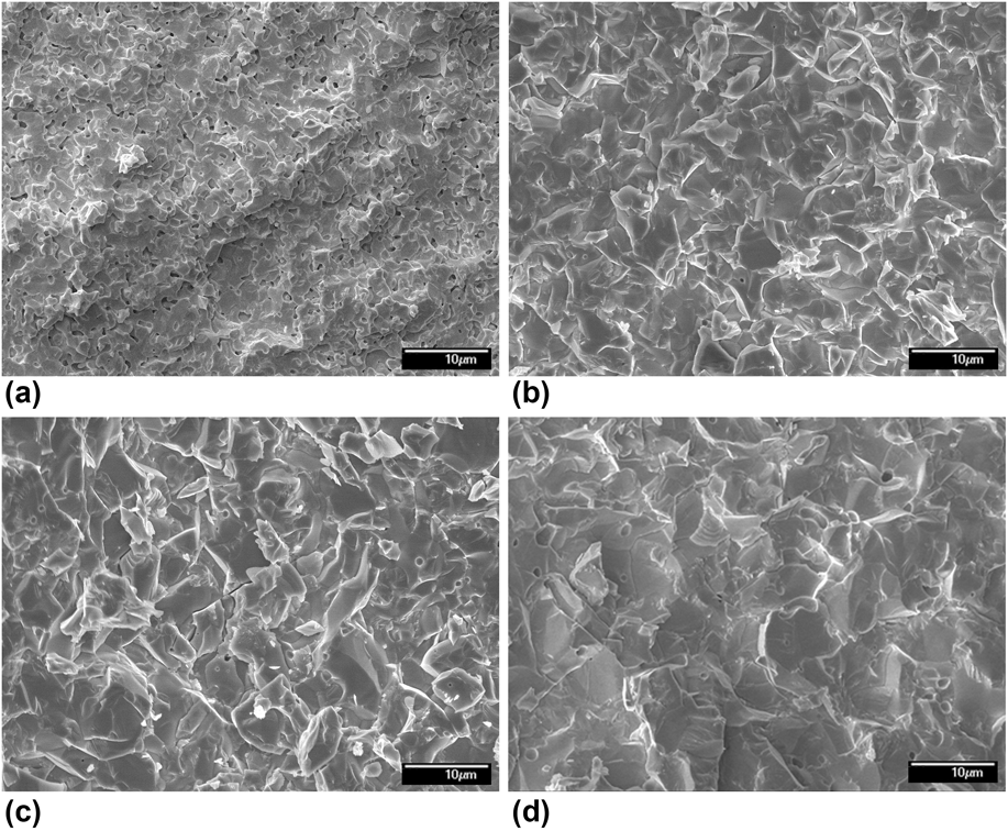
The XRD patterns in Fig. 4 illustrate the positions of diffraction peaks of LSO powder and as-sintered LSO:Ce ceramics which are compatible well with the standard diffraction data (ICSD #279584) of monoclinic LSO phase. No other second phase is found, indicating that LSO phase remains stable and no decomposition occurs during the sintering course. Morphology of fracture surfaces of LSO:Ce ceramics sintered at different sintering conditions is observed by SEM to investigate microstructure variation of polycrystalline LSO:Ce ceramics during sintering process. SEM micrograph shown in Fig. 5(a) revealed that the grain size in the sample was only around 2–3 μm, suggesting that grains growth of LSO:Ce ceramics sintered at 1650 °C for 4 h is not complete, and considerable residual pores at the grain boundaries were observed, hindering achievement of the fully dense polycrystalline LSO ceramics. Figure 5(b) shows the fracture micrograph of LSO ceramics sintered at 1720 °C for 4 h. It shows that grain size reaches about 5 μm and that residual pores have been eliminated completely as sintering temperature reached 1720 °C. This microstructure is quite beneficial to achieve good optical transmittance in polycrystalline sample, which has been proved by following photograph of LSO ceramics under visible light. From fracture micrograph of LSO ceramics sintered at 1780 °C for 4 h shown in Fig. 5(c), it is obvious that more big pores appear in grains and grain boundaries relative to the LSO ceramics sintered at 1720 °C for 4 h. This phenomenon may indicate that excessively fast grain growth due to high sintering temperature causes pores growing and secondary recrystallization, which result in reduce of transparency of obtained LSO ceramics. Figure 5(d) presents the fracture micrograph of LSO ceramics sintered at 1720 °C for 8 h. When holding time was extended to 8 h at the sintering temperature of 1720 °C, LSO ceramics undergo remarkable grain coarsening. As-attained translucent LSO ceramics sintered at 1720 °C for 4 h was mechanically polished on both sides. SEM micrograph of polished surface of the polished LSO ceramics presents in Fig. 6(a). It further manifests that no minor second phases or decomposition of the LSO was found in the sintered LSO ceramics. Corresponding energy-dispersive x-ray spectrum (EDS) of rectangle range of the SEM micrograph is shown in Fig. 6(b). The EDS results suggest that the atom ratio of Lu and Si in the sintered LSO ceramics is in accord well with chemical formula of LSO.
XRD patterns of the synthetic LSO powder and as-fabricated ceramics.
SEM micrographs of fracture surface of LSO ceramics obtained at different sintering conditions: (a) 1650 °C for 4 h, (b) 1720 °C for 4 h, (c) 1780 °C for 4 h, (d) 1720 °C for 8 h.
SEM micrograph (a) and corresponding EDS (b) of polished surface of LSO ceramics obtained at 1720 °C for 4 h.
Appearance of the polished LSO ceramics is presented in Fig. 7(a). The polished specimen with a diameter of 14.5 mm and a thickness of 1.0 mm shows a translucent state, and the characters under the LSO ceramics can be observed. Unlike YAG, LuAG, and Lu2O3 matrix with isotropical cubic crystal structure, LSO has a monoclinic structure leading to an optical anisotropy in different crystal orientation. Owing to existence of birefringence, it will bring about light scattering when light cross grain boundaries. Hence, it is much more difficult to obtain transparent polycrystalline LSO ceramics by sintering under normal pressure. Figure 7(b) shows fluorescent photograph of the LSO ceramics under UV light of 365 nm, which emit strong blue luminescence demonstrating good luminescent property of the LSO ceramics.
Photographs under (a) visible light and (b) UV light of 365 nm of the translucent LSO ceramics sintered at 1720 °C for 4 h.
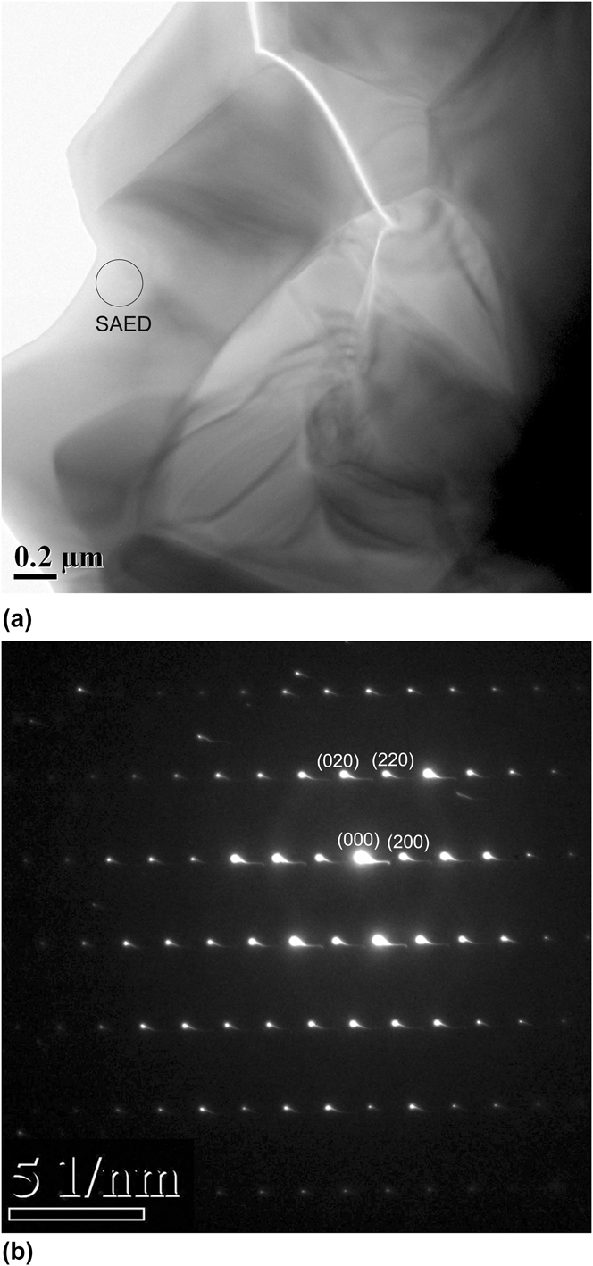
TEM micrograph of the LSO ceramics sintered at 1720 °C for 4 h is shown in Fig. 8(a). The grain boundary was clean and no intergranular second phase was observed between adjacent grains. The selected area electron diffraction (SAED) pattern is shown in Fig. 8(b).
TEM micrograph (a) and SAED pattern (b) of the translucent LSO:Ce ceramics.
C. Luminescent properties of the fabricated LSO:Ce ceramics
Figure 9(a) presents a VUV-UV excitation spectrum of the LSO:Ce ceramics sintered at 1720 °C for 4 h by monitoring the emission band at 420 nm. There are four strong excitation bands respectively centered at 189, 262, 297, and 356 nm in the excitation spectrum. The excitation band centered at 189 nm is considered to be the host absorption band, which correspond to the band gap (E g = 6.5 eV, ∼190 nm) of LSO matrix.Reference Cooke, Bennett, McClellan, Roper, Whittaker and Portis18 Moreover, 4f → 5d electron transition of Ce3+ is responsible for the excitation bands centered at 262, 297, and 356 nm.Reference Shi, Liu, Zhang, Qi, Ren, Tao and Xu19 Emission spectra of the LSO:Ce ceramics under 6.58 eV (189 nm) and 3.49 eV (356 nm) excitation both shows a broad emission band with the peak centered at about 2.96 eV (420 nm) as shown in Fig. 9(b), but the emission spectrum under 189 nm excitation has a broader emission band than emission spectrum under 356 nm excitation. Emission spectrum under 356 nm excitation can be divided into three components (red dot lines) centered at 3.14 eV (396 nm), 2.97 eV (419 nm), and 2.74 eV (454 nm) by Gaussian fitting, which is closely related to the Ce occupation in LSO lattice.Reference Cooke, Bennett, McClellan, Roper, Whittaker and Portis18,Reference Suzuki, Tombrello, Melcher and Schweitzer20 The emission peaking at 396 nm and 419 nm ascribes transition of 5d lowest level to 4f ground state (2F5/2 and 2F7/2) of Ce1 (Ce3+ located in one of two independent lattice sites), whereas the emission of peaking at 454 nm originates from Ce2 (Ce3+ located in the other site in LSO crystal) emission.Reference Suzuki, Tombrello, Melcher and Schweitzer20,Reference Liu, Shi, Yin, Fu, Zhang and Ren21 Percentage of Ce2 emission intensity reaches 27.1% in emission spectrum obtained from 356 nm excitation, which is much lower than 36.5% in emission spectrum obtained from 189 nm excitation. It may indicate that efficiency of host absorbing (189 nm) transfer to Ce2 is higher than Ce1 absorbing (356 nm) transfer to Ce2.
(a) Excitation and (b) emission spectra of the translucent LSO:Ce ceramics.
XEL spectra of the LSO:Ce ceramics are depicted in Fig. 10(a), exhibiting a broad emission band in the range of 350–650 nm. Radioluminescence intensity of LSO:Ce ceramics can reach twice of that of BGO single crystal, suggested that the as-sintered LSO:Ce ceramics has been provided with good light yield property. Figure 10(b) shows decay profile of the LSO:Ce ceramics under excitation of 356 nm and emission of 420 nm. The decay profile contains only a fast decay component of 21.2 ns by a single exponential function fitting, which is obviously faster than that (28.7 ns) of LSO:Ce single crystal,Reference Ren, Qin, Lu and Li22 Lempicki et al. proposed that this phenomenon was due to that the ceramics fabrication process had shifted more of the emitters into the faster site than is normally found in the single crystal,Reference Lempicki, Brecher, Lingertat, Miller, Glodo and Sarin12 but we deem it is due to oxygen vacancies and grain boundaries existed in LSO:Ce ceramics, both of them will bring about nonradiative transitions of Ce3+ in LSO lattice, which is much faster than radiative transitions.
(a) XEL spectra of the translucent LSO:Ce ceramics and BGO single crystal and (b) luminescence decay curve of the LSO:Ce ceramics sintered at 1720 °C for 4 h under excitation of 356 nm and emission of 420 nm.
Photopeak measurement data of the samples are depicted in Fig. 11. It shows that the light yield of the fabricated LSO ceramics is 21,300 ph/MeV under the 137Cs excitation source, which reaches 91% that of LSO single crystal. The energy resolutions of the ceramics and crystal are 19 and 12%, respectively. Both light yield and energy resolution of the fabricated LSO ceramics are comparable to that of bulk LSO single crystal and the previously reported results.Reference Wang, Loef, Rhodes, Glodo, Brecher, Nguyen, Lempicki, Baldoni, Higgins and Shah14,Reference Roy, Lingertat, Brecher and Sarin23
Comparison of the light yield of LSO:Ce ceramics with BGO and LSO bulk single crystals.
IV. CONCLUSION
Polycrystalline LSO:Ce powder synthesized by a sol–gel processing is employed to investigate the fabrication of LSO:Ce ceramics by pressureless sintering. The thermal shrinkage behaviors determination reveals that remarkable thermal shrinkage is achieved when temperature exceeds 1400 °C. The LSO:Ce scintillation ceramics fabricated by pressureless sintering in flowing H2 atmosphere at 1720 °C for 4 h achieved a translucent state. Its relative density is 99.5%, and average grain size was about 5 μm without residual pores within grains or at grain boundary. A broad emission spectrum of the LSO:Ce ceramics at room temperature ascribes the 5d → 4f transition of Ce3+ under VUV and UV excitation. Radioluminescence of obtained the LSO:Ce ceramics can attain double overall emission intensity with respect to BGO single crystal by x-ray excitation at room temperature. The luminescent decay time contains only a fast decay component, which is 21.2 ns. The light yield of the LSO ceramics is calculated to be 21,300 ph/MeV, which reaches 91% of that of LSO single crystal. The good luminescent and scintillation properties indicate that LSO:Ce ceramics is a promising candidate for a new polycrystalline scintillator for radiation detection.
ACKNOWLEDGMENT
The authors gratefully acknowledge Pro. Guobin Zhang and Junyan Shi of NSRL for their very useful help in VUV spectra measurements. This work is supported by the National Science Foundation of China (Grant No. 51172139).