Introduction
Intrauterine growth restriction (IUGR) describes a condition where the fetus fails to achieve its genetic growth potential in utero with a drop-off in weight across growth centiles. In Australia, the 2014 census showed that 6.4% of live born babies were of low birth weight (defined as birth weight of less than 2.5 kg). 1 Infants with IUGR born at term, without prematurity, have worse respiratory outcomes. In particular, IUGR and low birth weight are associated with an increased risk of developing asthma in later life. Reference Källén, Finnström, Nygren and Otterblad Olausson2,Reference Villamor, Iliadou and Cnattingius3
In an attempt to understand the association between growth restriction and asthma, we have previously examined functional and structural changes to the airways and lungs in an animal model of IUGR. Reference Wang, Larcombe and Berry4–Reference Wang and Noble7 We found that airway responsiveness to contractile stimuli is altered in an age- and sex-dependent manner in mice following maternal hypoxia-induced IUGR. Reference Wang, Larcombe and Berry4 The above functional findings in IUGR mice were not explained by changes in gross wall structure, including thickness of the airway smooth muscle (ASM) layer. Reference Wang, Larcombe and Berry4,Reference Wang, Morton and Davidge5 While increased heterogeneity of airway dimensions also affects bronchoconstrictor response, Reference Wang, Morton and Davidge5 an alternative possibility is that changes in airway responsiveness are mediated by abnormal airway epithelial function.
The airway epithelial layer presents an initial interface between the host and environment and is the frontline of defense against aero-pathogens, microorganisms, and gases. This is achieved through a physical, chemical, and immune barrier and is supported by a network of junctional proteins including tight junctions (TJs), adherens junctions, gap junctions, desmosomes, and connexions Reference Farquhar and Palade8 which act synergistically and dynamically to constitute a protective mechanism against external aggressions. Airway epithelial cells express many pattern recognition receptors to allow for rapid detection and response to pathogen or damage-associated patterns. The activation of epithelial pattern recognition receptors subsequently releases cytokines, chemokines, and antimicrobial peptides to attract and activate the innate and adaptive immune cells. Reference Hammad, Chieppa, Perros, Willart, Germain and Lambrecht9,Reference Nathan, Peterson, Chakir and Wills-Karp10
Damage to the epithelial barrier, including disruption of TJs proteins in response to environmental pollutants, increases the paracellular traffic of pathogenic molecules (e.g., aeroallergens) into the lung interstitium. Reference Sajjan11 Importantly, the epithelium also modifies contraction of the ASM and the nature of this regulation is different between males and females, Reference Van Winkle, Gunderson, Shimizu, Baker and Brown12,Reference Degano, Prevost and Berger13 potentially contributing to the aforementioned sex-dependent changes in airway responsiveness after IUGR. Compared with healthy Controls, the airway epithelium is intrinsically different in children with asthma Reference Kicic, Sutanto, Stevens, Knight and Stick14,Reference Looi, Buckley and Rigby15 which is consistent with a developmental origin.
In view of growing evidence for in utero priming of the developing immune system Reference Wilcox and Jones16 that may be sex-specific, Reference Wooldridge, Bischof and Meeusen17 our overarching hypothesis is that susceptibility to asthma after IUGR is driven by intrinsic changes to epithelial barrier properties and/or a shift to a proinflammatory phenotype. Using our established maternal hypoxia-induced IUGR mouse model, Reference Wang, Larcombe and Berry4,Reference Noble, Kowlessur, Larcombe, Donovan and Wang6,Reference Francis, Pinniger, Noble and Wang18 we characterized epithelial TJs protein expression and inflammation in male and female offspring.
Materials and methods
Ethics statement
Our experimental approach was to subject pregnant mice to a hypoxic or normoxic environment and, in both male and female offspring, assess lung-specific and systemic inflammation and airway epithelial TJs protein expression. This project was approved by the Telethon Kids Institute Animal Ethics Committee (Project Number #264) and was carried out in accordance with the Australian Code of Practice for the Care and Use of Animals for Scientific Purposes (7th Edition).
Maternal hypoxia-induced IUGR mouse model
Pregnant female BALB/c mice at gestational day (GD) 7 were obtained from Animal Resources Centre (Murdoch, WA, Australia) and were housed under hypoxic conditions (10.5% O2) from GD 11 to 17.5 (i.e., pseudoglandular–canalicular stage in the mouse and spanning the period of airway development). Following hypoxic exposure, mice were returned to a normoxic environment (21% O2) until birth (GD 21). Reference Wang, Larcombe and Berry4,Reference Noble, Kowlessur, Larcombe, Donovan and Wang6,Reference Wang and Noble7,Reference Francis, Pinniger, Noble and Wang18 A Control group of pregnant mice was housed under normoxic conditions throughout pregnancy. Reference Wang, Larcombe and Berry4,Reference Noble, Kowlessur, Larcombe, Donovan and Wang6,Reference Wang and Noble7,Reference Francis, Pinniger, Noble and Wang18 Mice were maintained on a 15:9 h light:dark cycle at 23 ± 1 °C and supplied with an allergen-free diet (Specialty Feeds, Glen Forrest, WA, Australia) and water ad libitum. Each dam was housed individually with their litter until weaning at 3 weeks of age when the offspring were separated to different cages based on sex. Body weights of offspring were recorded at birth and again at 2 or 8 weeks of age, at which point mice were euthanized by an intraperitoneal injection of ketamine (240 mg/kg) and xylazine (12 mg/kg).
Bronchoalveolar and peritoneal lavage
In one group of mice, bronchoalveolar lavage (BAL) fluid was collected at 2 weeks (Control male, n = 7; IUGR male, n = 9; Control female, n = 12; IUGR female, n = 8) or 8 weeks (Control male, n = 8; IUGR male, n = 8; Control female, n = 11; IUGR female, n = 10) of age by washing 0.2 or 0.5 ml of saline in and out of the lungs three times via the tracheal cannula of each mouse. Lungs were not weighed or analyzed further after BAL fluid collection. As peritoneal cavity fluid may reflect a systemic inflammatory profile, Reference Wiest, Leidl and Kopp19,Reference Glitz, Souza and Rodini20 peritoneal cavity fluid of the mouse was also collected at 2 weeks (Control male, n = 7; IUGR male, n = 8; Control female, n = 12; IUGR female, n = 8) and 8 weeks (Control male, n = 10; IUGR male, n = 8; Control female, n = 11; IUGR female, n = 10) to assess systemic inflammation. Reference Ray and Dittel21 Briefly, 2 or 5 ml of saline were injected intraperitoneally and mice were shaken gently for 30 s. Lavage samples were processed for total and differential cell counts as previously described. Reference Larcombe, Janka, Mullins, Berry, Bredin and Franklin22
Cytokine assay
The Bio-Plex Pro™ Mouse Cytokine 23-plex Assay (Bio-Rad, Hercules, CA) was used to measure the concentration of 23 cytokines concentration in BAL fluid supernatant. Reference Larcombe, Janka, Mullins, Berry, Bredin and Franklin22 The cytokines measured were eotaxin, granulocyte-macrophage colony-stimulating factor (GM-CSF), interferon (IFN)-γ, tumor necrosis factor (TNF)-α, monocyte chemoattractant protein (MCP)-1α, macrophage inflammatory protein (MIP)-1α, MIP-1β, regulated on activation, normal T cell expressed and secreted (RANTES), interleukin (IL)-1α, IL-1β, IL-2, IL-3, IL-4, IL-5, IL-6, IL-9, IL-10, IL-12 (p40), IL-12 (p70), IL-13, IL-17A, keratinocyte chemoattractant (KC), and granulocyte colony-stimulating factor (G-CSF).
Immunofluorescence of tight junction proteins
Paraffin-embedded, formalin-fixed sections (Control male, n = 10; IUGR male, n = 12; Control female, n = 12; IUGR female, n = 12) were initially deparaffinized, rehydrated, and subjected to manufacturer-recommended heat-induced antigen retrieval for claudin-1, claudin-18, and occludin; protease-induced antigen retrieval for zonula occluden (ZO)-1. Post-antigen retrieval, slides were washed three times in 1× tris-buffered saline (TBS) containing 0.1% (v/v) saponin. Sections were then blocked in 5% (w/v) bovine serum albumin, 10% fetal bovine serum (v/v), 0.1% (v/v) Triton X-100, and 0.1% (v/v) saponin in 1× TBS for 1 h at room temperature (RT) followed by a second series of washes. Sections were incubated with the primary antibodies to claudin-1 (1:100), claudin-18 (1:100), occludin (1:100), and ZO-1 (1:100) (Invitrogen, Thermo Fisher Scientific, California, USA), diluted in the blocking buffer solution and added to the slides and were incubated overnight at 4 °C. The following day, sections were washed in 1× TBS with 0.1% (w/v) saponin (3 × 15 min/wash). Fluorescent secondary antibodies (AlexaFluor-488 (1:1000), Life Technologies) were prepared in blocking buffer and added to the slides which were further incubated overnight at 4 °C. Following that, slides were washed in 1× TBS with saponin and the nucleus of the cells counterstained with 4′, 6-diamidino-2-phenylindole, (1:50,000) in 1× TBS for 10 min and then washed in 1× TBS (3 × 15 min/wash). Fluorescent mounting media was used to minimize fading and slides were visualized using a Nikon epifluorescence microscope.
Assessment of tight junctions protein expression by Western blot
From a separate group of 8-week-old mice (growth outcomes have previously been published Reference Noble, Kowlessur, Larcombe, Donovan and Wang6 ), lungs were dissected and weighed, and right lungs snap-frozen (Control male, n = 11; IUGR male, n = 12; Control female, n = 12; IUGR female, n = 12). Protein was collected from cells by cell extraction buffer, quantitated by BCA assay and stored at -80 °C. Prior to Western blot analysis, protein samples were thawed but placed on ice to prevent degradation. A 10-µg protein sample was mixed with NUPAGE® Lithium Dodecyl Sulphate buffer (Invitrogen, Thermo Fisher Scientific, California, USA), NUPAGE® reducing agent, and ddH2O to make up a final volume of 20 µl. Samples were then heated for 10 min at 70 °C on a heating block for optimal denaturation before being loaded into a pre-cast Novex BOLT® 4–12% 1.0 mm Bis-Tris Plus polyacrylamide gel (Invitrogen, Thermo Fisher Scientific, California, USA). Samples were then electrophoresed using a Novex BOLT® Western Blot apparatus (Life Technologies, California, USA) in MES SDS running buffer at a constant 200 V for 35 min at RT. A pre-stained protein ladder was run on all gels in addition to samples for reference purposes. After separation, proteins were transferred onto a polyvinylidene fluoride (PVDF) membrane using a semi-dry transfer method on iBlot (Invitrogen, Thermo Fisher Scientific, California, USA) system at 200 V for 7 min or a wet transfer method at a constant 230 mA for 2 h at 4 °C.
At the completion of protein transfer, the PVDF membrane was blocked for nonspecific staining using the LI-COR Odyssey Blocking Buffer (LI-COR Biosciences, Nebraska, USA) for 60 min at RT. Membranes were then incubated overnight while gentle rocking at 4 °C with primary antibodies to claudin-1 (1:100), claudin-18 (1:100), occludin (1:125), and ZO-1 (1:125) prepared in LI-COR Odyssey Blocking Buffer. Membranes were then washed 3 × 15 min/wash in 0.2% (v/v final) Tween-20 in TBS solution at RT. After washing, membranes were incubated in the dark with respective IRDye® secondary antibodies made up in a solution of LI-COR Odyssey Blocking Buffer with 2% (v/v final) Tween-20 diluent for 2 h at RT with gentle rocking. The membranes were then washed 3 × 15 min/wash in 0.2% (v/v final) Tween-20 in TBS solution followed by two times (10 min per wash) in TBS alone. The membranes were scanned using the LI-COR Odyssey infrared scanner at 680 nm and 800 nm channels. Bands of protein expression were quantified using the LI-COR Odyssey v.3.0 software. The integrated intensity (I.I) of each band was normalized to the I.I of the house-keeping protein, β-actin.
Statistical analysis
Before statistical evaluation, all results were tested for population normality (Shapiro-Wilk test) and homogeneity of variance and transformed where necessary. Birth weights were compared by t-tests (Control vs. IUGR). The analyses for the other measurements were performed via two-way ANOVA with prenatal treatment (Control or IUGR) and sex (male or female) as factors and Holm–Sidak post hoc tests. Graphical and statistical analyses were performed using SigmaPlot version 13.0. Data are reported as mean ± SEM. *P < 0.05 was considered significant.
Results
Growth outcomes
IUGR offspring had lower birth weight compared with Control offspring (Control, 1.43 ± 0.02 g; n = 60; IUGR, 1.37 ± 0.02 g; n = 58; P = 0.047). At 2 weeks of age, body weight was comparable between Control and IUGR groups (P = 0.35, F = 0.89; Table 1) and sexes independent of prenatal treatment (P = 0.53, F = 0.4; Table 1). While there was evidence of “catch-up growth” by 2 weeks of age, IUGR offspring were again smaller at 8 weeks of age compared with Control offspring (P = 0.006, F = 8.54; Table 1). At 8 weeks of age, female offspring were smaller than male offspring (P < 0.001, F = 68.90; Table 1).
Body weights (g)

Data are mean ± SEM.
* Compared with Control (P < 0.05).
# Compared with Males (P < 0.05).
IUGR, intrauterine growth restriction.
At 8 weeks of age, lung weight was smaller in the IUGR group compared with Control group (P = 0.004, F = 9.11; Table 2) and smaller in female offspring compared with male offspring (P = 0.006, F = 8.26; Table 2). Lung weight relative to body weight was comparable between Control and IUGR groups (P = 0.186, F = 1.81; Table 2) and there was no effect of sex (P = 0.635, F = 0.23; Table 2).
Lung weights at 8 weeks of age

Data are mean ± SEM.
* Compared with Control (P < 0.05).
# Compared with Males (P < 0.05).
IUGR, intrauterine growth restriction.
Lung and peritoneal inflammation
The IUGR offspring had higher total inflammatory cells in the BAL fluid at 2 weeks (P = 0.005, F = 8.89; Fig. 1a) and 8 weeks (P = 0.004 F = 9.42; Fig. 1b) of age. Changes were independent of sex at both ages (2 weeks old, P = 0.74, F = 0.11; 8 weeks old, P = 0.22, F = 1.54). Macrophages, neutrophils, lymphocytes, and eosinophils were detected in Control and IUGR groups, but there was no difference in the proportion of these inflammatory phenotypes. There was no difference in total inflammatory cells in the peritoneal cavity fluid at 2 weeks (P = 0.07, F = 3.63; Fig. 1c) and 8 weeks (P = 0.184, F = 1.84; Fig. 1d) of age between Control and IUGR groups. There was also no effect of sex (2 weeks old, P = 0.22, F = 1.59; 8 weeks old, P = 0.13, F = 2.43) on the total inflammatory cells in the peritoneal cavity.
Total inflammatory cells in BAL fluid from Control and IUGR male and female offspring at 2 weeks (a) and 8 weeks (b) of age. Total inflammatory cells in the peritoneal cavity from Control and IUGR offspring at 2 weeks (c) and 8 weeks (d) of age. Data are mean ± SEM. *Significantly different from Control (P < 0.05). Male, open bars; Female, closed bars. IUGR, intrauterine growth restriction; BAL, bronchoalveolar lavage.

Cytokine concentration in BAL fluid
There were 12 cytokines with levels below the assay detection limit in the majority of mice (TNF-α, MCP-1α, MIP-1β, RANTES, IL-1β, IL-3, IL-6, IL-9, IL-10, IL-12 (p70), IL-17A, and G-CSF). GM-CSF factor was not detected in any mice. All other cytokines were compared between Control and IUGR groups, in both male and female mice; see below.
At 2 weeks of age, IL-13 (female, 101.7 ± 11.5 pg/ml; male, 49.8 ± 12.9 pg/ml; P = 0.006, F = 9.06), IFN-γ (female, 1.9 ± 0.2 pg/ml; male, 1.2 ± 0.3 pg/ml; P = 0.04, F = 4.76), and eotaxin (female, 3.8 ± 0.3 pg/ml; male, 2.9 ± 0.3 pg/ml; P = 0.03, F = 5.54) concentrations were higher in females compared with males. None of the cytokines in the BAL fluid were affected by prenatal treatment at this time point. At 8 weeks of age, IL-2 (P = 0.006, F = 8.51; Fig. 2a), IL-13 (P = 0.01, F = 6.76; Fig. 2b), and eotaxin (P = 0.04, F = 4.64; Fig. 2c) concentrations were higher in male IUGR offspring compared with male Control offspring, but there was no difference in females. Independent of IUGR, the concentration of IL-1α (female, 1.4 ± 0.1 pg/ml; male, 1.82 ± 0.1 pg/ml; P = 0.01, F = 7.59), IL-5 (female, 2.6 ± 0.2 pg/ml; male, 3.1 ± 0.2 pg/ml; P = 0.003, F = 10.95), IFN-γ (female, 1.9 ± 0.1 pg/ml; male, 2.2 ± 0.1 pg/ml; P = 0.03, F = 5.23), and KC (female, 40.5 ± 2.7 pg/ml; male, 56.5 ± 3.1 pg/ml; P < 0.001, F = 15.45) were greater in males compared with females.
Cytokines concentration (IL-2 (a), IL-13 (b), and eotaxin (c)) in Control and IUGR male and female offspring. Data are mean ± SEM. *Significantly different from Control (P < 0.05). Control, open bars; IUGR, closed bars. IUGR, intrauterine growth restriction; IL, interleukin.

Tight junctions protein expression
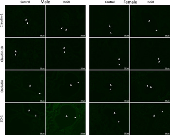
To investigate the direct effects of prenatal treatment on barrier integrity, we examined TJs protein expression by immunofluorescence staining and Western blotting using lung tissue from animals at 8 weeks of age. No marked differences in staining intensities of claudin-1, claudin-18, occludin, and ZO-1 were observed in immunofluorescence staining of lung tissues as shown in Fig. 3. Protein expression for claudin-1 (P = 0.34, F = 0.92; Fig. 4a), claudin-18 (P = 0.90, F = 0.017; Fig. 4b), occludin (P = 0.64, F = 0.22; Fig. 4c), and ZO-1 (P = 0.76, F = 0.09; Fig. 4d) were not altered by IUGR or sex (P > 0.05).
Immunofluorescence staining of tight junction proteins (claudin-1, claudin-18, occludin, and ZO-1) in Control and IUGR male and female offspring. IUGR, intrauterine growth restriction; ZO-1, zonula occludens-1; A, airway; arrows (→) indicate epithelium.

Tight junction protein abundance (claudin-1 (a), claudin-18 (b), occludin (c), and ZO-1 (d)) in Control and IUGR male and female offspring. Data are mean ± SEM. Control, open bars; IUGR, closed bars. IUGR, intrauterine growth restriction; ZO-1, zonula occludens-1; AU, arbitrary unit.

Discussion
Given the fundamental role the airway epithelium plays in the development of asthma, Reference Holgate23 and in view of associations between asthma and IUGR Reference Källén, Finnström, Nygren and Otterblad Olausson2,Reference Sonnenschein-van der Voort, Gaillard, de Jongste, Hofman, Jaddoe and Duijts24 and the sex dependence of these diseases, Reference Myers25,Reference Schatz and Camargo26 we considered the possibility that developmental abnormalities of the epithelium may alter the risk of asthma development and that this may be different in males and females. Our approach was to use a validated model of IUGR (discussed further below) and to probe barrier and inflammatory function of the airway epithelium across multiple time points in both male and female mice. Findings indicate that while barrier properties are unaltered, an inflammatory phenotype exists in the lungs of IUGR offspring, particularly in males, which may increase susceptibility to future disease as a result of this relatively primed immune defense.
Maternal hypoxia-induced IUGR offspring had lower birth weight compared with Control offspring, exhibited “catch-up” growth by 2 weeks of age, before becoming smaller again at 8 weeks of age (relative to Control), all of which are observations documented in previous studies. Reference Wang, Larcombe and Berry4,Reference Noble, Kowlessur, Larcombe, Donovan and Wang6,Reference Francis, Pinniger, Noble and Wang18 These data are evidence of a reproducible model of IUGR which has value in helping us understand the mechanisms of disease. Additionally, we report new data on lung weight and show that lungs were smaller in the IUGR offspring compared with Control offspring, indicating that lung sparing does not occur in this IUGR model, consistent with some IUGR models, Reference Wignarajah, Cock, Pinkerton and Harding27,Reference Soo, Orgeig, McGillick, Zhang, McMillen and Morrison28 but not others. Reference Lipsett, Tamblyn and Madigan29 Differences between studies may relate to the duration, timing, and cause of IUGR which may have varying effects on lung development.
Past studies, as summarized in leading reviews, highlight the importance of airway hyperresponsiveness in asthma. Reference Hargreave, Dolovich, O’Byrne, Ramsdale and Daniel30–Reference O’Byrne and Inman32 Data from our group have accumulated evidence demonstrating that IUGR affects respiratory system development in an age- and sex-dependent manner, which may modify the risk of developing asthma in later life. Reference Wang, Larcombe and Berry4–Reference Wang and Noble7,Reference Francis, Pinniger, Noble and Wang18 Given the vital role that the airway epithelium plays in providing a physical barrier and being a central modulator of inflammatory responses, the focus of the present study was to examine the impact of IUGR on lung cytokine response and epithelial TJs protein expression. To our knowledge, this is the first study to assess the interrelationship between IUGR and airway epithelial responses in mice.
It has been recognized for many years that abnormalities of the epithelium may underlie the pathophysiology of airway hyperresponsiveness, Reference Fahy, Corry and Boushey33 and recent interest in this area has demonstrated that the airway epithelium is dysfunctional in asthma and is characterized by defective repair and dissociated TJ formation. Reference Looi, Buckley and Rigby15,Reference Ling, Sutanto and Iosifidis34 In addition to its physical barrier properties, the airway epithelium is a potent source of proinflammatory cytokines. Reference Lambrecht and Hammad35 Any damage to the epithelium initiates a cascade of inflammatory and cell signaling events that lead to regeneration or repair. It has been postulated that epithelial susceptibility to environmental insults may lead to inappropriate activation of immune cells. In asthmatic airways, damaged epithelium results in localized Th2-type inflammation, Reference Fahy and Locksley36 which could increase cytokine levels. Cytokines have been shown to have the ability to directly and indirectly interfere and damage the epithelial TJ. Reference Coyne, Vanhook, Gambling, Carson, Boucher and Johnson37 An epithelium-driven inflammatory response can also have direct effects on ASM contraction Reference Morrison, Gao and Vanhoutte38 and therefore airway responsiveness, although the ASM layer itself is a source of cytokines. Reference Xia, Harris, Stewart and Mackay39 Considering that in the mouse, epithelial differentiation occurs during the transition from pseudoglandular to canalicular phases of lung development, Reference Rackley and Stripp40 it is therefore a possibility that any insults during this period of fetal development will have an effect on epithelium structure or function.
We first compared the total inflammatory cells in BAL fluid from juvenile and adult offspring as it is established that BAL fluid cell patterns reflect inflammatory cell profiles in affected lung tissues. Reference Costabel and Guzman41 The heightened immune response was observed in early life, which persisted in adulthood. The finding of increased inflammatory cells in the BAL fluid of adult IUGR offspring in mice is consistent with our earlier observations in rats. Reference Wang, Morton and Davidge5 The only difference, after differential analysis was performed, was that there was no change in the proportion of inflammatory cells types in mice, while neutrophils were detected only in the IUGR and not Control offspring of rats. Neutrophil enrichment in the lungs is an early indicator of more chronic inflammation. Reference Wittekindt42 Therefore, the presence of neutrophils in the IUGR rat offspring indicates the first signs of a primed inflammatory state that could promote an asthma-like inflammatory cascade, Reference Pascoe43 which may subsequently impact epithelial TJs in the IUGR offspring. Even without a shift in the cell proportions, an increase in the number of inflammatory cells in the lungs likely drives an increase in the production of inflammatory cytokines in IUGR offspring. Additionally in this present study, we found that the altered immune response is only isolated to the lungs, since inflammation was not altered in the peritoneal cavity (liver, spleen, most of the gastrointestinal tract, and other viscera Reference Ray and Dittel21 ) between groups at both ages.
In the context of IUGR, increased cytokine levels have been detected in various tissues, such as peripheral blood lymphocytes, Reference Raghupathy, Al-Azemi and Azizieh44 metrial glands, Reference Chakraborty, Islam, Saha and Ain45 and neurons. Reference Wixey, Lee and Miller46 In the lungs, a maternal undernutrition-induced IUGR rat study has found increased expression of inflammatory cytokines associated with profibrotic processes. Reference Alcazar, Östreicher and Appel47 In view that hypoxia is associated with the production of proinflammatory cytokines Reference Whyte and Walmsley48,Reference Eltzschig and Carmeliet49 and that we have used this as a means to induce IUGR, it is therefore not surprising that we found several elevated cytokines in the BAL fluid. Specifically, this study demonstrated an effect of IUGR on lung cytokines concentrations (IL-2, IL-13, and eotaxin) only in male offspring during adulthood, which would suggest a sex-dependent vulnerability toward hypoxia in the lungs of IUGR males, as is observed in the heart. Reference Rueda-Clausen, Morton and Davidge50 Hypoxia has been shown to increase levels of eosinophilic inflammation and is also associated with increased levels of eotaxin in the lungs, Reference Baek, Cho, Rosenthal, Alexander, Nizet and Broide51 which is in agreement with the present study. Interestingly, we also show increased levels of IL-13, a T-helper type-2 cytokine, which is involved in the upregulation of eotaxin and subsequently, eosinophilia, and allergy. Reference Matsukura, Stellato and Georas52 Moreover, previous studies have demonstrated the role of IL-2 and IL-13 in inducing airway hyperresponsiveness Reference Wills-Karp, Luyimbazi and Xu53–Reference Nag, Lamkhioued and Renzi55 and ASM contraction Reference Chiba, Nakazawa, Todoroki, Shinozaki, Sakai and Misawa56 after allergen challenge.
As shown in childhood, males are consistently found to be at an increased risk of asthma pathogenesis due to the different growth rate of their lungs, airways, and immunological differences such as increased levels of total immunoglobulin-E. Reference Almqvist, Worm and Leynaert57 After childhood, the effect of sex on asthma is reversed, with females exhibiting an increased risk of developing asthma. Reference Almqvist, Worm and Leynaert57 Thus, we would expect that proinflammatory cytokine concentrations would be higher in males during the juvenile period and higher in females in adulthood. Reference Uekert, Akan and Evans58–Reference Becerra-Díaz, Strickland, Keselman and Heller61 In contrast, independent of IUGR, we found that some of the cytokine concentrations (IL-13, IFN-γ, and eotaxin) were in fact higher in females during the juvenile period, and some others higher in males at adulthood. These data therefore highlight the dynamic homeostasis of airway immunity with aging and sex. Reference Barrat, Lesourd and Boulouis62,Reference Matha, Shim, Steer, Yin, Martinez-Gonzalez and Takei63
It is useful to explicitly discuss how these new data advance our understanding on previously observed physiological phenomena after IUGR; bronchoconstriction in vivo, Reference Wang, Larcombe and Berry4 ASM contraction in vitro, and airway stiffness. Reference Noble, Kowlessur, Larcombe, Donovan and Wang6 The present study shows that one of the key consequences of IUGR is an increase in airway inflammation despite no other environmental challenge. Evoking an inflammation-dependent shift in respiratory physiology is logical, however, when looking more closely at the nature of these changes, the picture appears more complicated. For example, while adult male mice after IUGR are hypo-responsive, Reference Wang, Larcombe and Berry4 this cannot be explained by increased cytokine release which instead favors increased bronchoconstriction. Reference Barnes64 Rather it is possible that a cytokine-mediated increase in ASM contraction partly offsets the pronounced decrease in ASM contractile capacity observed in adult male IUGR offspring. Reference Noble, Kowlessur, Larcombe, Donovan and Wang6 Increased stiffness could similarly either contribute to reduced bronchoconstriction as a result of a greater mechanical load on the ASM Reference Noble, Turner and Mitchell65 or enhance bronchoconstriction by blunting the beneficial bronchodilatory effects of lung inflation. Reference Cairncross, Noble and McFawn66,Reference Noble, McFawn and Mitchell67 Despite this complexity, we have now reported the findings of six studies examining the effect of IUGR on respiratory structure and function in rodents, and on every occasion it was apparent that the IUGR-affected animal is physiologically unique. The clinical consequences of these changes are not yet apparent, though increased basal inflammation alone (specific to the lungs) places an IUGR-affected individual at greater risk of an excessive immune response to inhaled allergen, virus, or bacterium, that is independent of epithelial barrier properties, as discussed below.
The airway epithelial barrier prevents the transport of pathogens, cells, and fluids from crossing into the airway lumen and vice versa and is also involved in the active transport of ions and solutes, a process essential for mucocilliary clearance. This is achieved through the inter-related networks of TJ proteins, which are comprised of approximately 40 different proteins. The three main TJ proteins are occludin, the claudin family of 27 proteins, and junctional adhesion molecules, which are associated with the cytoplasmic peripheral membrane protein family, ZO-1 for anchorage to the actin filaments within the cytoskeleton. Reference Anderson and Van Itallie68 It has been shown that, during severe lung injury, the airway epithelium undergoes apoptosis and barrier function becomes compromised. However, in mild to moderate lung injury, cell death is not pronounced Reference Berthiaume and Matthay69 and barrier integrity is maintained. As demonstrated in previous studies, the airway epithelium in children with mild asthma is intrinsically and biochemically different Reference Kicic, Sutanto, Stevens, Knight and Stick14 as well as having an impaired airway epithelial barrier with dissociation of TJ proteins. Reference Looi, Buckley and Rigby15 Moreover, LaFemina et al. also demonstrated that deficiencies in claudin-18 resulted in epithelial barrier dysfunction as well as impairment of alveologenesis. Reference LaFemina, Sutherland and Bentley70 The regulation of epithelial TJs, such as claudins, occludin, and zonula occludens and the critical role they have in affecting innate and adaptive immunity has also been summarized in a concise review by Georas and Rezaee. Reference Georas and Rezaee71 We have previously shown that epithelium thickness was comparable between the Control and IUGR offspring Reference Wang, Larcombe and Berry4 and in the present study, we found that there were no differences in any of the four TJ proteins that we measured, which suggests that the airway epithelial barrier is resistant to conditions associated with IUGR. The reason behind the apparent resilience of the epithelium is unclear; however, a potential candidate could be activation of hypoxia-inducible factors (HIFs), in particular, HIF-1, which has been previously shown to confer protection to the airway epithelium against loss of barrier integrity through oxidative stress. Reference Olson, Hristova, Heintz, Lounsbury and Van Der Vliet72 The potential compensatory effect in response to a hypoxic environment during fetal development which persists in adulthood further highlights the dynamism of the airway epithelium in maintaining epithelial homeostasis.
In conclusion, the present study demonstrates fetal growth disruption during the pseudoglandular–canalicular phase increased lung, but not systemic, inflammation profile in mice after IUGR in the absence of any changes to airway epithelial barrier integrity in IUGR offspring. While increased lung inflammation was common to males and females, cytokine release was only affected in the former after IUGR.
Acknowledgments
The authors thank Teagan Wagner with her assistance in the collection of peritoneal cavity fluid, Carolyn J. Wang for her assistance with various technical aspects of this work, and Darshinee Kowlessur for her assistance in animal handling and monitoring.
Author Contributions
K.L., K.C.W.W., and P.B.N. conceived and designed the experiments. K.L. and K.C.W.W. performed the experiments. K.L. and K.C.W.W. analyzed the data. K.L. and K.C.W.W. drafted the manuscript. K.L., A.K., P.B.N., and K.C.W.W. critically revised the manuscript. All authors have read and approved the manuscript.
Financial Support
This project was funded by the National Health and Medical Research Council (NHMRC) of Australia Project Grant (1120128) and The University of Western Australia Early Career Researcher Fellowship Support to K.C.W.W. K.L. was supported by a TSANZ/AstraZeneca Early Career Respiratory Research Fellowship. K.C.W.W. was supported by a NHMRC Early Career Research Fellowship (1090888) and a Western Australian Department of Health – New Independent Researcher Infrastructure Support. P.B.N. was supported by a Western Australian Department of Health – Merit Award and a Medical and Health Research Infrastructure Fund. A.K. is a recipient of a Rothwell Family Fellowship.
Conflicts of Interest
The authors report no conflicts of interest. The authors alone are responsible for the content and writing of the paper.
Ethical standards
The authors assert that all procedures contributing to this work comply with the ethical standards of the relevant national guides on the care and use of laboratory animals (Australian Code of Practice for the Care and Use of Animals for Scientific Purposes (7th Edition)) and has been approved by the institutional committee (Telethon Kids Institute Animal Ethics Committee (Project Number #264)).