Introduction
Representatives of the family Bucephalidae possess a number of traits that set them apart from the other digeneans. Adult flukes lack suckers and have an anterior rhynchus disassociated from the digestive system. The latter consists of a pharynx that opens ventrally, an oesophagus and a sac-like, unbranched caecum. Parthenogenetic generations are represented by a single cercariae-producing sporocyst. These sporocysts are almost immobile ‘giant’ organisms which have branching bodies that penetrate the internal organs of the first intermediate hosts, bivalve molluscs. The precise status of bucephalid sporocysts remains obscure. The fact that they produce cercariae has led James and colleagues (James et al., Reference James, Bowers and Richards1966; James & Bowers, Reference James and Bowers1967) to conjecture that they represent a daughter parthenogenetic generation. That scenario presupposes the existence of one more parthenogenetic generation (the mother sporocyst) in the life history of bucephalid digeneans, but, so far, these mother sporocysts have not been observed. It is true that they may exist and, due to their small size and brief life span, have remained undiscovered. Gibson (Reference Gibson1987), who also did not rule out the possibility that bucephalids may have two sporocyst generations, pointed to the fact that mother sporocysts are frequently overlooked by researchers. Nevertheless, most authors tend to think that branching bucephalid sporocysts develop directly from miracidia and are, in fact, the mother parthenogenetic generation (Tennent, Reference Tennent1909; Ciordia, Reference Ciordia1956; Overstreet & Curran, Reference Overstreet, Curran, Gibson, Jones and Bray2002; Galaktionov & Dobrovolskij, Reference Galaktionov and Dobrovolskij2003). This issue can only be clarified by thorough examination of the early developmental stages of bucephalid parthenitae in experimentally infected molluscs. Such studies, however, are lacking.
The ultrastructure of bucephalid sporocysts remains poorly investigated. The only study of bucephalid sporocyst body wall ultrastructure was carried out on Bucephalus (Labratrema) minimus, the tegumental structure of which was shown to be unusual for digeneans (James et al., Reference James, Bowers and Richards1966; James & Bowers, Reference James and Bowers1967). Podvyaznaya (Reference Podvyaznaya2007) described the fine structural organization of the germinal elements in sporocysts of Prosorhynchoides borealis (syn. P. gracilescens), but the ultrastructure of differing portions of bucephalid sporocyst branches has never been analysed. The present study aims to fill this gap.
Sporocysts of P. borealis Bartoli, Gibson & Bray, 2006 were chosen for this study. Adult flukes of this species are specific parasites of angler fish Lophius piscatorius L. in the northern North-East Atlantic (Bartoli et al., Reference Bartoli, Gibson and Bray2006). In coastal waters of Iceland, where the material for the present study was collected, metacercariae of P. borealis (syn. P. gracilescens) have been reported from the nervous system of gadoid fishes (Eydal et al., Reference Eydal, Bambir, Helgason and Ólafsdóttir1998, Reference Eydal, Helgason, Kristmundsson and Bambir1999), and their sporocysts have been identified in the mollusc Abra prismatica (Montagu, 1808) (see Eydal et al., Reference Eydal, Helgason, Kristmundsson, Bambir and Jónsson2000).
Materials and methods
Specimens of the mollusc A. prismatica infected with sporocysts of P. borealis were collected at a depth of 60–80 m off the southern coast of Iceland, near the Westman Islands, in September 2000.
For transmission electron microscopy, pieces of P. borealis sporocysts were dissected from the molluscs together with the surrounding host tissues (of the digestive gland and the gonads) and fixed using 3% glutaraldehyde in 0.1 m cacodylate buffer (pH = 7.4) containing sucrose (760 mosm/l in total). After 7–10 days, the samples were rinsed in cacodylate buffer containing sucrose (760 mosm/l in total), postfixed at 4°C in 1% osmium tetroxide (3–4 h), then rinsed in the buffer again, dehydrated in an ascending alcohol series and acetone, and embedded in Epon-Araldite. Before being placed in flat embedding moulds, the samples were carefully separated into smaller pieces, so that the shape of the sporocyst branches and their terminal portions were visible. Semi-thin and thin sections of the sporocysts were cut on an LKB-III ultramicrotome (LKB, Sweden). Thin sections were stained with aqueous uranyl acetate solution and Reynold's lead citrate, and viewed in JEM 1200EX (JEOL, Japan) and LEO 900 (Leo, Oberkochen, Germany) electron microscopes at an accelerating voltage of 60 kV. Semi-thin sections were stained with toluidine blue and viewed under a Leica DMLS light microscope (Leica, Germany). Altogether, 13 different portions of sporocyst branches were studied in a series of alternating thin and semi-thin sections. Of these, six terminal areas were studied as a series of longitudinal sections.
Results
Light microscopy
The body of the P. borealis sporocyst is a mass of intertwined branched yellowish tubules. Their inner cavities are continuous through the junctions to form the common body cavity of the sporocyst. The body cavity contains numerous cercarial embryos, germinal elements and large amounts of ‘cellular debris’. The diameter of the tubules varies from 40 to 160 μm. Distended areas with a spacious cavity and a thin wall (the so-called brood chambers) alternate with narrow areas (constrictions) with a relatively thick body wall and a poorly developed lumen. Terminal portions of the tubules consist of a brood chamber with an adjacent tip region. The tip regions consist of a mass of densely packed cells and are devoid of a lumen. In fixed sporocysts, the shape of the tip regions varies: some are conical, some elongate and narrow, and some beaded.
Electron microscopy
General structure
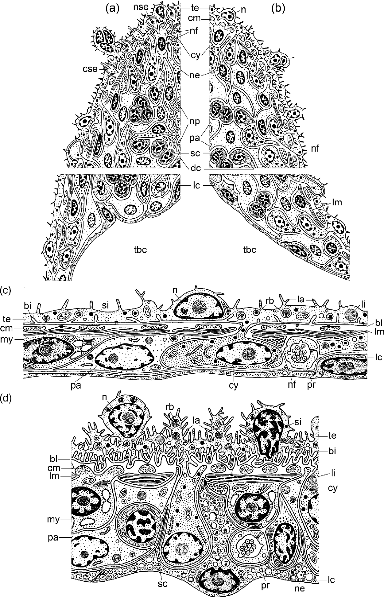
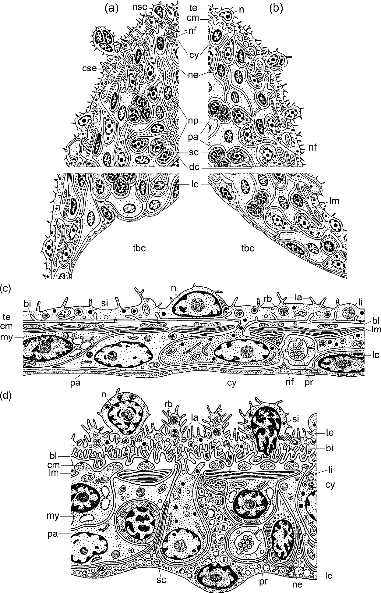
The structure of distended areas, constricted portions and terminal regions of the sporocyst branches, based on analyses of transmission electron microscopy (TEM) results, are presented in fig. 1 and illustrated by electron micrographs in figs 2–5. Two distinct types of terminal region can be identified and these are referred to as type A and type B. In type A terminal regions, the cell mass comprising the branch tips contains many nerve elements, and numerous sensory endings are found in the overlying tegument (figs 1a, 4a and 5a–f). Terminal brood chambers associated with these tips mainly contain late developmental stages of cercariae. Type B terminal portions are characterized by a lack of sensory receptors and the presence of very few nerve elements in the tip region (fig. 1b). Brood chambers of type B terminal portions contain germinal masses, germinal cells and, for the most part, early developmental stages of cercariae.
Schematic drawing of the different regions of a Prosorhynchoides borealis sporocyst: (a, b) terminal portions of sporocyst branches of the type A (a) and type B (b); solid tip region and distal part of the terminal brood chamber are shown; (c, d) body wall ultrastructure in a distended region (c) and in a constricted region (d) of the sporocyst tubule. Key to the lettering of the figures: bb, basal bodies; bc, body cavity; bi, invaginations of basal plasma membrane; bl, basal lamina; ca, excretory capillary cell; cd, cellular debris; ce, developing cercaria; ci, cilia; cm, circular muscles; cse, ciliated sensory ending; cy, cyton; dc, weakly differentiated cell; dm, electron-dense material; dr, electron-dense rings; dv, dense-cored vesicles; em, extracellular matrix; fc, flame cell; gc, Golgi complex; hd, hemidesmosome; la, lamellae; lc, body cavity lining; li, lipid inclusions; lm, longitudinal muscles; lu, lumen; mt, mitochondria; mu, muscle fibre; my, myocyte; n, nucleus; ne, nerve cell; nf, nerve fibre; np, neuropile; nse, non-ciliated sensory ending; nu, nucleolus; pa, parenchymal cell; pr, elements of protonephridial system; r, rootlet; rb, residual bodies; rer, rough endoplasmic reticulum; sc, stem cell; si, secretory inclusions; sj, septate junction; t, neurotubules; tbc, terminal brood chamber; te, tegument; tu, excretory tubule cell; v, vesicles; va, vacuoles.
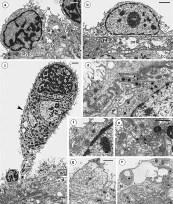
Tegument of a P. borealis sporocyst: (a) section showing tegument of a constricted region of tubule; (b) tegument covering brood chamber; (c) section through a tip region showing large outgrowth of tegument (arrowhead) containing several nuclei; (d, e) sections through cytoplasmic bridges (arrowheads) connecting cytons with the outer tegumental layer in constricted portions of tubules; (f) section showing Golgi complex and presumed pinocytotic vesicles (arrows) in outer tegumental layer; (g, h) sections showing ingrowths (asterisk) of cyton cytoplasm into outer tegumental layer in terminal regions of sporocyst tubules. Scale bars = 1 μm. See fig. 1 for key to labelling.

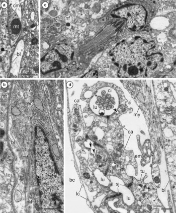
Ultrastructure of the subtegument in a P. borealis sporocyst: (a) micrograph of degenerating cyton with numerous pleomorphic vesicles and residual bodies in the cytoplasm; (b) section showing a myocyte with myofilaments, Golgi complex, RER and mitochondria; (c) section through a constricted region of a tubule showing nerve fibres lying close to the muscles, a nerve cell, a differentiating flame bulb (in longitudinal section) and a parenchymal cell; (d) section through a brood chamber showing a capillary cell, a tuft of flame cell cilia in the capillary lumen and a tubule cell; note pits (arrowheads) and short ridge-like projections (arrows) on the capillary luminal surface. Scale bars = 0.5 μm. See fig. 1 for key to labelling.

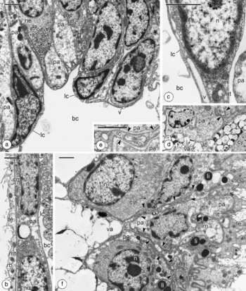
Ultrastructure of the parenchymal cells and body cavity lining in the different regions of the sporocyst body of P. borealis: (a) section of a type A terminal region; note the cells lining the lumen of a brood chamber adjacent to a solid tip region where parenchymal and nerve cells are seen; (b) sporocyst body wall in a distended region of the tubule; (c) detail of the body cavity lining of a terminal brood chamber; (d) section of a constricted region of tubule showing a parenchymal cell, cyton and lumen lining cell filled by numerous vacuoles; thin extensions of this cell are marked by arrowheads; (e) micrograph of an analogous extension passing between parenchymal cells; note the layer of electron-dense material underlying the body cavity lining; (f) cross-section through a constricted region of a tubule; note a parenchymal cell, differentiating and mature cytons and lumen lining cells with large vacuoles and extensions projecting towards the tegument (arrowheads). Scale bars = 1 μm. See fig. 1 for key to labelling.

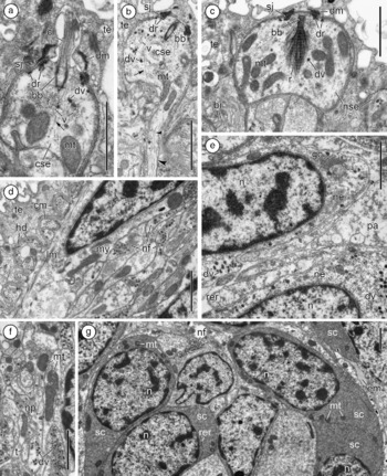
Ultrastructure of the sensory endings, nerve elements and stem cells of a P. borealis sporocyst: (a, b) longitudinal and oblique sections of breviciliated sensory endings; note a rudimentary rootlet, electron-dense material in the tegument surrounding the base of the cilium, small dense vesicles, lucid vesicles and neurotubules (arrows) in the sensory bulb and dendrite (arrowhead); (c) non-ciliated sensory endings with a cross-striated, broom-like rootlet; (d) section showing a bundle of nerve fibres lying under the muscles in a type A terminal region; (e, f) sections through the central area of a type A tip region showing nerve cells (e) and a neuropile (f); (g) micrograph showing stem cells in the tip region. Scale bars = 1 μm. See fig. 1 for key to labelling.

The sporocyst body wall in the distended areas of the tubules is composed of the tegument, underlying basal lamina and a layer of flattened subtegumental cells (figs 1c, 4b). The latter are represented by myocytes, tegumental cytons, parenchymal cells, protonephridial system elements and cells lining the sporocyst body cavity. In recently formed areas of the sporocyst, all these cells are closely adjoined to each other (figs 1c and 4b), whereas in ‘old’ brood chambers they are more loosely arranged and surrounded by large quantities of extracellular matrix (fig. 3d). In the constricted portions of the tubules, the subtegumental part of the body wall consists of two, or more rarely three, layers of densely packed cells (figs 1d, 4f). These areas differ from brood chambers in having a better developed musculature and presence of stem cells and cell bodies of neurons in the subtegument.
Tegument and tegumental cytons
Tegument of the P. borealis sporocyst is represented by an external nucleated syncytium connected by cytoplasmic bridges with tegumental cytons (figs 1c). The tegument apical surface bears short irregular lamellae, which are less frequent in the distended areas of the tubules (figs 2b and 4b) than in the constricted regions (fig. 2a and d). The basal plasma membrane of the tegument forms invaginations, which are most numerous in the narrow portions of the tubules and in tip regions (figs 2d and 5c). Tegumental nuclear structure varies considerably. Nuclei differ in shape and size, in nucleoplasm electron density, in size of nucleoli and in the proportion of condensed chromatin (fig. 2a–c). In the thin external syncytium covering brood chambers, nuclei are few and far between and, as a rule, slightly flattened (fig. 2b). In narrow areas of the branches, and especially in their terminal regions, the nuclei are more numerous and often located in the tegumental outgrowths (fig. 2a and c). The tegumental cytoplasm contains mitochondria, Golgi complexes, rounded electron-dense secretory granules, numerous residual bodies often represented by multilamellar structures (fig. 2a–g) and occasional lipid inclusions. Local agglomerations of electron-transparent vesicles of an obscure nature sometimes occur in the cytoplasm, and occasional coated vesicles resembling pinocytotic vesicles can be seen near the apical plasma membrane (fig. 2f).
Only one type of the tegumental cyton was observed throughout the sporocysts examined, even though cytons at various stages of development can be clearly distinguished by TEM. The cytoplasm of the most recently formed cytons contains a lot of free ribosomes, a loose network of rough endoplasmic reticulum (RER), small mitochondria, poorly developed Golgi apparatus and infrequent electron-dense secretory granules analogous to those in the external tegumental layer. More mature cytons also possess electron-transparent vesicles (fig. 2d), pleomorphic vesicles resembling secondary lysosomes, residual bodies and a few lipid inclusions (fig. 2e). The cytoplasm of some cytons (probably old and degenerating) is packed with pleomorphic vesicles and residual bodies (fig. 3a). Secretory granules, residual bodies and lipid droplets occur frequently in the cytoplasmic bridges connecting cytons with the external tegumental layer (fig. 2d and e). In terminal regions of the tubules, alongside the cytoplasmic bridges, rather deep, extensive ingrowths of the cyton cytoplasm into the external tegumental layer were observed (fig. 2g and h).
Myocytes
Beneath the outer syncytium and underlying basal lamina there are two layers of muscle fibres which represent the contractile parts of myocytes. The external layer consists of circular muscle fibres, and the internal layer is composed of longitudinal ones (figs 1c, d# 2d, 5d). The muscle fibres are connected to the basal lamina by hemidesmosomes; their cytoplasm contains both thin and thick myofilaments and solitary mitochondria (figs 2d, 5d). Myocyte perikarya contain RER with distended cisternae, mitochondria and well-developed Golgi apparatus (fig. 3a).
Elements of the protonephridial system
Flame cells, cells of capillaries and excretory tubules are regularly observed in the body wall, both in the distended and narrow regions of the tubules (figs 1c and ). No excretory system elements were found in the cell masses at branch tips. Each flame bulb consists of a flame cell, bearing a tuft of cilia, and part of its associated capillary cell (fig. 3c and d). They are similar to those of the cercariae of P. borealis described in detail under the name Bucephaloides gracilescens by Podvyaznaya & Galaktionov (Reference Podvyaznaya and Galaktionov2004). The structure of the flame cells, capillaries and excretory tubules is typical for digeneans (fig. 3d). No large collecting tubes were identified in cross-sections of sporocyst branches.
Parenchymal cells
Parenchymal cells are present in all regions of sporocysts (figs 1 and ). They have long, variously directed processes, filling the spaces between other types of cell. Much parenchymal cell cytoplasm has a low electron density (figs 2b, 5e). Cell organelles, such as free ribosomes, mitochondria and RER cisternae, are few. Some lipid inclusions, pleomorphic vesicles and residual bodies are also found in the cytoplasm of parenchymal cells (figs 2b, 5e).
Cells lining the sporocyst body cavity
The body cavity of the P. borealis sporocyst is lined with a layer of specialized cells (figs 1 and 4). In brood chambers, the lining cells are strongly flattened (figs 3d and 4a–c). Their thin layer of electron-dense cytoplasm contains infrequent small electron-transparent vesicles and, in perinuclear regions, a few small mitochondria and RER cisternae are present (fig. 4a and c). Cells lining the body cavity in narrow regions of the tubules are relatively large and their cytoplasm is filled with numerous large electron-transparent vacuoles, small pleomorphic vesicles and residual bodies (figs 1d and ). Large electron-transparent vacuoles often contain lipid-like material and, sometimes, multilamellar structures (fig. 4d). Some vesicles and residual bodies were observed apparently merging with large electron-transparent vacuoles. In some constricted regions, the strongly vacuolated cytoplasm of the lining cells is so extensive that it fills the body cavity, making it barely identifiable in sectioned material. The vacuolated cytoplasm also penetrates deep between the subtegumental cells in the direction of the outer syncytium (fig. 4f). The lining cells and their extensions are separated from the other subtegumental cells by a thin, but distinct layer of dense extracellular material (figs 1c and 4e).
Sensory receptors and nerve cells
Two kinds of sensory endings are found in the tegument of type A terminal regions of sporocysts (figs 1a and 5a–c). Sensory bulbs bearing a short cilium have a closed basal body and a very small conical rootlet (fig. 5a). The cytoplasm of these sensory bulbs contains small vesicles (some electron-dense and some electron-lucid), neurotubules and several large mitochondria (fig. 5a and b). Each ciliated sensory ending is connected to the tegument by an apical septate junction, adjoined from the internal side by a broad electron-dense ring with some inclusions of less dense material (fig. 5a). A narrower electron-dense ring is situated proximally (fig. 5a and b). In the tegument surrounding the base of the cilium and septate junction, a dome-shaped agglomeration of electron-dense material is observed (fig. 5a). Non-ciliated sensory endings are completely submerged into the tegument and attached to it by septate junctions (fig. 5c). Each of these sensory bulbs has a distinct basal body and a powerful cross-striated, broom-like rootlet, and its cytoplasm contains numerous mitochondria, as well as neurotubules and a few small electron-dense vesicles (fig. 5c). Non-ciliated sensory endings have two electron-dense rings, the larger apical one being located at the level of the septate junction (fig. 5c). A discernible thickening of the electron-dense material is also observed in the external syncytium adjacent to the septate junction.
In the type A terminal portions of the tubules, numerous neurons and nerve fibres were observed (figs 4a and 5d–f). In the central part of their tip region nerve fibres interweave to form a dense feltwork (neuropile) (fig. 5f). Bundles of nerve fibres and solitary nerve fibres are abundant near the subtegumental muscles (figs 2b and 5d). Nerve cells are located both in the central part of the tip region (around the neuropile) and at its periphery. Other regions of sporocysts possess solitary nerve fibres in close proximity to the muscles (fig. 1). Solitary nerve cell bodies were observed in the type B terminal portions of the tubules (fig. 1b) and in the walls of constrictions (fig. 3c). Perikarya of nerve cells have a finely granular cytoplasm filled with small dense-cored vesicles (fig. 5e). The cytoplasm also contains a well-developed Golgi apparatus, sparse RER, mitochondria and neurotubules (figs 3c and 5e). Secretory vesicles, mitochondria and neurotubules are also found in the nerve cell processes (figs 3c and ).
Stem cells
A considerable pool of stem cells was revealed in all sporocysts studied. They were mostly concentrated in the cell masses of branch tips (figs 1a and 5g). Single stem cells were also noted in the walls of constrictions (fig. 1d). Comparatively small and compact undifferentiated cells are characterized by a high nuclear/cytoplasmic ratio. Their nuclei lack nucleoli (fig. 5g). Condensed chromatin forms average sized aggregates at the centre and periphery of each nucleus. Electron-dense cytoplasm contains numerous free ribosomes, a few small mitochondria and occasional RER cisternae (fig. 5g). In both types of terminal region, stem cells usually occur in small groups (figs 1a and 5g). Both in the tip regions and in the body wall, stem cells occur adjacent to the cells that, although still poorly differentiated, have already embarked upon the cell differentiation process. Such cells have a larger proportion of less dense cytoplasm; their nuclei contain nucleoli (fig. 4f) and some have extensions projecting from them. In type A terminal portions, stem cells and poorly differentiated cells are especially numerous.
Discussion
Although the body wall ultrastructure of P. borealis sporocysts varies in each of the regions chosen for study, the component parts are, in general, arranged similarly in each body area. The overall structure is similar to that described in Cercaria buccini (see Køie, Reference Køie1971), Cercaria helvetica XII (see Reader, Reference Reader1975) and Diplostomum pseudospathaceum (see Klag et al., Reference Klag, Niewiadomska and Czubaj1997). However, some ultrastructural details of our sporocysts set them apart from the parthenogenetic generations of other digeneans.
The most remarkable feature of the tegument of mature P. borealis sporocysts is the presence of nuclei in the external syncytium. Nucleated tegument was first described in sporocysts of another bucephalid species, Bucephalus minimus (James et al., Reference James, Bowers and Richards1966; James & Bowers, Reference James and Bowers1967). According to James et al. (Reference James, Bowers and Richards1966), in B. minimus sporocysts, numerous nuclei are located in the ‘middle region’ of a rather thick external syncytium, at a considerable distance from the basal plasma membrane. Later, Popiel & James (Reference Popiel and James1978a, Reference Popiel and Jamesb) and Popiel (Reference Popiel1978) reported two-layered tegument in the daughter sporocysts of representatives of the family Microphallidae, with the outer layer (region) having epithelial cellular structure and containing nuclei. The idea that the tegument consisted of two layers (regions) was extended to all daughter sporocysts without a birth pore, including those of bucephalids (Popiel & James, Reference Popiel and James1978b). Later on, however, detailed ultrastructural research carried out on different digeneans, including the Microphallus species studied by Popiel & James (Reference Popiel and James1978a, Reference Popiel and Jamesb), showed that these authors had mistakenly applied the term ‘outer nucleated epithelial region of the tegument’ to a layer of molluscan host haemocytes surrounding the daughter sporocysts (Dobrovolskij et al., Reference Dobrovolskij, Galaktionov, Muhamedov, Sinha and Tihomirov1983; Køie, Reference Køie1987; Galaktionov, Reference Galaktionov1993; Galaktionov & Dobrovolskij, Reference Galaktionov and Dobrovolskij2003). In many Plagiorchioidea and Microphalloidea, host haemocytes closely adjoin the external tegumental layer and its projections. As a result, daughter sporocysts are surrounded by an epithelial-like cellular layer of host origin, the so-called ‘paletot’, which can be easily mistaken for an outer nucleated region of the tegument (for review see Galaktionov & Dobrovolskij, Reference Galaktionov and Dobrovolskij2003). Our results on P. borealis tegument provide clear evidence that the ‘nucleated region’ of the tegument of bucephalid sporocysts and the ‘outer nucleated epithelial region’ of the tegument of microphallid sporocysts are not identical structures, as proposed by Popiel & James (Reference Popiel and James1978b). The latter, as mentioned above, is formed by host tissue, while the former is made up of tegumental outgrowths containing nuclei analogous to those that we observed in the constrictions and terminal portions of the P. borealis sporocysts. In P. borealis, however, the tegumental nuclei are often situated directly above the continuous basal plasma membrane of the external syncytium, indicating that they are undoubtedly part of the tegument. James et al. (Reference James, Bowers and Richards1966) interpreted the presence of nuclei in the tegument of B. minimus sporocysts as embryonic features that had been retained in their body covering. It is now known that the nucleated syncytium stage is characteristic of early tegument development in daughter parthenogenetic generations (for a review see Galaktionov & Dobrovolskij, Reference Galaktionov and Dobrovolskij2003). In mother sporocysts, the external tegumental layer, formed by the overgrowth of hypodermal ridges above the basal lamina, does not contain nuclei at early stages of its development. Thus, the conjecture of James et al. implies acknowledgement that the branching bucephalid sporocysts are the daughter generation arising from reproduction of the mother sporocysts. However, the existence of two generations of bucephalid sporocysts remains unproven and the interpretation of the origin of the nuclei in the external syncytium of the bucephalid sporocysts suggested by James et al. is left open to question. Our alternative proposition is based on the observations of Korneva & Poddubnaya (Reference Korneva and Poddubnaya1999) who showed the migration of nuclei from tegumental cytons into the initially anucleate tegumental layer in developing procercoids of the cestode Triaenophorus nodulosus. It is quite likely that a similar phenomenon occurs during formation of the body covering in other Neodermata, including the sporocysts of P. borealis and B. minimus. Our observations suggest that recently formed tegumental cytons are actively involved in the growth of the outer tegumental layer in the terminal regions of P. borealis sporocysts. The cytoplasm of the tegument is continuous with that of the cytons which possess all the apparatus for its production. The nuclei of some cytons might well migrate into the growing epithelial covering together with the translocating cytoplasm.
An important function of tegument in sporocysts, which lack a gut, is the absorption of nutrients from the host organism. Køie (Reference Køie1971), Reader (Reference Reader1975) and Al-Salman & James (Reference Al-Salman and James1989) proposed that pinocytosis plays a significant role in the feeding of daughter sporocysts in Cercaria buccini, C. helvetica XII and Meiogymnophallus minutus, respectively. We also observed micro-invaginations of the apical plasma membrane and small, coated vesicles in the tegument of P. borealis sporocysts, but the paucity of these structures indicates a low level of pinocytotic activity. Prosorhynchoides borealis sporocysts, localized in the haemocoel of the affected organs, appear to actively assimilate low-molecular-weight organic compounds dissolved in molluscan haemolymph. These substances may pass across the apical plasma membrane of the tegument, the absorbing surface of which is increased by the presence of lamellae, by means of simple and/or facilitated diffusion. The well-developed system of tegument basal invaginations is another indication of active transport of nutrients to the internal tissues of P. borealis sporocyst. The fact that these features are best developed in the narrow parts of the sporocyst tubules testifies to more intense transport processes in these regions.
The structure of tegumental cytons and muscles in P. borealis sporocysts is essentially similar to that described in sporocysts of other digeneans (Køie, Reference Køie1971; Gibson, Reference Gibson1974; Reader, Reference Reader1975; Fournier & Théron, Reference Fournier and Théron1985; Pojmanska & Machaj, Reference Pojmanska and Machaj1991; Klag et al., Reference Klag, Niewiadomska and Czubaj1997; and others). Parenchymal cells described in this paper are, in our opinion, equivalent to the ‘vacuolar’ cells of D. pseudospathaceum sporocysts (Klag et al., Reference Klag, Niewiadomska and Czubaj1997) and irregular-shaped cells, containing lipids and glycogen, in the body wall of C. buccini, Postharmostomum gallinum and Schistosoma mansoni sporocysts (Køie, Reference Køie1971; Žd'árská & Soboleva, Reference Žd'árská and Soboleva1985; Fournier & Théron, Reference Fournier and Théron1985). Parenchymal cells of sporocysts, like those of adult flukes, appear to have diverse functions, including the transport of various substances and the participation in carbohydrate and lipid metabolism.
According to James et al. (Reference James, Bowers and Richards1966), in B. minimus sporocysts, numerous flame cells are united in small groups, each group opening into the body cavity through its own excretory pore. Matthews (Reference Matthews1973) described at the light microscope level numerous excretory pores also opening into the sporocyst body cavity in another bucephalid species, Prosorhynchus crucibulum. We failed to observe any evidence of excretory pores opening into the sporocyst body cavity in P. borealis. Nevertheless our conclusion that P. borealis sporocysts lack large collecting tubes agrees well with the observations of James et al. (Reference James, Bowers and Richards1966) and Matthews (Reference Matthews1973) that bucephalid sporocysts do not possess a common system of collecting tubes uniting all the flame cells.
The body cavity of P. borealis sporocysts clearly has a well-developed lining formed by specialized subtegumental cells. Analogous structures were noted in sporocysts of other digeneans, including C. helvetica XII, M. minutus and D. pseudospathaceum (see Reader, Reference Reader1975; Russell-Pinto et al., Reference Russell-Pinto, Bowers and James1996; Klag et al., Reference Klag, Niewiadomska and Czubaj1997). However in P. borealis sporocysts, the structure of body cavity lining differs in each of the body regions studied. It is thinnest in the brood chambers which contain developing cercariae and its characteristics would appear to render it easily permeable for nutrient transportation for their support. In the constricted portions, the cytoplasm of the lining cells is hypertrophied and contains many large vacuoles and small vesicles, the contents of which are typical residual material that may accumulate from metabolic activity. The enlarged lining cells almost completely block the lumen in some narrow portions, thereby effectively isolating and separating distinct sections of the P. borealis sporocyst body cavity.
Cells identified as germinal cells have been reported in the body walls of B. minimus, Gymnophallus choledochus (syn. Cercaria dichotoma), M. minutus and D. pseudospathaceum sporocysts by James et al. (Reference James, Bowers and Richards1966), James & Bowers (Reference James and Bowers1967), Al-Salman & James (Reference Al-Salman and James1989) and Klag et al. (Reference Klag, Niewiadomska and Czubaj1997). In P. borealis sporocysts, germinal cells were found by Podvyaznaya (Reference Podvyaznaya2007) only in the body cavity. Additionally, we observed a few undifferentiated cells in the wall of constricted portions of P. borealis sporocysts. The nuclei in these cells differ distinctly from those of the germinal cells. The cytoplasm of the stem cells does not contain nuage, which is a characteristic of germinal cells (Podvyaznaya, Reference Podvyaznaya2007). The presence of differentiating cells (e.g. cytons) in constricted regions points to the fact that the stem cells participate in renewal of somatic structures in the sporocyst body wall. In P. borealis sporocysts, stem cells are much more numerous in the cell mass of the branch tips. Tennent (Reference Tennent1906), Kniskern (Reference Kniskern1952), James & Bowers (Reference James and Bowers1967) and other researchers all concur that the growth foci of bucephalid sporocysts are located at these points. According to Kniskern (Reference Kniskern1952), James & Bowers (Reference James and Bowers1967) and Podvyaznaya (Reference Podvyaznaya2007), development of germinal elements is also associated with terminal portions of branches in Rhipidocotyle septpapillata, B. minimus and P. borealis. Our data suggest that, in the case of P. borealis, these processes (growth and germinal development) are to some extent separated in space, being associated with the different types (A and B) of the terminal region identified in this study. The reproductive function of the sporocyst is mostly confined to type B terminal portions containing germinal masses and germinal cells and having little nerve tissue. Unfortunately, we failed to track early development of the germinal masses (Podvyaznaya, Reference Podvyaznaya2007), and consequently we can now only conjecture that they are formed with the participation of stem cells contained in the cell mass at branch tips. Type B terminal regions seem to have a limited growth potential. It would appear that the main growth zones of P. borealis sporocysts are located in type A terminal portions that have well-developed nervous and sensory apparatuses and whose tips contain especially numerous stem and differentiating cells. The considerable concentration of nerve tissue near the proliferation foci of stem cells would indicate that the growth of sporocysts is controlled by the nervous system. The numerous sensory endings in branch tips are likely to be involved in the determination of optimal growth directions. The non-ciliated sensory endings are similar to the receptors described in cercariae of Echinostoma revolutum (type-2 non-ciliated receptors) (Žd'árská, Reference Žd'árská1992) and juvenile flukes of Fasciola hepatica (see Bennett, Reference Bennett1975) in having two electron-dense rings, a basal body and a well-developed cross-striated rootlet. It is commonly thought (Rohde & Watson, Reference Rohde and Watson1990; Czubaj & Niewiadomska, Reference Czubaj and Niewiadomska1996; Žd'árská & Nebesářová, Reference Žd'árská and Nebesářová2003) that non-ciliated sensory endings, which have no direct contact with the environment, are receptive to mechanical stimuli. Sensory endings with one short cilium and an absent or weakly developed rootlet, similar to those observed in the P. borealis sporocysts, have been recorded in various life cycle stages of many digenean species, such as cercariae of E. revolutum (Žd'árská, Reference Žd'árská1992), daughter sporocysts and cercariae of D. pseudospathaceum (Czubaj & Niewiadomska, Reference Czubaj and Niewiadomska1991, Reference Czubaj and Niewiadomska1996), adults of Crepidostomum metoecus (Žd'árská & Nebesářová, Reference Žd'árská and Nebesářová2003, Reference Žd'árská and Nebesářová2004). Such sensory endings are usually believed to be mechano- or chemoreceptors.
Even the most detailed morphological descriptions of bucephalid sporocysts (Tennent, Reference Tennent1906; Kniskern, Reference Kniskern1952; James et al., Reference James, Bowers and Richards1966) do not provide any information about the nervous system. This study has revealed nervous system elements in all of the body segments examined. Tips of actively growing sporocyst branches possess large accumulations of neurons and nerve fibres but no evidence of ganglia was observed. Elsewhere in the body the nervous system is represented by a loose submuscular network of nerve fibres with integrated single nerve cells.
In conclusion, our study showed that the branching P. borealis sporocyst is organized into modules consisting of brood chambers, constricted areas and terminal portions of two types, which are repeated many times. It demonstrates that each module has ultrastructural features consistent with its functions. The bulk of the body is composed of brood chambers in which cercariae develop. Their thin body walls are compatible with the transport of monomers from the haemolymph of the molluscan host and also facilitate the rupture necessary for cercarial release.
Constricted regions are specialized for absorption of nutrients and their subsequent transport to other areas of the sporocyst. The constrictions also isolate individual brood chambers, making them more or less autonomous, so that degeneration or the damage of an area (resulting, for instance, from body wall rupture prior to cercarial release) does not bring about the death of the whole organism. Consistent with the autonomy of the different parts of the sporocyst are the absence of a common system of protonephridial collecting tubes and the presence of multiple centres of concentrated nerve elements and multiple foci of growth and reproduction.
Continuous growth of the sporocyst, which necessitates its penetration into various organs of the molluscan host, is ensured by the type A terminal regions containing numerous stem cells and cells in early stages of differentiation. The numerous sensory endings and nerve elements concentrated in these regions may well direct the orientation of growing sporocyst branches in the tissues of the molluscan host. Finally, the reproductive function is mostly confined to the brood chambers of the type B terminal portions, where germinal cells are formed and the early stages of cercarial development take place.
Due to its modular organization, the branching bucephalid sporocyst is capable of long-term existence in its molluscan host, ensuring a level of cercarial production comparable with, if not exceeding, that of infrapopulations of rediae or daughter sporocysts in other digeneans.
Acknowledgements
We thank Dr M. Eydal (University of Iceland, Reykjavik) for help in the collection and initial identification of the specimens studied and Mr A.K. Levit (Oncological Institute, St. Petersburg) for excellent technical assistance. We are grateful to Dr S.W.B. Irwin for his helpful comments and improvement of the English version of the manuscript. We are also indebted to an anonymous referee for valuable corrections. This work was supported by the Russian Foundation for Basic Research (N 09-04-01309 and N 10-04-00430).