Tetralogy of Fallot is characterised by the presence of anterior malaligned ventricular septal defect and valvar and/or subvalvar right ventricular outflow tract obstruction.Reference Van Praagh and Etienne-Louis 1 Surgical repair usually includes patch closure of the ventricular septal defect and relief of the right ventricular outflow tract obstruction with/without preservation of the pulmonary valve. During transventricular repair, a patch material is typically used to enlarge the right ventricular outflow tract. Preservation of right ventricular function is important in improving the long-term outcome of tetralogy of Fallot.Reference Geva 2 Various patch materials have been used to enlarge the right ventricular outflow, and these include bovine pericardium, autologous pericardium, and polytetrafluoroethylene patch; however, these materials are inert and have no potential to lead to a repair that has functional myocardial tissue in the area of the right ventricular outflow tract patch. Aneurysm formation in right ventricular outflow tract, fibrosis, infection, and thrombogenicity are known to be associated with these patch materials.Reference Parks, Ngo and Plauth 3 These complications can affect right ventricular systolic performance adversely. Thus, there is a growing need for a patch material that can provide a matrix for viable myocardial tissue to rejuvenate at the area of injury. As a result, over time, the right ventricular outflow tract myocardium should be “seamless” between the ventriculotomy and the uninjured myocardium. Such patch material would potentially avoid complications and may lead to preserved right ventricular systolic performance. Regenerative medicine represents the future in which some of these obstacles to surgical repair of CHD can be overcome. Recently, interest has accrued in the use of porcine small intestinal submucosal extracellular matrix (CorMatrix®Cardiovascular Inc., Roswell, Georgia, United States of America) as the right ventricular outflow tract patch, hoping that it would allow migration of neighbouring myocardial cells. Cells that migrate into the bio-scaffold may have the potential to undergo proliferation and architectural organisation that lead to viable tissue.Reference Badylak, Freytes and Gilbert 4 , Reference Zhao, Puskas and Xu 5 Extracellular matrix was first used for non-cardiac surgeries such as vascular graft, dura mater substitute, oesophageal graft, and musculotendinous scaffold.Reference Mirensky and Breuer 6 – Reference Chen, Yoo and Atala 9 The first use of extracellular matrix for cardiac surgery has been reported for pericardial repair.Reference Boyd, Johnson and Sultan 7 Owing to its theoretical potential of tissue integration, isolated reports of cardiac and non-cardiac repairs using CorMatrix patch material have increased, even though currently there is scant evidence to support this contention. If the assumption of tissue regeneration is indeed true, then histological changes should translate into improvement of right ventricle function as evidenced by right ventricular functional echocardiographic indices.
Evaluation of right ventricular function on echocardiography has not been as straightforward as evaluation of left ventricular function because of its geometry. More than one echocardiographic index has been always relied upon to reflect true right ventricular function. In the last decade, more promising echocardiographic indices such as tricuspid annular plane systolic excursion, three-dimensional ejection fraction, and global longitudinal strain have been developed. Tricuspid annular plane systolic excursion reflects longitudinal right ventricular function, whereas the other two indices reflect global function.
To evaluate the impact of extracellular matrix patch on right ventricular function, we designed a study to compare echocardiographic characteristics of the right ventricle after non-transannular patch repair of tetralogy of Fallot with extracellular matrix versus bovine pericardium.
Materials and methods
The echocardiography indices were measured prospectively. The Institutional Review Board of University of Tennessee Health Science Center and Le Bonheur Children’s Hospital approved the study. Our paediatric cardiac surgery database identified patients who met the following inclusion criteria: cardiac surgery with cardiopulmonary bypass for non-transannular patch repair of tetralogy of Fallot. All repairs were performed via a transventricular approach to relieve right ventricular outflow tract obstruction. Subjects who had surgeries performed between 01 January, 2009 and 31 December, 2012 were included. Patients with significant associated CHD such as pulmonary atresia or atrioventricular septal defect or those with extracardiac defects or debilitating genetic abnormalities were excluded. Patients with previous palliative procedures were also excluded. Patients who had an extracellular matrix patch constituted the study group, whereas patients with glutaraldehyde-treated bovine pericardium served as the control group. Patients were contacted and were enrolled to the study postoperatively. An informed consent was obtained before each echocardiography procedure.
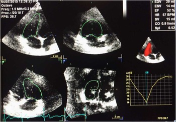
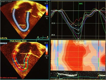
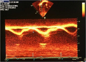
A comprehensive transthoracic echocardiogram – two-dimensional and three-dimensional – was obtained for all patients meeting inclusion criteria; three-dimensional right ventricular ejection fraction, right ventricular global longitudinal strain, and tricuspid annular plane systolic excursion were used to assess right ventricular function.Reference Mor-Avi, Lang and Badano 10 , Reference Lang, Badano and Mor-Avi 11 The studies were performed using General Electric vivid 7 BT08 echocardiography machines (General Electric Company, Boston, Massachusetts, United States of America). We obtained six-segment right ventricle longitudinal strain values from the apical four-chamber view. Strain analysis and three-dimensional ejection fraction calculation were performed using Echopac software as demonstrated in Figures 1 and 2. Tricuspid annular plane systolic excursion was measured from the apical four-chamber view with M-mode as demonstrated in Figure 3. These data were interpreted by a paediatric cardiologist in a blinded manner.
Right ventricular ejection fraction using three-dimension techniques.

Right ventricular global longitudinal strain. Top right: strain curve of each of the six segments. Bottom left: segmental strain.

Tricuspid annular plane systolic excursion.

Descriptive statistics such as mean and standard deviation were used to summarise all continuous variables. All continuous variables were assessed for normality. A two-sample t-test was used to compare differences between study and control groups for each of the continuous outcomes. All analyses were performed using Statistical Package for the Social Science (SPSS) software. A p-value<0.05 was considered significant.
Results
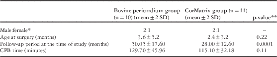
A total of 21 patients who met inclusion criteria were enrolled to the study, and received a right ventricular outflow tract patch of either extracellular matrix (n=11) or bovine pericardium (n=10). The male-to-female ratio, age at operation, and cardiopulmonary bypass times were similar between study and control groups (Table 1). The follow-up period was 28±12 months in the CorMatrix group and 50±17 months in the bovine pericardium group (p=0.001).
Comparison of demographic and operative characteristics between the groups.

CPB=cardiopulmonary bypass
* Rounded values
**p-value of<0.05 was considered as significant
Echocardiographic findings
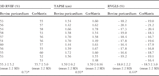
The mean three-dimensional right ventricular ejection fraction in the extracellular matrix group was 55.7±5.0% and 55.3±5.2% in the bovine pericardium group (p=0.7). The average right ventricular global longitudinal strain in healthy, age-matched children from our echocardiography laboratory was −25.6±2%. Both the study group and the control group had similar but lower right ventricular global longitudinal strain compared with healthy children: −18.0±2.2% in the extracellular matrix group compared with −18.5±3.0% in the bovine group (p=0.4). There was no difference in tricuspid annular plane systolic excursion values between the study group and the control group: 1.59±0.16 cm and 1.59±0.2 cm, respectively (p=0.9). Left ventricular systolic function was normal in all patients. No patient had right ventricular outflow tract obstruction or aneurysm. None of the patients had more than mild pulmonary valve insufficiency or stenosis. No patient required surgical or catheter-based re-intervention. Echocardiographic characteristics of both the groups are reported in Table 2.
Comparison of echocardiographic characteristics between two groups.

3D RVEF=right ventricular three-dimensional ejection fractions; RVGLS=right ventricular global longitudinal strain; TAPSE=tricuspid annular plane systolic excursion
*p-value of<0.05 was considered as significant
Discussion
Right ventricular size and function are important factors determining outcome and performance after repair of tetralogy of Fallot.Reference Bacha, Scheule and Zurakowski 12 Long-term goals of tetralogy of Fallot repair are aimed at limiting deterioration of right ventricular function, encouraging right ventricular remodelling after a competent pulmonary valve is placed, and eliminating aneurysm formation in the right ventricular outflow tract.Reference Oosterho, Straten and Vliegen 13 Although this study has focussed on only non-transannular patch repair, ideal patch material may aid in achieving long-term goals regardless of type of repair.
Bovine and autologous pericardium are easily available and have low thrombogenicity and low antigenicity, making them the more preferred patch material; however, they are associated with calcification, retraction, and thickening. Similarly, polytetrafluoroethylene is also associated with progressive calcification.Reference Us, Sungun and Sanioglu 14 Aneurysms and rupture are sequalae of patch calcification; furthermore, formation of fibrosis around the patch may serve as a nidus for ventricular arrhythmia. All these effects may lead to decreased right ventricular systolic performance over time. Ideal patch material should have the potential for growth and the ability to remodel. A strategy to achieve viable myocardium after repair is to infuse progenitor cells in a targeted area or biologically active molecules such as growth factors or enhance the gene expression controlling such biologically active molecules.Reference Li, Jia and Weisel 15 – Reference Taylor, Atkins and Hungspreugs 17 Although there were some favourable results for this approach, it never became popular. The second strategy was to promote constructive remodelling of tissue by providing supportive microenvironment. Porcine small intestinal extracellular matrix is thought to provide viable extracellular scaffold, which has the potential to be populated and replaced by viable myocardial cells; this could potentially lead to better right ventricular function and fewer long-term complications.Reference Badylak, Freytes and Gilbert 4 It also has the advantage of avoiding sensitisation such as homograft materials. These postulates have some basis in animal experiments involving tricuspid valve repairs, aortic valve repairs, and intracardiac patch repairs, although clinical evidence has not yet been validated.Reference Scully, Fan and Grigoryan 18 – Reference Lopes, Cabrita and Ilharco 20 Extracellular matrix had shown promise in terms of less graft failure in congenital heart surgery; however, no study has demonstrated tissue integration or myocardial cell regeneration. Woo et alReference Woo, Fishbain and Reemtsen 21 examined 12 explanted extracellular matrix patches from cardiac valves and outflow tracts but failed to demonstrate re-cellularisation of patch material. Explanted extracellular matrix patches from hemi-Fontan and cardiac valves demonstrated no cell growth and no calcification.Reference Nelson, Haider and Si 22 , Reference Zaidi, Nathan and Emani 23 Rosario-Quinones examined six explanted extracellular matrix patches out of which two were from the right ventricular outflow tract. All the explanted patches demonstrated intense inflammatory reactions without fibrosis.Reference Rosario-Quinones, Magid and Yau 24 With less fibrosis and calcification seen in the extracellular matrix, it may affect right ventricular function positively. No study till date has examined its effect on right ventricular function.
At our institution, the standard repair for tetralogy of Fallot is via a limited transventricular approach. Our use of a transannular patch represents <10% of our patients. We selected patients with non-transannular patch because use of extracellular matrix as a right ventricular outflow tract patch in the infundibular region of the right ventricle where the patch border is surrounded by viable contracting myocardium theoretically provides the maximum chance of myocardial cell generation over it, and hence may contribute positively to right ventricular systolic function over a period of time.
In the extracellular matrix group, the follow-up period was shorter than in the bovine pericardium group, as we started using extracellular matrix at a later stage; however, we chose a follow-up period longer than 12 months to allow sufficient time for any benefit in the use of extracellular matrix to be realised. Several echocardiographic indices of right ventricular function have been identified in the literature.Reference Rudski, Lai and Afilalo 25 We selected reliable, reproducible, and sensitive indices that take in to account right ventricular outflow tract function for this study.Reference Lang, Badano and Mor-Avi 11 These included three-dimensional right ventricular ejection fraction, tricuspid annular plane systolic excursion, and right ventricular global longitudinal strain. Out of all three, ejection fraction is the last one to get negatively affected after tetralogy of Fallot repair; three-dimensional right ventricular ejection fraction is the most reliable echocardiography method to assess ejection fraction. It has a very good correlation with ejection fraction measured by cardiovascular magnetic resonance. Furthermore, three-dimensional reconstructions of the images include the right ventricular outflow tract in ejection fraction calculation, allowing evaluation of the effect of the patch material on ejection fraction in repaired tetralogy of Fallot. Both the groups in our study had normal three-dimensional right ventricular ejection fractions with no statistically significant difference. Several reasons can be considered for this. First, there may not be myocardial cell regeneration on the extracellular matrix patch; however, the mean follow-up period of 28 months in our study may not be sufficient to either have myocardial regeneration or have enough contributing towards ventricular systolic function that can be demonstrated by echocardiography. On the contrary, because of the fact that three-dimensional right ventricular ejection fraction was normal in the bovine pericardium group, longer time may be required for right ventricular dysfunction to set in at which point positive effect of extracellular matrix patch may be unmasked.
Tricuspid annular plane systolic excursion is a marker for longitudinal right ventricular function. Tricuspid annular plane systolic excursion simply measures annular displacement of lateral tricuspid annulus during systole. It has a good correlation with right ventricular global function as measured by radionuclide-derived right ventricular ejection fraction.Reference Lang, Badano and Mor-Avi 11 It is more reproducible and has high specificity and negative predictive value for detecting abnormal right ventricular systolic performance. The normal value of tricuspid annular plane systolic excursion is age dependant. For the age cohort in our study, the normal value is 1.7 cm and above.Reference Koestenberger, Nagel and Favekes 26 Ours is the first study to demonstrate tricuspid annular plane systolic excursion in repaired tetralogy of Fallot with non-transannular patch. We observed a borderline tricuspid annular plane systolic excursion in both groups; however, we were unable to demonstrate difference between the groups. Interestingly, the tricuspid annular plane systolic excursion values in both our groups are very similar to the published data of tricuspid annular plane systolic excursion of 1.5–1.6 cm in age groups of 2–5 years after tetralogy of Fallot repair in children by Koestenberger et al.Reference Koestenberger, Nagel and Favekes 26 In that study, all 131 patients had transannular patch with autologous pericardium with ages ranging from birth to 28 years. The average tricuspid annular plane systolic excursion value from 2 years and above in that cohort was 1.6 cm. This demonstrates an interesting point that tricuspid annular plane systolic excursion reaches its nadir for the paediatric population at 2 years of age after repair of tetralogy of Fallot regardless of the type and extension of the patch.
Strain is the percentage of systolic shortening of the myocardial segment. It can be measured as longitudinal or circumferential. Circumferential strain would not include the right ventricular outflow tract, and thus we analysed only longitudinal strain for our cohort. Right ventricular global longitudinal strain has established prognostic values in heart failure, pulmonary hypertension, etc. Studies have demonstrated lower strain values with normal ejection fraction indicating preserved ejection fraction earlier in the disease.Reference Solarz, Witt and Glascock 27 Thus, strain can be considered a sensitive early indicator of deteriorating ventricular function. Normal right ventricular global longitudinal strain values have been reported as −29±3% from the meta-analysis.Reference Levy, Sanchez-Mejia and Machefsky 28 The right ventricular global longitudinal strain value in the age-matched control cohort obtained at our echocardiography lab was −25.6±2%. We observed lower strain in both the groups compared to healthy controls. We did not find any difference in right ventricular strain values among the groups at the follow-up interval selected.
This study has several limitations. The relatively small cohort of patients may have decreased the power to detect differences in characteristics or outcomes between the study groups. The modest follow-up period may not have allowed adequate time for realising potential myocardial re-population of the extracellular matrix patches; perhaps with further time in vivo, the extracellular matrix patches would have demonstrated appreciable contractility or augmentation of right ventricular function. Finally, as the Institutional Review Board did not approve the use of cardiac MRI as an evaluation tool for this study due to requirement of sedation, the ability to optimally characterise right ventricular and patch performance may have been limited by the omission of this imaging modality.
Conclusion
In a small cohort of the patients after repair of tetralogy of Fallot followed-up for more than 1 year, the average right ventricular global longitudinal strain was lower in both the bovine pericardium patch group and the porcine small intestinal extracellular matrix group compared with the healthy population without significant difference among the groups. There was no significant difference in three-dimensional right ventricular ejection fraction and tricuspid annular plane systolic excursion values between the groups.
Acknowledgements
None.
Financial Support
This research received no specific grant from any funding agency, commercial or not-for-profit sectors.
Conflicts of Interest
None.