Introduction
Medical implantology is a major challenge for health and is a field enjoying continuous growth. The dynamics of this market are driven by many factors including population aging, new knowledge in pathology treatment and new demands in terms of quality of life requiring more successful medical devices. Titanium is a widely used biomaterial because of its mechanical properties, corrosion resistance and biocompatibility: it does not provoke a rejection reaction. However, titanium is inert, meaning it is then necessary to wait three to six months before obtaining full bone tissue integration around titanium implants. This delay depends on bone quality and other patient conditions.
Several works have shown that surface properties such as roughness, wettability, electric charge or chemical composition may modulate adhesion, proliferation and cell differentiation [Reference Ponsonnet, Reybier, Jaffrezic, Comte, Lagneau, Lissac and Martelet1, Reference Gittens, Olivares-Navarrete, Cheng, Anderson, McLachlan, Stephan, Geis-Gerstorfer, Sandhage, Fedorov, Rupp, Boyan, Tannenbaum and Schwartz2, Reference Le Guehennec, Soueidan, Layrolle and Amouriq3, Reference Stanford4, Reference Junker, Dimakis, Thoneick and Jansen5]. Since the beginning of the 1980s, studies have focused on titanium and alloy surface treatments as a means of enhancing the osseointegration of metal implants. The initial surface of an implant is usually turned or machined. This surface is relatively smooth and only has a few micrometric machining streaks. The polishing process (i.e., electropolishing) makes it possible to reduce the machining traces resulting in polished mirror surface roughness at the range of 0.1 µm. The main advantage of polished surfaces is the aesthetic surface and the inhibition of plaque accumulation [Reference Amarante, Chambrone, Moreira Lotufo and Lima6].
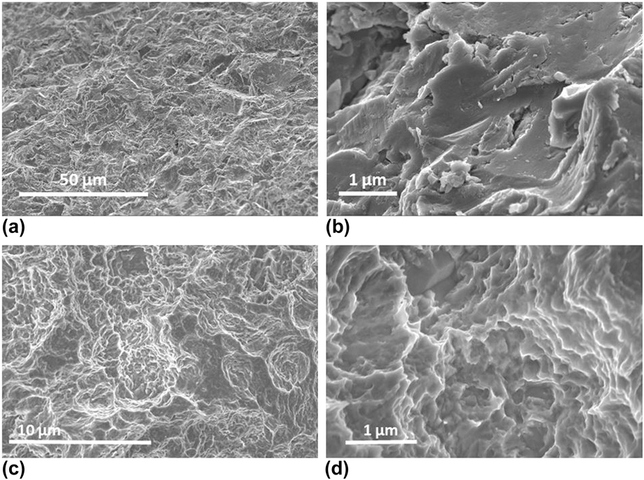
The grit-blasting process creates micrometric roughness in the range of 0.5–6 µm depending on the type of material and particle sizes used, as well as other treatment conditions [Reference Krishna Alla, Ginjupalli, Upadhya, Shammas, Krishna Ravi and Sekhar7] (i.e., the TiOblast™ surface from Astra Tech Implant; see Fig. 1).
SEM observation of the morphology of grit-blasted surfaces of TiOblast™ at two magnifications: (a) ×1000 and (b) ×20,000; SEM morphology of an SLA surface (Straumann) (c) ×5000 and (d) ×20,000.

The combination of both grit-blasting and dual acid-etching is the standard surface method used in dental implantology. Grit-blasting using Al2O3 particles 250–500 µm in diameter followed by a warm hydrochloric and sulfuric acid attack makes it possible to both remove sandblasting residue and produce 1–2 µm random micrometric cavities. These surfaces are developed by Straumann SLA [Figs. 1(c) and 1(d)].
Anodic oxidation is also widely used and mastered on metal to increase its oxide layer. It may be either additive or subtractive, depending on the electrochemical parameters used. Currently, the use of this technique on titanium surfaces makes it possible to produce a compact metal oxide layer. The thickness of this oxide layer on the titanium substrate leads to a color-coding surface treatment, through a light constructive interference phenomenon. This colored surface can be used for the identification of medical instrumentation. Under high electrochemical conditions (i.e., 100–300 V), dielectric breaks with sparks occur at the titanium surface causing the melting of the oxide layer [Reference Elizabeth, Baranwal, Krishnan, Menon and Nair8]. This method produces 1–10 µm porosity. This treatment is called spark anodization and is commercialized by Nobel Biocare under the name of TiUnite.
The titanium plasma spray process, also called the plasma torch process, is one of the main additive surface treatments leading to coatings in the range of 50 to 200 µm in thickness. This process makes it possible to create a globular and porous coating increasing the roughness of the surface. It could also increase the bioactivity of the surface by using a coating such as hydroxyapatite (Ca10(PO4)6(OH)2).
Laser ablation, also called laser peeling, is a subtractive technology that consists of laser beam bombardment of the surface causing small indentations or dimples. These microstructures greatly increase hardness and corrosion resistance and provide a high degree of purity with a standard roughness and thicker oxide layer on the titanium surface. This surface treatment is currently manufactured for dental applications such as Laser-Lok by BioHorizons (Birmingham, Alabama).
Most surface treatments presented above produce random, micrometric roughness and can only play a role at the cellular scale. With the development of new nanotechnologies, surface treatments that modify the surface at the protein scale are being proposed. In this way, proteins in the extracellular matrix (ECM), such as integrins, may control focal cell adhesion and alter a number of metabolic changes and cellular morphology. Several studies prove this mechanotransduction phenomenon and have shown higher cell attachment and MSC differentiation into osteoblasts using nano-surfaces [Reference Le Guehennec, Soueidan, Layrolle and Amouriq3, Reference Stanford4, Reference Junker, Dimakis, Thoneick and Jansen5, Reference Li, Xiong, Zhang, Gao, Guan, Cheng, Fu and Li9, Reference Oh, Brammer, Li, Teng, Engler, Chien and Jin10, Reference Lavenus, Ricquier, Louarn and Layrolle11]. These nano-surfaces may be created by different means, but the simplest and most cost-effective process is anodization. The biological effects of nano-porous surfaces have already been studied in our laboratory and others [Reference Lavenus, Trichet, Le Chevalier, Hoornaert, Louarn and Layrolle12, Reference Brammer, Frandsen and Jin13, Reference Durmus and Webster14]. However, only a few publications have applied this surface treatment directly to medical devices [Reference Descamps, Awitor, Raspal, Johnson, Bokalawela, Larson and Doiron15, Reference Sul16, Reference Lee, Choi, Jang and Choi17]. Here, we have focused the discussion on the preparation of nanostructuration of titanium implants by anodization, and their physicochemical and biological characterization. Much of the characterization of the titania nanotubes was carried out using nano or surface techniques, analysis of the chemicals and structural composition. Osseointegration on custom-made titanium implants with our nano-surface was then compared to other types of surfaces.
Results
We present the experimental results obtained on titania nanostructures synthesized following the Sandrine Lavenus’ protocol, presented in the methodology section below. Anodization was performed at 20 V (potentiostatic mode) for 20 min. The electrolyte solution used was composed of 1 wt% hydrofluoric acid and 1 M acetic acid at room temperature.
Nanostructured surface formation
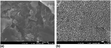
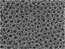
From a macroscopic point of view, typical coloration was observed on the anodized surface. The initial surface is grey-mat colored while the anodic treated surface presented a glossy blue color. Notice that the colors result from the thickness of the oxide layer, as explained by Van Gils et al. [Reference Mor, Carvalho, Varghese, Pishko and Grimes18]. Submicroscopic changes are presented using SEM images in Fig. 2. This figure shows titanium surfaces before and after anodic treatment. The initial surface used was a machined and sand-blasted surface. It presented randomly microscopic roughness. The surface processed by our anodizing protocol showed the regular size and geometry of nanoporosity. Diameters were measured around 60 nm with a minimum of 25 nm and a maximum value of 115 nm.
SEM images (×50,000) of the specimen before and after the anodization process: (a) initial sand-blasted surface presenting randomly microscopic large roughness, (b) TiO2 nanostructure formed under 20 V and presenting a nanotubular morphology.

Effect of anodic conditions
The anodic parameters such as voltage, time of treatment and stirring were studied in order to determine their effect on the nano-surface and in order to optimize the process. Many publications have already dealt with these anodic condition effects, such as electrode material, voltage, time of treatment, agitation, solution etc. and have been used to discuss the results.
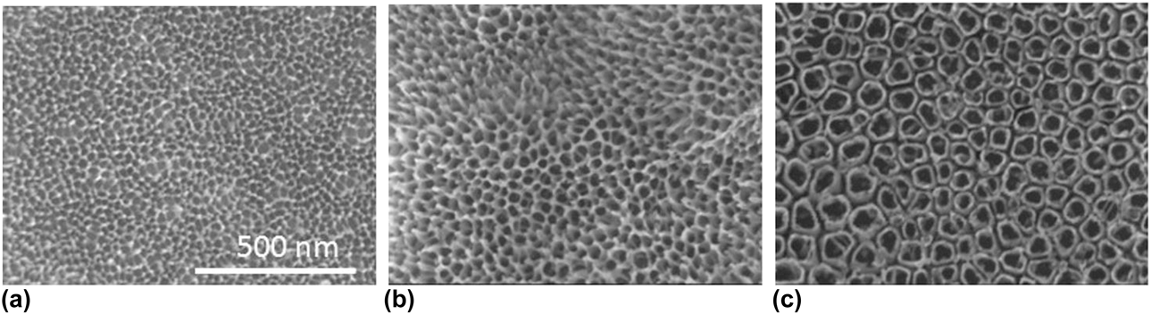
First of all, anodization voltage was studied. It varied from 2 to 25 V. SEM images presented in Fig. 3 clearly show that the voltage applied affects the surface morphology. By applying a voltage of 5 V, nano-pits were created on the surface while nanopore arrays were generated at a voltage of 10 V. Self-organized nanotubes with regular patterns were obtained at a high voltage of 20 V. Slightly above this value (22–25 V), the nanotubes were damaged. At the same time, voltage that was too low led to acid-etching of the surface. This observation is consistent with the results of other publications [Reference Mor, Varghese, Paulose, Shankar and Grimes19, Reference Anitha, Menon, Nair and Prasanth20]. Our main interest and focus for the continuity of this work was a nanostructure array composed of nanopores or nanotubes.
SEM micrographs (top view) of a TiO2 nanostructure with significantly different morphologies and diameters created at potentials 5 V (a), 10 V (b) and 20 V (c) (magnification ×100,000).

Measuring wettability is a basic characteristic when carrying out a surface treatment. In our case, we calculated the surface wettability by sessile drop. The initial surface prior to anodization was hydrophobic with a contact angle of 81°. In the same position, nanotubular structures give a higher contact angle of 105°. This drop behavior resembles that observed for the lotus leaf. Unlike the other two surfaces, the nanopores led to a higher hydrophilicity with a contact angle down to 62°. Thus, these two surfaces, whose manufacturing processes are very similar, have different properties. This is interesting for biological applications. The wettability of the surface has often been studied in cell and tissue integration and also as an antibacterial surface [Reference Rosales-Leal, Rodríguez-Valverde, Mazzaglia, Ramón-Torregrosa, Díaz-Rodríguez, García-Martínez, Vallecillo-Capilla, Ruiz and Cabrerizo-Vílchez21, Reference Abbasi-Firouzjah and Rezaei22].
The final parameter studied was solution stirring. Stirring is generally a fixed parameter in many studies. Lavenus et al. used a stirring speed of 350 rpm. We studied the effect of increasing or decreasing stirring speed, from 0 to 500 rpm. Without stirring, the current drops, while higher currents were obtained with vigorous agitation. Mixing the solution made it possible to increase the ion exchange and thus accelerate the reaction. As seen previously, current density is important for nano-formation, and stirring plays an important role in the electrochemical process.
Effect of annealing on crystal structure
The main disadvantage of titania nanostructuration by means of the anodic process is the formation of an amorphous structure. This structure is thermodynamically unstable. Annealing treatment is used to convert the amorphous phase into a crystalline phase of TiO2: anatase, rutile or brookite. Brookite is a metastable structure like anatase which was less explored in the literature until recently. Note that the composition of the crystalline structure may play a role in surface biocompatibility in comparison to a non-crystalline surface.
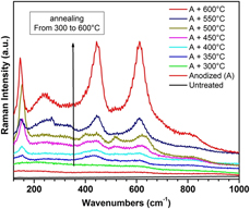
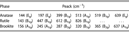
For instance, we present Fig. 4 showing the Raman data measured TA6V samples heated from 300 to 600 °C in air for 0.5–2 h. Titania nanotubes were observed by SEM to determine the effect of the annealing temperature on nanotube morphology and by Raman spectroscopy (Fig. 4 and Table I) to determine the resulting crystal structure.
Raman spectroscopy showing evolution in the titania crystalline structure in relation to the annealing temperature obtained on nanopores formed at 10 V for 20 min.

Raman vibrational modes for TiO2 phases.

First of all, these results show an amorphous structure for the TiO2 before and after anodization without annealing. Heat treatment with varying temperature and time parameters made it possible to obtain different structures. At low temperatures (from 300 to 450 °C), the conversion of amorphous titania nanotubes into a mixture of anatase and rutile phases was observed. As reported in the literature, anatase is known to progressively transform into rutile. At temperatures of more than 550 °C, the TiO2 crystal phase was composed of only the pure rutile form.
Chemical composition of the nanostructure
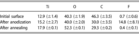
The anodization process makes it possible to increase the natural oxide layer from 2 to 6 nm to further micrometer thicknesses with specific architecture. This process uses electrolytes containing acidic solutions such as hydrofluoric acid. Therefore, for quality control, it is necessary to control the absence of any trace of acid.
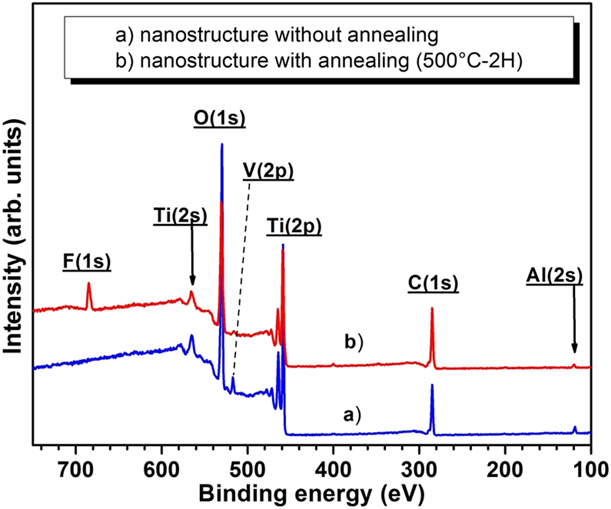
For instance, wide-scan energy spectrum using X-ray photoelectron spectroscopy (XPS) is presented Fig. 5. High-resolution XPS was recorded before and after anodizing. Decomposition of the spectrum of each sample and quantification are reported Table II.
XPS wide-scan energy spectrum of a nanostructured surface with and without annealing.

Chemical surface compositions of specimens.

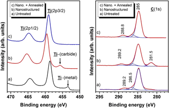
High-resolution C (1s) spectra (Fig. 6) revealed high carbon peaks at 284.8 eV. This carbon is due to absorption of atmospheric contamination. Indeed, titanium, especially TiO2, is known to have the particularity of adsorbing any element or compound around it. The analyses also revealed the presence of carbide (281–283 eV) which disappeared with the anodic process. Acid traces such as acetic acid (CH3COOH) could be characterized by the carboxyl links (–COOH) visible at 289.2 eV. An at.% of 4–5% was observed in the reference sample at 8–12% in the sample without the cleaning and annealing step. Rinsing or annealing reduced the presence of –COOH links on the surface.
High-resolution Ti 2p and C 1s spectra obtained on untreated surface, anodized at 20 V for 20 min, and both anodized and annealed at 500 °C for 2 h.

Concerning the two characteristic peaks of Ti 2p and Ti 2p, they were usually pointed at 465 eV and at 458.2 eV, respectively. According to the literature, the peak at 453–454 eV can be assigned to the Ti metal (Ti0) originating from the substrate. Titanium is naturally covered with a 2–6 nm oxide layer, and XPS analysis shows that it is about 2–10 nm deep.
These results are in agreement with other XPS analyses of synthesized self-aligned titanium dioxide nanostructures found in the literature [Reference Antony, Mathews, Dash, Tyagi and Raj23]. In the case of titanium alloys such as TA6V, aluminum (Al 2s) at 118 eV and vanadium at 512 eV could also be detected in the oxide layer.
Biological characterization of the nanostructured surface
It is known that nano-modified surfaces can interact with the biological environment at the scale of proteins. Honeycomb titania surfaces may induce cell elongation, cytoskeletal stress and selective differentiation into osteoblastic cells by mechanotransduction as already presented. Recent in vitro studies have shown higher osteoblastic differentiation and mineralization. These results correlates with the in vivo studies in rabbits and other animals, in which better anchorage or bone apposition were observed [Reference Sul16, Reference Kang, Park, Choi, Oh, Lee, Choi and Shim24]. However, many of these studies were performed using titanium disks or wire geometry samples and compared to a non-treated surface.
We have biologically characterized the honeycomb titania surface in a rabbit femur model. This study was performed using cylindrical custom-made implants with a growth chamber geometry. The osseointegration of three different types of surface were compared: machined surface (MA), nanostructured (NANO) implants and “homemade” standard alumina grit-blasted and acid-etched (MICRO) implants, also called SLA-like (see above). The specimens were implanted into the femoral condyles of New Zealand white rabbits. After 4 weeks, a pull-out strength test and histomorphometry analysis with the calculation of both bone-to-implant contact and bone growth values were performed.
The results showed that after 4 weeks, the anchorage of the NANO surface was higher than for the other groups while its roughness was three times lower than that of the MICRO surface (0.5 versus 1.5 µm). Histology analysis showed a direct apposition of bone tissue on to the NANO surface. Histomorphometric calculations were in accordance with the bone-to-implant contact and bone growth values. Both displayed higher values for the NANO than for the other implant surfaces. This study thus showed by using “homemade” samples in a rabbit model that the nanostructured surface improved the osseointegration of titanium implants and may be an alternative to conventional surface treatments. Details of these results can be found in Refs. Reference Salou, Hoornaert, Louarn and Layrolle25 and Reference Salou, Hoornaert, Stanovici, Briand, Louarn and Layrolle26.
Conclusion and prospects
This article first centered on briefly presenting the titanium material as well as its applications in the field of medicine. The processes of micrometric surface modifications currently commercialized were then described and made it possible to introduce a new approach to surface treatment by means of nanoscale modification. Nano-modification of surfaces mimics bone architecture and may modulate cell behavior by the phenomenon of mechanotransduction.
The article then gave details on anodizing surface treatments, which despite their simplicity in terms of equipment reveal wide complexity in terms of the influences of parameters on the nanostructure formation and morphology. As titanium is used as a biomaterial, it required multi-scale characterization from both materials science—for the surface characterization techniques used (SEM, RAMAN, XPS, AFM, etc.)—and biology.
Indeed, anodizing surface treatment makes it possible to propose colorful titanium implants with an oxide layer presenting nanopore or nanotube architecture, with a controllable crystalline structure (anatase, rutile, or brookite). Annealing treatments at 500 °C reduce fluoride insertion and eliminate acid traces adsorbed on to the surface but may risk modification of the titania nanostructure’s morphology due to the crystal growth of rutile. In vivo studies performed in rabbits have shown that our TiO2 honeycomb surface may enhance bone healing quality than non-treated surfaces. Thus, nano-surfaces could be an alternative to rough surfaces which are difficult to clean up for bacterial decontamination as in the case of peri-implantitis on dental implants [Reference Mellado-Valero, Buitrago-Vera, Soli-Ruiz and Ferrer-Garcia27, Reference Dennison, Huerzeler, Quinones and Caffesse28]. However, it should be stressed here that the new European regulation regarding medical devices including nanomaterials could be a restriction for the use of this treatment in medical applications.
Methodology
Here we introduce the experimental methods for nanotubular titanium dioxide formation using an electrochemical anodization process. The protocol is described in detail in Ref. Reference Salou, Hoornaert, Stanovici, Briand, Louarn and Layrolle26.
The anodizing protocol was adapted from Lavenus et al. [Reference Salou, Hoornaert, Louarn, Layrolle, Makhlouf and Scharnweber29] who were inspired by the Seughan Oh’s publication in 2009 [Reference Oh, Brammer, Li, Teng, Engler, Chien and Jin10]. This process used a first-generation electrolyte composed of diluted HF based on an aqueous solution. The thickness of the titania nanostructure is relatively limited (<500 nm) and may thus minimize the risk of delamination (separate titania layers from the titanium substrate).
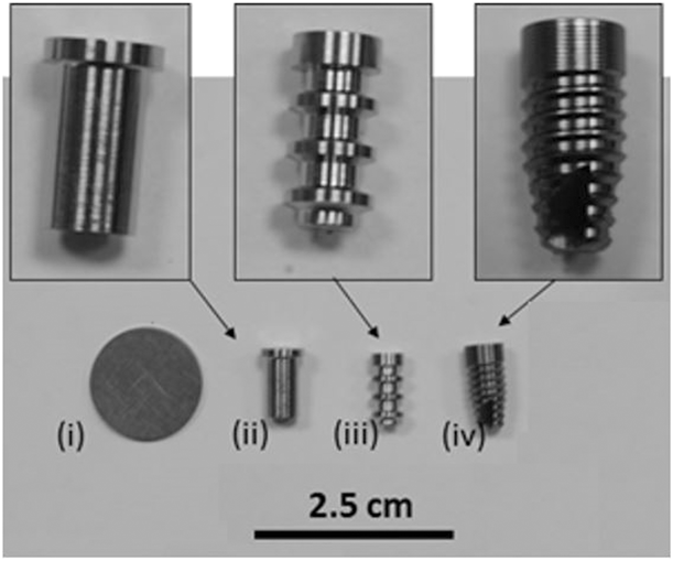
Various geometries were used in our work: coin-shaped, cylindrical-shaped and dental implants. All the samples used are displayed in Fig. 7.
Macroscopic images of samples used in this work: (i) titanium disks (Ti cp and TA6V, machined and blasted); (ii) cylindrical implants with an apical bone growth chamber (Ti cp, machined) used for an in vivo study published in [Reference Salou, Hoornaert, Stanovici, Briand, Louarn and Layrolle26]; (iii) cylindrical implants in the shape of a circular bone chamber (TA6V, machined and blasted) used for surface characterization; (iv) dental implants (Tip, TA6V, machined and blasted) used for process validation and in an in vivo study published in [Reference Salou, Hoornaert, Louarn and Layrolle25].

In addition to the different chemical compositions, these alloys present different structural compositions. Pure titanium is composed only of an alpha phase, while the TA6V microstructure is a mixture of alpha and beta phases. This beta phase is present in the range of 5–20%. The titanium microstructure was revealed by surface etching using Kroll’s reagent.
Concerning the substrate preparation, the original surfaces were either machined, slightly polished or sand-blasted using alumina particles. In order to obtain a homogeneous and adherent deposit, the substrate must be free of impurities (physical debris or fat traces) from manipulations. Thus, titanium pellets were cleaned by successive immersion in acetone, 70% alcohol and distilled water.
The anodization process consists of applying a potential (potentiostatic method) or a current (galvanostatic method) between a standard two- or three-electrode process as detailed in Ref. Reference Salou, Hoornaert, Louarn, Layrolle, Makhlouf and Scharnweber29. The treated metal plays the role of an anode and a platinum inert electrode is a cathode or working electrode.
Notice that the effect of applying current, bath temperature, annealing or other synthesis conditions on nanopore architecture (diameters, length, and wall thickness) have already been published in several papers [Reference Mor, Varghese, Paulose, Shankar and Grimes19, Reference Peng Xiao30, Reference Saji, Cheol Choe and Brantley31]. These parameters play a critical and sensitive role in the synthesis conditions which regulate TiO2 formation. In addition, the biological characterization of the titania nanoporous array is widely performed using in vitro studies or flat and non-complex samples but in vivo studies are required to assess the bone tissue integration of implants.