Introduction
Schistosomiasis is a parasitic disease caused by digenean trematodes living in the vascular system of vertebrates. These worms are members of the family Schistosomatidae, and they are unusual since they are dioecious. Hybridization between closely related species is possible. The genus Schistosoma includes 21 species; one-third are responsible for human schistosomiasis, four of which exist in Africa: Schistosoma mansoni, S. haematobium, S. intercalatum and S. guineensis. Both S. mansoni and S. haematobium have a large geographical distribution in Africa, whereas S. intercalatum is limited to the Democratic Republic of the Congo and S. guineensis to the Lower Guinean Gulf (Cameroon, Equatorial Guinea, Saõ Tomé and Gabon) (Brown et al., Reference Brown, Sarfati, Southgate, Ross and Knowles1984; Pagès et al., Reference Pagès, Jourdane, Southgate, Tchuem Tchuenté, Combes and Jourdane2003).
Two species of human schistosomes exist in Gabon: S. guineensis and S. haematobium (Gilles, Reference Gilles1971). The hypothesis of a hybridization zone between S. guineensis and S. haematobium has been suggested in two provinces of western Gabon, Moyen-Ogooué (Burchard & Kern, Reference Burchard and Kern1985) and Estuaire (Richard-Lenoble, 1993). The aim of this study was to validate, or not, the presence of hybrids between S. guineensis and S. haematobium in Estuaire province by using egg morphometry and a powerful molecular tool in identification of schistosomes (Kane et al., Reference Kane, Bartley, Stothard, Vercruysse, Rollinson and Southgate2002) and their hybrids (Webster et al., Reference Webster, Tchuem Tchuenté, Jourdane and Southgate2005, Reference Webster, Tchuem Tchuenté and Southgate2007), the single-stranded conformational polymorphism (SSCP) analysis.
Materials and methods
Malacological and parasitological surveys
Malacological surveys were focused on the snail intermediate hosts of human schistosomiasis (Bulinus spp.). Two waterbodies of the Estuaire province were visited: Melen, situated in Libreville city (09°30′59.55″ East; 00°23′46.22″ North; 16 m altitude) and Ekouk (10°21′00.20″ East; 00°04′48.64″ South; 65 m altitude). Freshwater snails were collected for 1 h by one person using a stainless-steel strainer (18 cm wide, 8 cm deep, 20 cm handle, and < 1 mm wide holes) attached to a handle. Submerged and emergent vegetation, as well as floating plants, were screened. For each waterbody, collected snails were carefully stored in plastic boxes (70 mm high and 35 mm in diameter), and taken to the laboratory in Libreville, Gabon. The snails were kept in glass vials with 150 ml of well water, and exposed to daylight. The water was examined under a binocular microscope for cercarial emergence 24 h later, and then every 3 days for 40 days. The snails were fed ad libitum with dried lettuce.
The parasitological surveys were carried out in the same sites as the malacological surveys: Melen and Ekouk. Informed consent was obtained from families and the participation was voluntary. Each volunteer was given two pots (50 ml each), one for urine and one for stools. Samples were collected between 08.00 and 10.00 hours and taken to the laboratory for their examination. From each urine sample, an aliquot of 5 ml was centrifuged in a conical plastic tube for 3 min at 1500 rpm. Pellets were examined microscopically for schistosome egg presence. Stool samples were examined by the merthiolate-iodine-formaldehyde concentration (MIF-C) method. Prevalences (number of individuals infected by schistosomes divided by the total number of individuals examined) were calculated.
Egg morphometry
Eggs were extracted from urine samples in Melen and Ekouk and from faeces in Ekouk. The positive urine samples of each site were filtered together through a 45 μm sieve in saline solution (NaCl 0.85%). The positive stool samples were filtered through a series of four sieves 425, 180, 106 and 45 μm pore size in saline solution (NaCl 0.85%). The eggs were collected on the 45 μm sieve and maintained in saline solution (NaCl 0.85%). All eggs were randomly sampled, and measured under the microscope. For each egg, we measured the total length (including the terminal spine) and the maximum width.
Adult worm recovery
Miracidia were obtained only from eggs extracted from urine samples at Melen and at Ekouk. Bulinus globosus was used as the intermediate host since it is more compatible with S. haematobium compared to B. forskalii and since it is compatible with the hybrids of S. haematobium and S. guineensis (Southgate et al., Reference Southgate, Van Wijk and Wright1976; Mutani et al., Reference Mutani, Christensen and Frandsen1985).
For Melen, positive urine samples of nine patients were filtered together through a 45 μm sieve in saline solution (NaCl 0.85%). The eggs were collected on the 45 μm sieve and maintained in well water for miracidial hatching. Twelve positive B. globosus (exposed to five miracidia) were used to infect Meriones unguiculatus as definitive host.
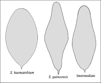
For Ekouk, positive urine samples of 14 patients (four of them also had eggs in the faeces) were filtered together as explained above. Among the collected eggs, we preferentially selected the S. guineensis, and intermediate morphotypes (see fig. 1) in order to get potential hybrid specimens. Nine positive B. globosus (exposed to five miracidia) were used to infect M. unguiculatus as definitive host.
Schistosoma haematobium, S. guineensis and intermediate morphotypes of schistosome eggs from human urine and faeces in Estuaire province, Gabon.

The rodents were anaesthetized with a solution of 2.5 ml of Rompun (20 mg/ml), 5 ml of Imalgène (100 mg/ml) and 42.5 ml NaCl by intraperitoneal injection of 0.1 ml/10 g of body weight. The abdomens were shaved and exposed to 300 cercariae per rodent. Five months post-exposure, the rodents were killed by lethal injection (0.5 ml) of Doléthal (Vétoquinol, France) and instantly perfused to collect adult worms. The perfusion was followed by supplementary examination to collect the worms trapped in the liver, in the superior and inferior mesenteric system, including rectum, and in the lungs. Worms were preserved in 95% alcohol for DNA analysis.
DNA extraction, ITS2 amplification
DNA was extracted from 80 adult worms from Gabon: 30 males from Melen (18, 2 and 10 from the mesentery–liver, rectum and lungs, respectively) and 50 adult worms from Ekouk (11, 2 and 13 males from the mesentery-liver, rectum and lungs, respectively, and 11, 2 and 11 females from the mesentery–liver, rectum and lungs, respectively). One male adult worm of each of S. haematobium (Benin and Niger), S. intercalatum (Democratic Republic of the Congo) and S. guineensis (Cameroon) were also used for comparison. A manufacture's protocol from the QIAamp DNA Mini Kit (Qiagen) was used.
Internal transcribed spacer-2 (ITS2) amplification was performed by polymerase chain reaction (PCR) using primers ITS2 single-stranded conformational polymorphism (SSCP) forward (5′-GCATATCAACGCGGG6-3′) and ITS2 SSCP reverse (5′-ACAAACCGTAGACCGAACC-3′), according to Kane et al. (Reference Kane, Bartley, Stothard, Vercruysse, Rollinson and Southgate2002). The cycling conditions included an initial denaturing step at 94°C for 5 min and then 40 cycles at 94°C for 30 s (denaturing), 66°C for 30 s (annealing) and 72°C for 1 min (elongation). There was a final elongation period at 72°C for 10 min. Each PCR product was checked by agarose gel electrophoresis of 5 μl of each product (Webster et al., Reference Webster, Tchuem Tchuenté and Southgate2007). PCR products were purified using a modified manufacture's protocol from Wizard SV Gel and PCR Clean-Up System kit (Promega, Madison, Wisconsin, USA).
Single-stranded conformation polymorphism ITS2 analysis
SSCP ITS2 analysis was carried out on the 80 adult worms on Bio-Rad PROTEAN® II xi Cell (Bio-Rad, Hercules, California, USA), adapting a protocol used elsewhere by Kane et al. (Reference Kane, Bartley, Stothard, Vercruysse, Rollinson and Southgate2002) with specific modifications and run conditions. Acrylamide 12% gel consisting of a mixture of: 21 ml of acrylamide/bis-acrylamide (37.5:1), 42 ml of 1 × Tris-borate-EDTA (TBE) buffer; 470 μl of 10% ammonium persulphate (APS), 117 μl of tetramethylethylenediamine (TEMED) and 7 ml of miliQ H2O was used. The resultant solution was poured into the assembled gel plates and stored at 4°C for 1 h, as well as 2 litres of 1 × TBE electrophoresis buffer. Sample preparation was as follows. Denaturing of PCR products was carried out by adding 10 μl of each PCR product to 22.5 μl of formamide and 2.5 μl NaOH (100 mm). This mixture was heated for 15 min in a thermocycler at 95°C and placed immediately on ice. The whole mixture of each sample added to 5 μl of charge buffer was loaded into the gel. Run conditions were as follows: system cooled 2 h before migration at 6°C, voltage 110 V and run time 17 h.
Statistical analyses
Yates's chi-square test was used to compare the percentages of eggs found in urine or faeces according to their morphotype. The Mann–Whitney, Kruskal–Wallis (KW) and Dunn's tests were used to compare egg sizes. All data were analysed using Microsoft Excel 2003 and GraphPad InStat, Version 3.05 (GraphPad Software, La Jolla, California, USA). Statistical significance was assessed at the P < 0.05 level.
Results
Malacological and parasitological surveys
Two species of Bulinus were found simultaneously in the two waterbodies, B. globosus and B. forskalii: 77 B. globosus and 15 B. forskalii in Melen and 21 B. globosus and 30 B. forskalii in Ekouk. All the collected snails were negative for schistosome infection.
Three hundred and thirteen persons were examined. The analyses of the urine samples gave the following prevalences: 16.8% (38/226) in Melen and 26.4% (23/87) in Ekouk. No patient showed eggs exclusively in the faeces. However, few patients harboured eggs both in urine and faeces: 3.1% (7/226) in Melen and 8.1% (7/87) in Ekouk.
Egg morphometry
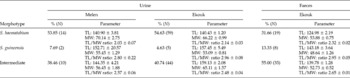
Whatever the studied site (Melen or Ekouk) and whatever the egg localization (urine or faeces), eggs exhibited three morphological types (fig. 1): (1) S. haematobium morphotype, predominantly oval and supplied with a well-visible terminal spine; (2) S. guineensis morphotype, predominantly spindle-shaped with two well-identified extremities, one of them supplied with a visible terminal spine and distinct shoulders; and (3) intermediate morphotype. The percentages of the different egg morphotypes and their mean egg sizes are presented in table 1 according to the studied sites and to the localization of the eggs (urine or faeces). The percentages of the eggs were significantly different between the three morphotypes in Melen (urine) and in Ekouk (urine and faeces) (Yates's chi-square test; χ2Yates = 8.70; χ2Yates = 43.19; χ2Yates = 15.74, respectively; P < 0.05): the percentages of eggs of S. guineensis morphotype were significantly smaller than the percentages of eggs of the two other morphotypes in Melen (urine) and in Ekouk (urine and faeces) (Yates's chi-square test; P < 0.05). In the faeces, the percentage of eggs of S. haematobium morphotype decreased in favour of those of the two other morphotypes.
Percentages of eggs, and mean egg sizes (±standard error) of the S. haematobium, S. guineensis, and intermediate morphotypes extracted from human urine and faeces in Estuaire province, Gabon.

N, number of eggs; TL, total length; MW, maximum width.
The statistical analyses on the mean egg sizes were made on the Ekouk data and showed that the total lengths of the eggs found in urine were significantly different between the morphotypes (KW = 48.77; P < 0.0001): S. haematobium morphotype eggs were significantly smaller than S. guineensis and intermediate morphotypes (Dunn's test, P < 0.001 and P < 0.05, respectively) but S. guineensis and intermediate morphotype eggs were not different (Dunn's test, P>0.05). Similarly, the total lengths of the eggs found in faeces were significantly different between the morphotypes (KW = 25.60; P < 0.0001): S. haematobium morphotype eggs were significantly smaller than the two other morphotypes (Dunn's test, P < 0.001) but S. guineensis and intermediate morphotype eggs were not different (Dunn's test, P>0.05). The eggs found in faeces were significantly smaller than those found in urine (U = 121, P < 0.0001; U = 7, P = 0.05; U = 144.5, P < 0.0001 for S. haematobium, S. guineensis and intermediate morphotypes, respectively). The eggs found in faeces had significantly higher ratios than those found in urine (U = 291.5, P < 0.0009; U = 388, P = 0.0005 for S. haematobium and intermediate morphotypes, respectively) but these ratios were not significantly different for S. guineensis morphotype (Dunn's test, P>0.05).
Single-strand conformation polymorphism
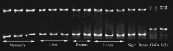
SSCP ITS2 analyses on some adult male worms from Gabon are presented in fig. 2 in comparison with S. haematobium from Benin and Niger, S. intercalatum from the Democratic Republic of the Congo (Zaïre) and S. guineensis from Cameroon. All the 80 individual worms (24 females and 56 males) gave the same pattern as that shown in fig. 2 (i.e. S. haematobium) whatever their localization in the circulatory system (mesentery, liver, lung or rectum).
Electronic image of the SSCP ITS2 profiles of 11 adult male worms from a Gabonese urine schistosome population (Melen, Estuaire province) according to their localization in Meriones unguiculatus. A comparison was performed with S. haematobium from Niger and Benin, S. guineensis from Cameroon (GuCa), and S. intercalatum from the Democratic Republic of the Congo (InZa).

Discussion
Egg morphology showed three morphotypes of eggs whatever their localization (urine or faeces): S. haematobium, S. guineensis and intermediate morphotypes. We could conclude that S. haematobium and S. guineensis coexist with their hybrids in our samples. Concerning the S. haematobium egg morphotype collected in urine, egg sizes were similar to those of S. haematobium (Loker, Reference Loker1983). The egg sizes of the S. guineensis morphotype collected in the faeces were much lower than those of S. guineensis for eggs also collected in the faeces (143.18 ± 3.64 instead of 172 ± 17 (Wright et al., Reference Wright, Southgate and Knowles1972)). Therefore, the egg size excludes the presence of S. guineensis in our samples but still maintains the possible presence of S. haematobium/S. guineensis hybrids, since the egg sizes of the intermediate morphotypes collected in urine (see egg lengths and length/width ratios in table 1) are similar to those of the hybrid eggs suspected by Richard-Lenoble et al. (Reference Richard-Lenoble, Kombila, Duong and Gendrel1993).
The SSCP results did not show any characteristic four-banded SSCP profiles which indicate hybridization between S. haematobium and S. guineensis. All the specimens (from Melen and Ekouk) were identical to S. haematobium from Benin and from Niger. No recombinants between S. haematobium and S. guineensis were present in Estuaire province in our samples, despite the fact that, for the last isolate, we preferentially selected eggs of S. guineensis or intermediate morphotypes. According to these results, we consider all these characteristics sufficient to conclude that the populations of schistosomes collected from urine in the studied areas belong to the S. haematobium species. These results are in line with those obtained in Cameroon by Webster et al. (Reference Webster, Tchuem Tchuenté, Jourdane and Southgate2005), who showed that 10 years were sufficient enough to S. haematobium to replace S. guineensis in areas of Cameroon through introgressive hybridization. The hypothesis according to which hybrids existed in Gabon was certainly true in the late 1980s and early 1990s but was not validated by the results presented in this paper, almost 20 years later, when the epidemiological study detected only S. haematobium.
Acknowledgements
This work received financial support from CNRS, University of Perpignan and Agence Universitaire de la Francophonie (grant no. 2092 RR 614). We thank Minguéba V for his technical assistance. We are grateful to J.R. Pagès and A. Rognon for providing us with some specimens. We are grateful to Callixte Nkoure Mba and Lambert Ndong Obiang from l'Institut d'Hygiène Publique of Libreville for their assistance during the sampling period.