In cattle, genetic susceptibility to mastitis has been documented in various studies around the world (see review by Detilleux, Reference Detilleux2002). However, as the world cattle population is dominated by one breed (Holstein-Friesian), researchers have been interested mostly in differences between individual animals. It has long been known that some individual cows within a herd can survive infections that cause serious clinical mastitis in other animals (Detilleux, Reference Detilleux2002).
In contrast to cattle breeding, there are many different breeds of sheep used around the world, including dairy ewes exploited in various parts of the world for their milk production. Animal trade and changes in breeding goals have resulted in improvements of local breeds; nevertheless, indigenous breeds of sheep still make up a significant proportion of sheep population around the world (Zygoyiannis, Reference Zygoyiannis2006).
In this paper we present the results of a study into the susceptibility of two breeds of dairy sheep to mastitis. We used an indigenous Greek sheep breed, Karagouniko, which has been used in previous studies of experimentally induced mastitis (Saratsis et al. Reference Saratsis, Alexopoulos, Tzora and Fthenakis1999; Mavrogianni et al. Reference Mavrogianni, Fthenakis, Brooks, Papaioannou, Cripps, Taitzoglou, Brellou and Saratsis2005, Reference Mavrogianni, Cripps, Papaioannou, Taitzoglou and Fthenakis2006b). Animals of this breed are medium milk-yielding (average milk production: 145 l over a 173-d lactation period), but are considered to be resistant to various diseases, e.g. annual incidence of respiratory infections is <2% (Christodoulopoulos & Fthenakis, Reference Christodoulopoulos and Fthenakis2005). We also included a sheep breed, Frisarta, which was established around 25 years ago, as the result of crossing between imported Friesian sheep and the local Arta-area sheep (Zygogiannis, Reference Zygoyiannis1999). These are high-yielding animals (average milk production: 245 l over a 205-d lactation period) but, according to our clinical experience, appear to be particularly susceptible to respiratory infections associated with Mannheimia haemolytica (Skoufos et al. Reference Skoufos, Mavrogianni, Tzora, Mavrommatis, Alexopoulos and Fthenakis2006). This organism is also a mammary pathogen (Bergonnier & Berthelot, Reference Bergonier and Berthelot2003). Therefore, a hypothesis was constructed that there may be differences in its pathogenicity for the mammary gland of the two breeds. This would have significant consequences, because Greek farmers increasingly use the Frisarta sheep for genetic improvement, with a view to increasing milk yield of replacement stock.
Materials and Methods
Experimental procedures: animals, inoculations, samplings and laboratory tests
In total, 16 lactating primiparous ewes were used; they were Karagouniko breed (group K, n=8) or Frisarta breed (group F, n=8). Animals of each breed were selected from two different sheep farms. Both farms were commercial enterprises with their own breeding programmes.
Immediately after lambing and every 2 d thereafter, including the day of inoculation (22 d after lambing, D0), a thorough clinical examination was carried out on the ewes. Special attention was paid to their mammary glands and teats, which were examined as described previously (Fthenakis, Reference Fthenakis1994; Mavrogianni et al. Reference Mavrogianni, Fthenakis, Brooks, Papaioannou, Cripps, Taitzoglou, Brellou and Saratsis2005). For sampling, a sterile plastic fine catheter 2 mm long (Abbocath®, Abbot), was inserted into the teat and moved from left to right, in order to obtain duct material samples (Mavrogianni et al. Reference Mavrogianni, Cripps and Fthenakis2006a). Then, mammary secretion samples were obtained. The first two squirts of secretion were discarded and then 10–15 ml was carefully collected into a sterile container.
Lambs of these ewes were weaned 18 d after lambing and subsequently the animals were hand-milked thrice daily. All animals were challenged with a Man. haemolytica strain (VSM08L) isolated in Greece and of known pathogenicity for the mammary gland.
For inoculation, the strain was grown on Columbia blood agar and checked for purity; then it was inoculated into Soy-broth and incubated aerobically at 37°C for 5 h. Serial dilutions of the broth culture into phosphate-buffered saline (PBS) were carried out; finally, 0·2 ml of the desired dilution was withdrawn with a syringe. Average inoculum for K ewes was 1250 c.f.u. (range: 1220–1300 c.f.u.) and for F ewes 1230 c.f.u. (range: 1200–1280 c.f.u.), as estimated by the method of Miles & Misra (Reference Miles and Misra1938). To ensure sterile conditions at inoculation, the hairs of the teats were clipped the day before, using fine scissors, and the skin of the udder and teats was scrubbed with chlorhexidine solution; then on the day of inoculation iodine povidone solution was used to clean the teat before challenge. A sterile plastic fine catheter 20 G, 2 mm long, was inserted into the teat; the syringe was attached to the catheter and the bacterial suspension was deposited inside the teat. The same technique was used to inject 0·2 ml of PBS into the respective site of the other teat of each ewe, used as control.
Subsequent to challenge, detailed examination of the mammary glands and teats was carried out 12 h later and then daily. Duct material and mammary secretion samples were collected at the same time.
All samples were cultured on Columbia blood agar; the media were incubated aerobically at 37°C for up to 72 h. The California Mastitis Test (CMT) was carried out on all secretion samples with five degrees of reaction (Fthenakis, Reference Fthenakis1995). Secretion films made by directly smearing 20 μl from each sample on a microscope objective plate, were stained by the Giemsa method; the percentage of leucocyte subpopulations was determined by counting at least 200 cells therein and distinguishing their type.
Four ewes of each group were euthanatized on D1 and the other four on D3 (bar one F ewe, which died on D2). Dissection of the mammary glands and the teats started immediately and was carried out by using the aseptic technique. The skin of the teats and the subcutaneous tissues were incised with a sterile blade; initially the mucosa of the teat cistern (sinus papillaris) was exposed and subsequently the teat duct (ductus papillaris) was incised and its mucosa was exposed. A new blade was used for scraping the mucosa of the teat cistern, whilst another one was used to scrape the mucosa of the teat duct. An electronic cutimeter was used to measure 2 mm from the teat orifice, in order to determine the precise site of the teat where the inoculum had been deposited. Longitudinal sections, involving all the structures of the teat, were taken out for histopathological examination. Then the mammary glands were dissected and samples were obtained for bacteriological and histological examination, as described before (El-Masannat et al. Reference El-Masannat, Jones and Scott1991). All samples were plated onto Columbia blood agar; the media were incubated aerobically at 37°C for up to 72 h.
All isolated bacteria were identified by using standard microbiological techniques (Barrow & Feltham, Reference Barrow and Feltham1993; Euzeby, Reference Euzeby1997). Tissue samples were fixed in 10% neutral-buffered formalin and embedded in paraffin wax, using conventional techniques. Haematoxylin and eosin (HE) standard staining procedures were performed for histopathological studies.
Data management and analysis
Clinical mastitis was defined as presence of abnormal gross findings systemically or in the mammary gland, including changes in secretion (Schalm et al. Reference Schalm, Carroll and Jain1971; Fthenakis & Jones, Reference Fthenakis and Jones1990a, Reference Fthenakis and Jonesb; Fthenakis, Reference Fthenakis1994; Bergonier & Berthelot, Reference Bergonier and Berthelot2003; Bergonier et al. Reference Bergonier, De Cremoux, Rupp, Lagriffoul and Berthelot2003).
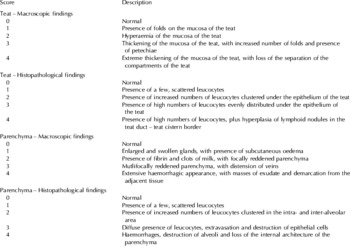
We used a scoring system that had been previously developed and described (Mavrogianni et al. Reference Mavrogianni, Fthenakis, Brooks, Papaioannou, Cripps, Taitzoglou, Brellou and Saratsis2005; Fragkou et al. Reference Fragkou, Mavrogianni, Cripps, Gougoulis and Fthenakis2007a); thus, numerical values were assigned for the pathological findings in the experimental animals. A separate score (0–4 scale) was given for macroscopic and for histological findings in the teat and in the mammary gland; these were then added to a 0–16 scale to produce a pathology score for the findings in each ewe euthanatized on a specific date. Maximum total pathology score summed up over all ewes of a group was 8×16=128. The system is detailed in Table 1.
Description of the scores given for pathological findings in teats and mammary parenchyma of experimental ewes

The difference between the challenged and unchallenged teats of the same animal was examined using McNemar's test. When all tissue samples from the same animal were bacteriologically positive, they were defined as ‘positive’ (as opposed to ‘not all positive’). Using this definition the Fisher Exact test was also used to compare the proportion of each breed that developed clinical mastitis after challenge and the proportion of secretion or tissue samples that were bacteriologically positive. Pathology scores obtained at either 1 d or 3 d after challenge were compared by using the Kruskal-Wallis test corrected for ties; a separate analysis was carried out for teat scores and parenchyma scores.
Statistical analysis was performed in Minitab 14 (Minitab Inc., State College, PA, USA) and Stata 9 (Stata Corp, College Station, TX, USA). Statistical significance was defined at P<0·05.
Results
Clinical, bacteriological and cytological findings
The mammary glands and the teats of all ewes were clinically healthy throughout the period from lambing to inoculation. The teats were soft with no external abnormalities. No bacteria were isolated from any duct material or secretion sample obtained. The CMT was always negative; in Giemsa-stained secretion films, no leucocytes were observed.
After challenge, no ewe in group K developed clinical mastitis. Man. haemolytica was isolated in pure culture from catheter and secretion samples; total isolation rate was 34/48 samples. The CMT increased (>‘1’); positive results were obtained in 17/24 samples (Table 2). Leucocytes were seen in Giemsa-stained secretion films: their majority (>80%) were neutrophils, with fewer macrophages and lymphocytes also present.
Recovery of Mannheimia haemolytica and results of CMT in samples of duct material (D) or mammary secretion (S) taken from ewes inoculated with the organism

Group F: Frisarta-breed ewes, group K: Karagouniko-breed ewes
† n/m=positive results out of total samples obtained
* differences between groups statistically significant (P<0.05)
After challenge, all ewes in group F became ill and developed acute clinical mastitis; rectal temperature up to 41·5°C and indifference were apparent; in 3/8 animals, mild diarrhoea was also observed. One ewe died 2 d after challenge. Mammary secretion was abnormal (serous or purulent, with flakes or clots); abnormal mammary signs (enlarged, hot, hard and painful mammary gland) were seen. Man. haemolytica was consistently isolated in pure culture from duct material and secretion samples; in total, bacteria were isolated from 46/46 samples. The CMT increased (>‘1’); positive results were obtained in 23/23 samples (Table 2). Leucocytes were seen in Giemsa-stained secretion films: their majority (>85%) were neutrophils, with fewer macrophages and lymphocytes also present.
No clinical changes were recorded in the contralateral teats and mammary glands of any ewe post-inoculation. No bacteria were isolated from any duct material or secretion sample collected from that side. CMT was negative for all samples obtained.
Within ewes of both breeds, bacterial deposition into the teat was significantly more likely to result in mastitis than the own within-ewe control teat (P<0·01). There was also a significant difference in the development of clinical mastitis among ewes of the two breeds; group K 0/8, group F 8/8 (P < 0·001).
Group K ewes had a significantly smaller proportion of positive bacteriological results than group F animals 12 h after challenge (P<0·001) and on D1 (P=0·026). However, there was no significant difference between the two groups on D2 (P=0·214) or D3 (P=0·571).
Pathological findings
Measurement of the length of the internal teat structures, after dissection of the teats of ewes, showed that the inoculum had always been deposited within the teat duct.
Group K: challenged side:
Man. haemolytica was isolated in pure culture from tissue samples; isolation rate was 12/24. The total pathology score summed over all ewes of the group was 41 (Table 3).
Recovery of Mannheimia haemolytica and total pathology scores in tissue samples from ewes inoculated with the organism

Group F: Frisarta-breed ewes, group K: Karagouniko-breed ewes
† n/m=positive results out of total sites sampled
‡ includes 3/3 samples from the ewe that died 2 d after challenge
§ figures include pathology score 7 (teat) and 8 (parenchyma) of samples from the ewe that died 2 d after challenge
* differences between groups statistically significant (P<0·05)
The teat duct appeared normal with an apparently smooth internal lining; the teat duct and the teat cistern were clearly distinguished as two different anatomical structures. Histologically, there was leucocytic infiltration (neutrophils, lymphocytes, plasma cells) prominent at the border between teat duct–teat cistern and at the teat cistern. Hyperplastic lymphoid nodules consisting of lymphocytes and plasma cells – with germinal activity –were observed at the border between teat duct–teat cistern.
No gross pathological findings were evident in the parenchyma. Mild neutrophilic infiltration was observed histologically in the mammary parenchyma.
Group F: challenged side:
Man. haemolytica was isolated in pure culture from tissue samples; isolation rate was 24/24. The total pathology score summed over all ewes of the group was 93 (Table 3).
Macroscopic findings in the inoculated teat were folds, hyperaemia and thickness of the mucosa in the duct; petechiae were seen; the teat duct and the teat cistern were clearly distinguished as two different anatomical structures. However, in the ewe that died, teat architecture was lost. Histologically, we recorded principally subepithelial neutrophilic infiltration, presence of some lymphocytes and some plasma cells, with exocytosis through the epithelium and into the lumen and lysis of neutrophils; no lymphoid nodules were observed.
Macroscopic lesions in the respective mammary parenchyma included marked enlargement and congestion of the gland, subcutaneous oedema and sanguineous fluid exuding from sections of the reddened parenchyma; the supramammary lymph nodes were enlarged. In the ewe that died, the mammary gland appeared extensively congested and enlarged, with significant subcutaneous oedema and discolouration of the overlaying skin. Histologically, conspicuous neutrophilic infiltration, extravasation, intra-alveolar live and exhausted neutrophils, destruction of epithelial cells, alveolar destruction and haemorrhages were evident.
Contralateral side:
Man. haemolytica was isolated from the mammary parenchyma of the contralateral side of the ewe that died (group F). Mild neutrophilic infiltration was evident histologically. No macroscopic or histological lesions were seen from the contralateral side of any other ewe.
Comparisons:
On D1, group K ewes had a significantly smaller proportion of positive bacteriological results from tissue samples (P=0·029). However, there was no significant difference between the two groups on D3 (P=0·071).
Furthermore, on D1 teat pathology scores were higher for group K ewes (P=0·04). However, no significant difference was evident on D3 (P=0·278). In contrast to that, parenchyma pathology scores were higher for group F than for group K ewes on both occasions (P<0·02).
Discussion
We investigated the susceptibility of two breeds of dairy sheep to deposition of Man. haemolytica into the teat. Both breeds of sheep are exploited in Greece for their milk. Karagouniko sheep are indigenous and although they have been subjected to genetic selection for their milk yield, their average milk production is still relatively average. They are considered to be well adapted to local conditions and are reputedly resistant to disease (Zygoyiannis, Reference Zygoyiannis1999; Christodoulopoulos & Fthenakis, Reference Christodoulopoulos and Fthenakis2005). Frisarta sheep are a cross between Friesian sheep and the local dairy breed in the Arta area (north-west Greece). They are increasingly used for their dairy abilities in Greece, but anecdotal evidence suggests that they are particularly susceptible to pneumonia associated with Man. haemolytica (Skoufos et al. Reference Skoufos, Mavrogianni, Tzora, Mavrommatis, Alexopoulos and Fthenakis2006).
The above was our stimulus to initiate the current project, given that the organism is also a confirmed mammary pathogen (Bergonier & Berthelot, Reference Bergonier and Berthelot2003). Possible differences between the two breeds have been evaluated under experimental conditions, by using the ‘teat-challenge’ model (Mavrogianni et al. Reference Mavrogianni, Fthenakis, Brooks, Papaioannou, Cripps, Taitzoglou, Brellou and Saratsis2005, Reference Mavrogianni, Cripps, Papaioannou, Taitzoglou and Fthenakis2006b). By means of this method, we tested for the pathogenic effects of the organism on the mammary gland of animals used in the present study. The results provide clear evidence that ewes of the Frisarta-breed (group F) were susceptible to clinical mastitis after Man. haemolytica deposition into the teat. All Frisarta animals developed clinical mastitis and had significantly severe pathological findings in the mammary parenchyma; this is in contrast to Karagouniko ewes which developed only subclinical mastitis, with less severe mammary lesions.
No lymphoid nodules were observed in the teats of Frisarta ewes. Since these structures have been identified as defensive mechanisms (Mavrogianni et al. Reference Mavrogianni, Fthenakis, Brooks, Papaioannou, Cripps, Taitzoglou, Brellou and Saratsis2005) located at the border between teat duct–teat cistern (Mavrogianni et al. Reference Mavrogianni, Cripps, Brooks, Taitzoglou and Fthenakis2007; Fragkou et al. Reference Fragkou, Mavrogianni, Papaioannou, Boscos, Cripps, Skoufos and Fthenakis2007b) it is reasonable to postulate that their lack contributed to the ascent of the organisms into the mammary parenchyma and development of acute clinical mastitis. In fact, the bacteriological findings (Table 2) indicate that although bacteria colonized the teat duct, they invaded the mammary parenchyma and caused mastitis only in Frisarta ewes. These findings are in line with our clinical observation regarding susceptibility of animals of the breed to respiratory infections, given that similar structures play a defence role in the lungs (Moyron-Quiroz et al. Reference Moyron-Quiroz, Rangel-Moreno, Kusser, Hartson, Sprague, Goodrich, Woodland, Lund and Randall2004).
In previous studies (Mavrogianni et al. Reference Mavrogianni, Fthenakis, Brooks, Papaioannou, Cripps, Taitzoglou, Brellou and Saratsis2005; Fragkou et al. Reference Fragkou, Mavrogianni, Cripps, Gougoulis and Fthenakis2007a) where Karagouniko-breed ewes have been used as model animals, we have consistently obtained similar results, i.e. deposition of Man. haemolytica into the teat duct of ewes did not result in clinical mastitis, although an inflammatory response was elicited. Direct inoculation of the same organism into the gland cistern resulted in acute clinical mastitis (El-Masannat et al. Reference El-Masannat, Jones and Scott1991; Mavrogianni et al. Reference Mavrogianni, Fthenakis, Brooks, Papaioannou, Cripps, Taitzoglou, Brellou and Saratsis2005). Moreover, in ewes with viral teat lesions where no lymphoid nodules had been observed, deposition of the strain into the teat duct resulted in acute clinical mastitis (Mavrogianni et al. Reference Mavrogianni, Cripps, Papaioannou, Taitzoglou and Fthenakis2006b).
Mucosa-associated lymphoid tissues are secondary lymphoid organs and represent the first encounter of antigens breaching mucosal surfaces with the immune system (Lydyard & Rossi, Reference Lydyard, Rossi, Roitt, Brostoff and Male2001). There is interest in inducible lymphoid structures developing de novo and in association with mucosae; formation of these lymphoid aggregates appears to be induced, and their mass expanded, in response to lumenal or mucosal surface stimuli, including bacterial invaders. Formation of induced bronchus associated lymphoid tissue is directly triggered by inflammatory responses to bacterial invasion (Moyron-Quiroz et al. Reference Moyron-Quiroz, Rangel-Moreno, Kusser, Hartson, Sprague, Goodrich, Woodland, Lund and Randall2004); similarly, the lymphoid nodules observed in the teat duct–teat cistern border are associated with bacterial presence therein. The bronchal epithelium is the interface between the lungs and the outside world; the teat duct plays a similar function for the mammary gland.
In cows, there is an abundance of information regarding genetic resistance to mastitis. Genetic differences in various defence determinants of susceptible/resistant animals have been reported, e.g. number of blood polymorphonuclear cells after calving (Detilleux et al. Reference Detilleux, Koehler, Freeman, Kehrli and Kelley1994), lactoferrin concentration (Schwerin et al. Reference Schwerin, Solinas-Toldo, Eggen, Bruner, Seyfert and Fries1994), production of immunoglobulins (Kelm et al. Reference Kelm, Detilleux, Freeman, Kehrli, Dietz, Fox, Butler, Kasckovics and Kelley1997), production of complement fragment C5a (Shuster et al. Reference Shuster, Kehrli, Rainard and Paape1997), production and mobilization of cytokines (McShane et al. Reference McShane, Gallagher, Newkirk, Taylor, Burzlaff, Davis and Skow2001). There is also information regarding genetic control of lymphocyte mobilization and role, e.g. heritability of T cell proliferation ranges between h 2=0–0·40 (Detilleux et al. Reference Detilleux, Koehler, Freeman, Kehrli and Kelley1994), genetic mechanisms have been identified for production of T cell and B cell receptor phenotypes (Saini et al. Reference Saini, Allore, Jacobs and Kaushik1999). Furthermore, special emphasis has been placed on the genetics of the Major Histocompatability Complex (MHC) of cattle, known as the bovine leucocyte antigen (BoLA), which has been linked to resistance to mastitis (Takeshima & Aida, Reference Takeshima and Aida2006). For example, prevalence of mastitis was higher in cows carrying the BoLA class II haplotype ‘b’ (Arriens et al. Reference Arriens, Ruff, Schallibaum and Lazary1994), whilst presence of the bovine lymphocyte antigens class I allele CA42 instead of EU28 increased susceptibility to Staphylococcus aureus infection (Schukken et al. Reference Schukken, Mallard, Dekkers, Leslie and Stear1994).
In sheep, significantly less work has been carried out in this area. Differences among individual ewes in MHC gene polymorphism have recently been reported (Swiderek et al. Reference Swiderek, Charon, Winnicka and Gruszczynska2006), but other results are conflicting. For example, Baro et al. (Reference Baro, Carriedo and SanPrimitivo1994) indicate small heritability to high somatic cell counts, whilst Barillet et al. (Reference Barillet, Rupp, Mignon-Grasteau, Astruc and Jacquin2001) suggest that selection for mastitis resistance could be based on somatic cell counting. Others (Watson et al. Reference Watson, Franklin, Davies, Kettlewell and Frost1990; Gonzalez-Rodriguez et al. Reference Gonzalez-Rodriguez, Gonzalo, San Primitivo and Carmenes1995; Burriel, Reference Burriel1997) also report differences in breed susceptibility to mastitis; Burriel (Reference Burriel1997) found that Mule ewes were more susceptible to intramammary infections and mastitis than Welsh Mountain ewes.
Conclusion
Our findings suggest that there are differences in the susceptibility of dairy sheep to the development of mastitis. In this case, defence mechanisms of the teat appeared to be inadequate against the invading organisms. The results further underline the importance of lymphoid nodules in the teat, as a defence structure, and provide an indication for the reduced susceptibility of Karagouniko animals. Whether these are true breed differences rather than line ones should be a subject of a further systematic investigation. Nevertheless, the results indicate the possibility for a genetic basis of susceptibility to mastitis in dairy sheep. This can be exploited in the development of future breeding strategies and resistance to mastitis may be incorporated into future goals for dairy sheep. In sheep husbandry systems where milk production is the primary target, lack of mammary diseases may be considered a trait of equal importance to high milk yield and be given appropriate weighting in their genetic improvement.
The project is co-funded by the European Social Fund & National Resources – EPEAEK II-ARCHIMIDIS II.