This article summarises some personal observations and suggestions regarding training in congenital cardiac surgery. Although the American Board of Thoracic Surgery has introduced an Accreditation Council for Graduate Medical Education (ACGME)-accredited pathway to board certification specifically in congenital cardiac surgery, the majority of congenital cardiac surgery programmes in the United States of America have chosen not to participate in this programme.Reference Calhoon, Shemin, Allen and Baumgartner 1 This allows non-participating programmes to design their own training structure for non-ACGME accredited fellows who in many cases are foreign medical graduates who have come to the United States of America for training in congenital cardiac surgery with a plan to return to their home country. Ideally, a global organisation should be developed to oversee and accredit training programmes to recognise the validity of both United States of America and international training programmes in producing well-qualified congenital cardiac surgeons.
Areas of competency
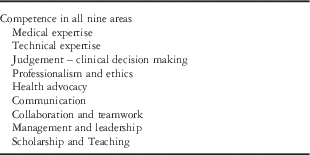
Surgical training is fundamentally an apprenticeship with a critically important relationship between the mentor and the trainee. By far, the most important component of surgical training is the example set by mentors, not just in the operating room but in all aspects of professional life. The colleges of surgeons in various countries have attempted to define all areas of competence that surgical mentors should strive to develop in every trainee. In the United States of America, the American College of Surgeons is a “scientific and educational association of surgeons that was founded in 1913 to improve the quality of care for the surgical patient by setting high standards for surgical education and practice”. In contrast to the colleges of surgeons in many other countries such as the United Kingdom, India, Australia, and New Zealand, the American College plays no direct role in professional certification. Nevertheless, the indirect oversight of education by the American College of Surgeons has led to creation of a list of competencies that are available through the American College of Surgeons website https://www.facs.org. There are six core areas of competence. In Australia and New Zealand, the Royal Australasian College of Surgeons has defined nine areas of competence that are listed in Table 1.Reference Crebbin and Watters 2
The goal of surgical training.

It is difficult to over-emphasise the critically important role that mentors play in setting an example of professionalism, careful judgement, collaboration, and teamwork in addition to technical expertise in the operating room. The author had an opportunity to discuss the critically important role his mentors played in his own professional development in an interview that is available through the webpage of the World Society for Pediatric and Congenital Heart surgery www.wspchs.org. Interviews are also available with two of his most important mentors – Dr Aldo Castaneda and Dr William Norwood.
Training in judgement and decision making
It is critically important that the trainee and mentor should work with comprehensive didactic materials such as a reputable textbook. There are many excellent textbooks available in the area of congenital cardiac surgery, including the author’s own textbook Comprehensive Surgical Management of Congenital Heart Disease.Reference Jonas 3 This textbook is now available in an electronic format for e-readers, including Ipads and Kindles as well as personal computers that allow viewing of streaming operative videos and the text and line drawings. The e-book is accessed through the publisher’s website www.bookshelf.vitalsource.com. For Apple devices, an app “Bookshelf” is available freely through the App store. Electronic access requires a user name and a password that are set up through a key that is supplied with the hardcover version of the book.
In order to make the most of a textbook for teaching fellows, a clear expectation is expressed at the beginning of the fellowship that the trainee will read the assigned textbook from cover to cover. There is also an expectation that the relevant operation and operative video will be reviewed the night before assisting at that procedure. The operative video includes audio narration that describes steps of the procedure, including small technical details that are helpful for both the assistant and the primary surgeon. At Children’s National Medical Center, we conduct a regular book club with discussion of a selected chapter. A list of questions is circulated 1 week before the meeting and answers are prepared. At the book club, there is more focus on embryology, pathological anatomy, pathophysiology, diagnostic studies, and indications for surgery rather than the technical details of procedures. The book club involves cardiology fellows as well as surgical fellows, so that trainee cardiologists can develop a sense of what is important to the surgical team – for example, in preoperative echocardiographic diagnoses.
Technical expertise: teaching in the operating room
Many of today’s senior mentors were trained under the Halsted tradition of “see one, do one, teach one”. In large part, this was a consequence of the rapidly expanding role of coronary artery bypass surgery at that time, which led to severe manpower shortages; however, it did mean that individual trainees were rapidly exposed to a huge volume of cases, and in this sink or swim environment it was soon apparent who was going to acquire the necessary skills and who would not. Whether it was the best way to achieve success in all areas of surgical competence in addition to emotional stability and happiness in life outside of the hospital – if there was any life at all outside the hospital – is less apparent.
The Halstead tradition is now just a distant memory that has been superseded for a number of reasons. Introduction of interventional catheter techniques has led to declining case numbers and increasing difficulty of even the easiest cases both for adult cardiac surgery as well as for paediatric cardiac surgery. There is a perception by senior mentors that the restriction of study hours is leading to a shiftwork mentality among trainees rather than a sense of responsibility to one’s “own” patient. Finally, increased scrutiny on a case-by-case basis including public reporting has made it increasingly difficult for programmes to accept any trainee “learning curve”. As a consequence, much of the technical training that used to be accomplished during a fellowship today takes place in the early years of supervised independent practice.
The Zwischberger model
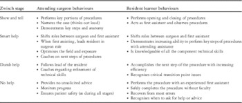
Table 2 depicts the so-called Zwischberger model that lists the common sense and practical steps for supervision of trainees as they progress in their surgical skills.Reference DaRosa, Zwischenberger and Meyerson 4
The “Zwischberger Model” for graduated levels of training in surgical skills in the Operating Room.

Different individuals progress through these stages at different rates. This has led some groups to call for competence-based graduation from surgical teaching programmes rather than adhering to a pre-determined standard duration. This is all the more relevant in the setting of restricted working hours and the new integrated 6-year residency programme for cardiothoracic training, which eliminates the all-important Chief Resident general surgery year of intensive operative experience and responsibility.
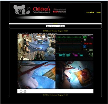
In the final stage of the Zwischberger model, the trainee performs procedures with an experienced first assistant other than the trainee’s mentor. The mentor is usually available in the operating room to ensure patient safety. In the modern era, however, where the trainee is already certified and independently operating, the mentorship can also be provided from a distance using modern technology. At Children’s National Medical Center, there is an internet-based system for operating room monitoring, which integrates four different screens including the full haemodynamic screen from the operating room as well as a steerable room camera that allows all members of the team to be observed including the perfusion team, the anaesthesia team, the circulating and scrub nursing team, as well as the surgical team (Fig 1). In addition, there is an overhead camera and a surgical headlight camera that is worn by the operating surgeon for all components of all cases. As the system is internet-based, it can be monitored wherever internet access is available. In order to comply with Health Insurance Portability and Accountability Act regulations, user name and password access are restricted to individuals who have a direct role in patient care. Records of individuals who have accessed the system at any given time are available to the system administrator.
An internet-based telemedicine system allows monitoring of the cardiac operating room from any location with internet access. The four screens include a steerable, zoomable room camera, the full haemodynamic screen, a camera mounted in one of the overhead surgical lights, and a headlight camera that is worn at all times by one of the members of the operating room surgical team. Password access is required, and compliance with HIPAA regulations is maintained.

The monitoring system for the cardiac operating room makes it possible to create video recordings of all operative procedures. Trainees have the opportunity to record procedures for their own viewing whether they are the operating surgeon or assistant. It was this system that allowed the recording of cases that have been edited, narrated, and incorporated in the textbook Comprehensive Surgical Management of Congenital Heart Disease, where ~50 operative videos are available through streaming via the internet.
Feedback for fellows
Surgical fellows need to be given regular feedback from the attending staff as to how they are progressing. Within the Halstead tradition, feedback was only given when egregious mistakes were made, but in the current era it is expected that attending staff will provide regular feedback on performance in the operating room. In addition, all areas of competence should be monitored, and feedback should be provided in all of these areas. Mentoring can extend beyond technical hints for the operating room and the officially recognised areas of competence to guidance in how to stay fit and avoid occupational work-related injury and how to achieve professional and personal life balance.
Surgical education using three-dimensional printing technology
In the past, a number of congenital heart programmes were able to establish impressively large cardiac pathology registries. These collections of hearts with congenital heart anomalies stored in formalin were an invaluable teaching resource. The author, for example, had the opportunity to attend twice weekly teaching sessions conducted by Drs Stella and Richard Van Praagh in the Cardiac Registry at Children’s Hospital Boston during rotations both as senior resident and chief resident. The ability to handle a heart specimen and to be asked to orient, describe, and identify all relevant pathological features was a remarkably useful exercise. The trainee learns that identification of landmark structures with a systematic approach is the only way to avoid becoming hopelessly disoriented and misidentifying even major structures within the heart. This skill is truly essential for the congenital surgeon who will undoubtedly at times be confronted by anatomy that has been misunderstood in preoperative studies.
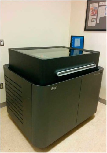
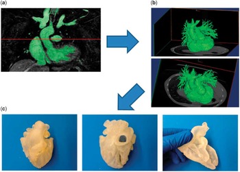
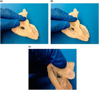
For a number of reasons, cardiac registries have become a preciously rare resource; however, a very helpful alternative is emerging in the form of three-dimensional printing. Children’s National Medical Center is fortunate to have a sophisticated three-dimensional printer within the Sheikh Zayed Bioengineering Research wing (Fig 2). This has allowed creation of life-size cardiac models that are developed using cardiac MRI (Fig 3). More recently, three-dimensional echocardiography is being applied as a user-friendly method for generating three-dimensional heart models (Fig 4). Dr Dilip Nath within the division of cardiac surgery at Children’s National Medical Center has documented that three-dimensional models are helpful in teaching medical students, cardiology trainees, as well as cardiac ICU trainees.Reference Costello, Olivieri and Krieger 5 It can also be helpful for cardiac ICU nurses who are caring for patients with complex intracardiac anatomy. Having the opportunity to view a model of the child’s heart problem immediately before caring for that child aids understanding of the procedure the child has undergone.
A three-dimensional printer at Children’s’ National Medical Center allows creation of life-size cardiac models including complex congenital cardiac anomalies.

Three-dimensional cardiac models were initially derived from three-dimensional MRI reconstructions, but more recently three-dimensional echocardiography is being used. (a) Area of interest for 3D model selected from MRI, (b) Rendering of 3D image by MRI, (c) 3D model printed using polymer.

Creation of heart models with flexible polymers allows realistic teaching of surgical techniques to surgical trainees, as well as the ability of users to view internal cardiac anatomy. (a) Exposure of VSD through retraction of flexible 3D model, (b) alternative exposure of intracardiac anatomy, (c) right atrial exposure of VSD.

Future plans for three-dimensional cardiac models include the development of polymers that closely recreate the fragile tissue consistency of the newborn and infant heart. These realistic models will be used for direct simulation of surgical procedures such as closure of a ventricular septal defect in the neonate.Reference Costello, Olivieri and Su 6 Simulation is also remarkably effective in the cardiac ICU for urgent procedures such as extracorporeal membrane oxygenation cannulation. A particularly helpful component of simulation exercises is to take advantage of the monitoring and storage of haemodynamic data as well as room video recording in the cardiac ICU. Reviewing a trainee’s decisions and actions together with simultaneous haemodynamic information collected as part of a simulation exercise is the best possible feedback for trainees. Furthermore, the same recording equipment is also extraordinarily helpful in post-event analysis of real-life situations such as resuscitation for cardiac arrests including extracorporeal membrane oxygenation cannulation.Reference Su, Spaeder and Jones 7
Training international fellows in congenital cardiac surgery in the United States of America
Background
Following two decades of very rapid expansion of cardiac surgery in the United States of America, the advent of catheter-based procedures to replace coronary, valve, and aortic surgical procedures led to an abrupt decline in the number of open-heart procedures being performed in the United States of America in the mid-to-late 1990s. Reduced job opportunities for graduates of cardiothoracic training programmes led to a decline in the number of applicants to cardiothoracic training programmes. Unfortunately, the declining number of applicants has persisted now for many years. Analyses of future manpower needs for the specialty of cardiothoracic surgery have projected that the reduced number of training positions and failure to fill even the reduced positions that are still available will almost certainly lead to a marked imbalance between the number of active cardiac surgeons and the number of cases that are likely to be performed in coming years.Reference Grover, Goerman and Dall 8 With the baby boom population rapidly entering the 7th, 8th, and 9th decades of life, the projected number of cardiothoracic cases is climbing steeply even with a reduced number of coronary artery bypass cases.
Congenital cardiac surgeons are required to have undertaken training in general cardiothoracic surgery before subspecialisation in congenital cardiac surgery. Thus, the declining numbers graduating from cardiothoracic programmes are likely to ultimately have an impact on the number of congenital surgeons graduating from ACGME congenital programmes in the United States of America. Fortunately, the popularity of congenital cardiac surgery as a subspecialty, which is no doubt a consequence of a number of factors including rising reimbursement rates and salaries, has meant that to date adequate numbers of graduates appear to be available for the job vacancies that have been available in the past few years. Furthermore, only a very small number of programmes in the range of 7–12 per year have taken ACGME congenital trainees, which appears to roughly match the number of new positions that become available annually; however, the small number of ACGME positions means that many programmes have training positions available that are not taken by ACGME trainees, which can be filled by international medical graduates.
Not all countries are in the same position as the United States of America where cardiothoracic surgery has been contracting for the last two decades. In India and China, in particular, cardiothoracic surgery is expanding rapidly; however, exposure of trainees in these countries to ultracomplex congenital cardiac surgery is quite limited. Thus, trainees from these countries have a great interest in taking a position in the United States of America for a period of up to 2–3 years. At the conclusion of their fellowship, there are usually excellent opportunities in their home countries, and this is where the majority of trainees return. By this time, they have benefitted from rigorous training in the United States of America. Of course, occasional outstanding fellows may be offered attending-level positions in the United States of America. This is reflected in the international flavour of many congenital cardiac surgery programmes in the United States of America. The same is true overseas, where the attending staff of several programmes in many countries are not native to that country. Furthermore, these staff surgeons have often trained in another country than the one in which they ultimately practise.
Challenges for international medical graduates coming to the United States of America for advanced training in congenital cardiac surgery
Most individuals who are thinking about undertaking a year or two of advanced training in congenital surgery in the United States of America have already completed general cardiothoracic surgical training in their home country. In fact, it is highly recommended to obtain home country certification in cardiothoracic surgery. This will be important to hospital privileging committees, as it will be looked upon as an equivalent of Board Certification in most cases. If an individual plans to come early in their cardiothoracic training, they should apply for an ACGME-certified position and work towards United States Board certification. Although in the past there was great competition for these positions among United States graduates, today many of these positions in traditional 2- or 3-year training programmes are unfilled, and therefore a qualified, motivated, and well-prepared foreign medical graduate will have a high probability of acceptance. General surgical training in the United States of America, however, has been a prerequisite, and General Surgery training positions in the United States of America are quite competitive. Either way, considerable advance planning is required whether one is planning to come to an ACGME-certified – often termed “categorical” – position or a non-certified fellowship. A position must be found at a training hospital, and there are a number of bureaucratic hurdles including obtaining a visa from the United States federal government, a licence from the appropriate state medical licensing board, and hospital privileges.
Visa optionsReference Jonas 9
The number of visas available for foreign physicians wishing to come to the United States of America varies from year to year and is set by the United States congress. At times, it has been more difficult for a physician to obtain a visa to work in the United States of America than almost any other vocation. In recent years, however, there seems to have been some relaxation of that barrier, perhaps because of the looming shortage of physicians that the United States of America with its ageing population is facing; however one thing has not changed and that is the seriousness with which ICE (Immigration and Customs Enforcement, now an arm of the Department of Homeland Security, formerly the INS) takes any attempt to work in the United States of America without the correct visa. Hospitals and state licensing boards are well aware of the importance of visa issues and are coordinated with ICE.
The most important resource in planning to obtain a visa is ECFMG, the Educational Council for Foreign Medical Graduates www.ecfmg.org. ECFMG provides a verification service that allows Graduate Medical Education programmes, state medical boards, hospitals, and credentialing agencies in the United States of America to obtain primary-source confirmation that their foreign medical graduate applicants are certified by ECFMG. ECFMG coordinates the application process for trainees to undertake the USMLE (United States Medical License Examination), which is undertaken in three steps and is administered by the National Board of Medical Examiners. The examination is the same as that taken by United States medical students. Successful completion of steps 1, basic science multiple-choice exam, and 2, clinical knowledge multiple-choice exam and clinical skills, which involves examination of simulated patients, is required to qualify for a restricted training licence. Step 2 clinical skills must be taken in one of five United States cities – Philadelphia, Chicago, Atlanta, Houston, or Los Angeles. Step 3 is a comprehensive 2-day examination that includes clinical case simulations, and similar to step 2 clinical skills it can only be taken in the United States of America. “ECFMG certification” is obtained when ECFMG has validated the applicant’s training in the home country and steps 1 and 2, both parts, have been completed and passed. This allows entry into a United States ACGME-accredited training programme but is also required for non-accredited positions. USMLE step 3 is usually taken by United States graduates at the end of their first post-graduate year. It is required for an unrestricted licence, which is usually required for a staff position. ECFMG certification and step 3 are required for some visas such as the H1B.
In summary, several years of planning, a considerable financial investment, as the exams are not cheap, and two trips to the United States of America are required just to qualify to apply for a visa. Moreover, a United States hospital is not likely to consider a foreign medical graduate applicant until ECFMG certification is complete. Ideally, steps one and two/clinical knowledge should be taken during the applicant’s medical school training years. Step two/clinical skills could be combined with a preliminary “scouting” trip to the United States of America for interview with potential programmes or during an observership at several prestigious institutions. Attendance at a national cardiothoracic surgery meeting such as the American Association for Thoracic Surgery (AATS), which is usually held in early May, or the Society of Thoracic Surgeons (STS), which is usually held in late January, may help improve the cost-effectiveness of the trip to the United States of America for exam taking.
J1 visa
When ECFMG certification and a job offer have been obtained from a hospital, the next step is to apply for a visa. There are many visa categories available to come to the United States of America in addition to the B visa that is typically obtained by tourists coming to the United States of America who do not intend to work. The J1 visa or “exchange visitor visa” is issued by the United States State Department to allow individuals to train in a specialist area with the provision that they will return to their home country for 2 years before being permitted to change their visa status. This “Foreign Residence Requirement” has for the most part been strictly enforced over the last 30 years, although occasionally waivers can be obtained if there is sufficient legal and financial support for the appeal. Large institutions affiliated with major medical schools are usually very familiar with this programme and are certified to participate in the J1 programme. They are likely to offer a J1 visa as the only option to an individual wishing to spend a year or 2 in a non-accredited congenital fellowship. Another disadvantage of the J1 visa in addition to the foreign residence requirement is that the individual’s spouse must enter the United States of America on a J2 visa, which is a non-working visa, although a legal challenge can be mounted to this. In addition, there is an overall time limit, which has been ~7 years. This may not be adequate for traditional complete training in cardiothoracic and congenital surgery.
H1B visa
A good alternative to the J1 visa is the H1B visa. There is year-to-year variability in the number of such visas available for physicians, although many non-profit institutions are not subject to the quota for H visas. This visa allows foreign workers in many specialties to work in the United States of America. The principal advantage of the H1B visa is that it does not have a foreign residence requirement or a time limitation. It also does not require an institution to be registered with the United States State Department as a sponsoring institution for the Exchange Visitor programme as is needed for a J1 visa; however, as noted above, it does require USMLE Step 3 to have been completed. There are also very specific requirements regarding working conditions and salary that must be competitive with the salary paid to United States workers. Institutions need to work with an experienced immigration attorney from a firm specialising in visas for foreign medical graduates – for example, Maggio and Kattar in Washington, District of Columbia. In the past, the cost and complexities for hospitals in obtaining a visa for foreign medical graduates have discouraged them from making the necessary investment; however, as the number of United States applicants even for ACGME-accredited training positions has declined with many positions being unfilled each year, it has become necessary for foreign medical graduates to be seriously considered. This is a win–win for trainees and the hospitals, as those individuals who are motivated to overcome the numerous bureaucratic hurdles listed above are generally outstanding team members for any cardiothoracic or congenital heart surgery programme.
Green card and citizenship
H1B visas must be renewed annually or if the individual moves from one position to another hospital. If a hospital is interested to offer an individual a longer-term staff position, it will want to sponsor that individual for a Permanent Resident visa, colloquially known as a “Green card”. Those who have enjoyed the movie of the same name by the Australian director Peter Weir should understand that the movie in which the character played by the French actor Gérard Depardieu attempts to obtain a green card fraudulently by marrying the character played by Andie MacDowell portrays some very real truths regarding the aggressiveness of the ICE. Marriage to a United States citizen, for example, does not reverse the foreign residence requirement of a J1 visa.
A Permanent Resident visa provides most of the benefits of United States citizenship, but like the other visas discussed it requires United States income tax to be paid; however, it does not allow voting, and there can occasionally be some limitations on obtaining loans from banks. There are no employment-related advantages.
Medical licence
Unfortunately, medical licensing in the United States of America is managed independently by each of the 50 states and the District of Columbia. Unlike visas that are managed through Federal agencies such as ICE and the Department of State and national non-profit organisations such as ECFMG, which have a uniform approach for candidates irrespective of where in the United States of America an individual will practise, there is great variability state-by-state to medical licensing. Fortunately, most state licensing boards now have websites that lay out the steps required to obtain a licence. Larger states such as New York, where many foreign medical graduates work, tend to be more familiar with processing licences for foreign medical graduates, whereas smaller states may not have a good understanding of the process. In the past, it was extremely difficult to speak with a live individual at a state board, and there were likely to be different answers to the same question from different individuals. State boards often require original documentation such as medical school transcripts, even though ECFMG certification has previously required this. They also may require a statement from every hospital that the physician has worked at regarding satisfactory performance of duties. This can be a problem for foreign medical graduates who are more senior and may have worked at many hospitals in non-English speaking countries. It is well worthwhile playing a very active role in contacting the relevant administrator and politely requesting that the necessary information be transmitted to the board. It is extremely important to begin the licensing process well before coming to the United States of America as soon as a job offer has been accepted and it is known which state licensing board will be processing one’s licence. It is not necessary to have a visa or to be in the United States of America to begin the process. In fact, it is an advantage to be in one’s home country to assemble the necessary documents. Some boards will require an in-person interview as a final step, but many do not require this at any point.
Medical licensing boards in most states require at least 2 years’ experience of working in a hospital with a limited training licence before they will issue a full licence. This is not a problem for a trainee who is restricted to practising in the training hospital; however, it can be a problem for a more senior surgeon who wishes to take a staff position but has never worked in the United States of America as a trainee. Generally, an agreement can be reached, but this varies state by state. If the licensing board will only issue a limited institutional licence, there may be restriction on the ability of the surgeon to bill for services.
Hospital privileges
Each individual hospital is responsible for ensuring that its trainees and staff are not only appropriately trained for the duties they are going to undertake, but in addition do not have a history of criminal activity, frequent malpractice suits, or drug or alcohol dependence. Penalties for hospitals who do not explore all of these areas in great detail before hiring a physician are huge. Although there is going to be a national computerised database that facilitates the review of a physician’s work history in the United States of America, there can be challenges for the hospital committee granting privileges to thoroughly review the foreign medical graduates’ work history in a foreign country. Not surprisingly, the process of hospital privileging is more rigorous for a staff position than for a training position; however, just as with the medical licence, it is essential to begin the process of assembling the needed documentation as soon as a job offer has been finalised, as even for a trainee the hospital privileging process may take several months. It will not be a good start for a foreign medical graduate to be unable to practise for several months after arrival in the United States of America because of lack of certain documents requested by the hospital.
Board certification
Board certification is not a legal requirement for practising in a speciality like congenital cardiac surgery in the United States of America. The wording is quite clear on the website of the American Board of Thoracic Surgery (ABTS): “Board certification in a medical specialty is evidence that a physician’s qualifications for specialty practice are recognised by his or her peers. It is not intended to define the requirements for membership on hospital staffs, to gain special recognition or privileges for its Diplomates, to define the scope of specialty practice, or to state who may or may not engage in the practice of the specialty. Specialty certification of a physician does not relieve a hospital’s governing body from responsibility in determining the hospital privileges of such specialist”. For foreign medical graduates, however, it is important to have finalised equivalent certification in one’s home country before coming to the United States of America unless the entire certification process is going to be undertaken. It is worth reiterating that little or no credit will be given by the ABTS for training or certification outside of the United States of America, so that essentially the entire board certification process must be completed over a period of 7–10 years. Although it is certainly not a legal requirement to be board certified to undertake a non-accredited congenital fellowship, hospital privileging boards are becoming increasingly rigid about requiring some form of certification in a specialty before granting privileges. Thus, a trainee who plans to come to the United States of America for a year or two of fellowship experience will be much better positioned to step into a staff position that may open at the training hospital if they have come to the United States of America after completing certification at home.
How does an interested trainee find a non-ACGME position?
As with many things in life, who you know is just as important as what you know; one of the surest tracks into a more prestigious programme in the United States of America is a personal relationship between a trainee’s chief in their home country and one or more surgeons in the targeted teaching programme. It is unlikely that this would lead to a categorical position in a training programme affiliated with a respected major medical school in a desirable geographic location; however, many of the major centres have several non-accredited positions in addition to their accredited positions. These positions are rarely advertised. It is also highly unlikely that there would be a response to a “cold-call” e-mail from an applicant. An initial contact from a senior surgeon on behalf of the applicant and extolling the intelligence, excellent surgical skills, and hard work ethic is probably the commonest way to get one’s foot in the door of the United States system.
Although many positions for fellowships in the United States of America used to be advertised in the speciality’s major journals such as the Journal of Thoracic and Cardiovascular Surgery and the Annals of Thoracic Surgery, most advertising of positions today takes place on the internet, especially through CTSnet. This website for the field of thoracic surgery was set up as a joint effort by the STS, AATS, and European Association for Cardiothoracic Surgery. The “careers” page at the CTSnet website www.ctsnet.org is the best method for staying informed about fellowship opportunities in the United States of America, some of which become available at short notice at times other than the traditional starting date of 1 July, which begins the training year for most ACGME-certified positions. Such short notices will favour the applicant who has planned ahead and educated himself or herself about the requirements for a United States visa and medical licensing.
Conclusions
In conclusion, it is important to avoid focussing exclusively on technical expertise and surgical judgement when training surgeons in cardiothoracic and specifically congenital cardiac surgery. All areas of competency need to be developed. Setting a good example as a mentor is the most reliable method to achieve this goal; however, training should also take advantage of current technology such as E-books, internet-based operating room videos, three-dimensional printing, and surgical simulation. Finally, in an era of inadequate numbers of United States applicants for training in cardiac surgery, centres should consider hiring international medical graduates. Programmes need to familiarise themselves with the paperwork challenges presented by hiring foreign medical graduates.
Acknowledgements
The author gratefully acknowledges the assistance of Laura Young in preparing the manuscript. He receives royalties from the publisher “Taylor and Francis” for sales of his textbook “Comprehensive Surgical Management of Congenital Heart Disease”.