Introduction
Lipoaspirate, an otherwise by-product of plastic surgery, has been shown to contain a putative source of stem cell population within the heterogeneous stromal cells of adipose tissue. This was first reported by Zuk et al. in 2001 (Ref. Reference Zuk1) and later termed ‘Adipose-derived stem cells (ASCs)’ by International Federation for Adipose Therapeutics and Science (Ref. Reference Gimble, Katz and Bunnell2). Since the discovery, ASCs have garnered considerable attention and scientific curiosity for their potentially boundless therapeutic applications, as they are abundant and readily accessible cell sources.
ASC exhibits properties similar to mesenchymal stem cell (MSC), an adult stem cell type (Ref. Reference Bourin3). According to the standard criteria set by the Mesenchymal and Tissue Stem Cell Committee of the International Society for Cellular Therapy, MSC must fulfil the following criteria: (i) plastic-adherent in standard culture conditions; (ii) express CD105, CD73 and CD90, but negative for the surface expression of CD45, CD34, CD14, CD79 and HLA-DR; and (iii) able to differentiate into osteoblasts, adipocytes and chondroblasts in vitro (Ref. Reference Dominici4).
For many years, bone marrow was considered as the major source of MSCs. However, traditional bone marrow procurement of MSCs is invasive, painful and inefficient because of low cell yield, which necessitates a subsequent ex vivo expansion step. A significant advantage of ASCs as an adult stem cell source, is that while bone marrow stem cells (BMSCs) are a rare population within the stromal cells, constituting only ~0.002%, ASCs constitute up to ~2% within the stromal vascular fraction (SVF) (Ref. Reference Strem and Hedrick5). Under appropriate conditions, ASCs have the ability to differentiate into multiple mesodermal lineages cell types, such as adipocytes, fibroblasts, myocytes, osteoblasts and chondrocytes. Furthermore, studies have shown that ASC has the ability to transdifferentiate into nonmesodermal cell types, such as neuronal cells, hepatocytes, endothelial cells and cardiomyocytes (Ref. Reference Schäffler and Büchler6).
Undeniably an attractive cell source for therapeutic applications, to date, clinical trials with ASCs have increased over years with assessments being made on safety and efficacy of ASC therapies across various medical disciplines. A decade since the discovery of ASCs, this paper seeks to provide a timely review of the advancement of ASCs in therapeutic use. The review highlights the clinical applicability of ASCs and its progress in various medical disciplines, as well as an outlook of the ASC therapy field.
The aim towards clinical-grade ASCs
Manual isolation and culture of ASCs
Adipose tissue consists of mature adipocytes and other heterogeneous cell populations, which upon isolation are termed SVF. The SVF is composed of preadipocytes, fibroblasts, vasculature cells, immune cells and most importantly, ASCs (Ref. Reference Lindroos, Suuronen and Miettinen7).
A typical manual isolation procedure of ASCs involves enzymatic digestion of adipose tissue, followed by centrifugation to separate floating mature adipocytes from the ASC-containing SVF in the pellet fraction (Ref. Reference Zuk1). SVF can be further passaged in expansion medium to isolate more homogeneous ASCs. Given that the multiple laboratory steps involved in the manual isolation of ASCs may have considerable effects on achieving clinical-grade ASCs, several groups have worked independently to develop and refine the procedures needed to isolate ASCs.
For clinical translation of isolated ASCs, it is essential that the collagenase used in liberating regenerative cells from adipose tissue are Good Manufacturing Practice (GMP)-manufactured and animal sources-free (e.g. Celase®) (Ref. Reference Fraser, Prockop, Bunnell and Phinney8), hence not posing risk of infectious diseases such as bovine spongiform encephalopathy/transmissible spongiform encephalopathy (BSE/TSE) to human recipients. Xeno-free collagenases have shown to have a similar efficiency in yield and proliferation of isolated cells as research-grade collagenases (Ref. Reference Carvalho9). Alternatively, the conventional protocol can be modified to omit the use of enzyme in the ASC isolation process. Bianchi et al. (Ref. Reference Bianchi10) have undertaken a nonenzymatic method of digestion of adipose tissue, by using mild mechanical forces to isolate a nonexpanded and ready-to-use fat product. According to the authors, ASCs isolated from this method exhibited a higher degree of vasculogenic potential. Shah et al. (Ref. Reference Shah11) demonstrated the use of manual vigorous shaking by hand and obtained ASCs that displayed enriched level of messenchymal markers and reduced level of haematopoietic markers, although there was reduction in yield. On the other hand, other groups have investigated the use of blood and saline portion of lipoaspirates as an enzyme-free source of ASCs, and isolated ASCs with differentiation and immunophenotypic characteristics comparable to those by enzymatic digestion, although the yield will need to be further optimised (Refs Reference Francis12, Reference Yoshimura13). If these novel methods are able to demonstrate similar levels in ASC yield, proliferation and differentiation as the enzymatic isolation protocols, it may eventually pave the way for using minimally manipulated ASCs in clinical trials.
Centrifugation is essential for concentrating SVF and ASCs, but excessive centrifugation may also lead to cellular damage. Hence, several groups have investigated an optimal centrifugal force (Refs Reference Kim14, Reference Kurita15) and currently, the recommendation is 1200 g for 5 min as an optimal centrifugation rate for both short- and long-term adipose transplantation (Ref. Reference Sterodimas, Illouz and Sterodimas16).
Another probable variability is the type of medium used for the culture expansion to achieve homogeneous ASCs. The classical basal medium used for ASCs are usually Minimum Essential Media (MEM), Dulbecco's Modified Eagle Medium (DMEM), Roswell Park Memorial Institute (RPMI)-1640 and DMEM:F12 (Ref. Reference Lindroos, Suuronen and Miettinen7). One study showed that the proliferation and differentiation of ASCs varied according to the type of culture medium used (Ref. Reference Lee17). Furthermore, the expression of genes in the cultured ASCs differed when different media were adopted (Ref. Reference Lee17). Therefore, the choice of an optimal medium for clinical trials using cultured ASCs would be one major consideration.
In order to achieve a high cellular dose of MSCs suitable for clinical trials, it is necessary to expand cells on the basal medium supplemented with serum. Typically, the standard in vitro cell expansion techniques utilise fetal bovine serum (FBS) to provide the cell with essential nutrients, attachment factors and growth factors (Ref. Reference Gstraunthaler18). However, serum of animal origins may pose several potential safety issues on patients, including risk of possible infections (Ref. Reference Will19) and severe immune responses (Ref. Reference Heiskanen20). In an effort to overcome the potential risk of introducing xenogeneic antigens into patients, human serum has been proposed as an alternative replacement for FBS. Examples of human serum derivatives include allogenic AB serum (Ref. Reference Kocaoemer21), thrombin-activated platelet-rich plasma (Ref. Reference Kakudo22) and human platelet lysate (Refs Reference Kocaoemer21, Reference Johansson23, Reference Mirabet24). Studies using such human serum derivatives have shown comparable or superior results to FBS in supporting proliferation and differentiation of ASCs (Ref. Reference Kocaoemer21). However, limited availability and batch-to-batch variability of human serum are potential barriers for large-scale production of clinical-grade ASCs (Refs Reference Lindroos, Suuronen and Miettinen7, Reference Kim25). Alternatively, a serum-free medium could be an optimal option for standardisation of cell culture as it provides defined and controlled culture conditions that are highly specific to cell types in terms of its stimulation and growth (Ref. Reference Gstraunthaler18). Existing studies have reported promising results of serum-free medium (Refs Reference Lindroos26, Reference Rajala27, Reference Santos28), although further pre-clinical safety and efficacy studies are required to test these culture formulations.
Another major challenge of producing clinical-grade cells is having a reliable, reproducible and safe method of preparing cells for administration to patients. The in vitro culturing and expansion process of ASCs still lacks an established and standardised protocol (Ref. Reference Lund29). Hence, the identification of an optimal basal medium and serum replacer will be an important step forward for both the implementation of a GMP and large-scale production of clinical-grade ASCs for therapeutic applications.
Industrial approach to clinical-grade ASCs
In order to achieve production of clinical-grade cells that avoids inherent contamination risk and intra/inter-operator variability associated with traditional techniques, scientists, engineers and clinicians have taken collaborative efforts to develop a sophisticated bioreactor-based system. The bioreactor-based device offers an attractive device that could possibly provide a stand-alone, fully automated and closed system that facilitates complicance to GMP in manufacturing ASCs aimed for therapeutic use (Ref. Reference Martin, Smith and Wendt30).
Preparation of clinical-grade ASCs for immediate use to patients involves three steps: (i) Harvesting, (ii) Processing and (iii) Injection of SVF or ASCs. Several automated devices have been developed to handle the most cumbersome step among these – (ii) Processing. Examples of such systems include, Cytori Celution® systemFootnote 1, Tissue Genesis Icellator Cell Isolation System®Footnote 2, Medi-Khan Maxstem®Footnote 3 and Sepax-based automated isolation system (Ref. Reference Güven31).
An example of such integrated systems is Celution® system developed by Cytori Therapeutic Inc (San Diego, CA, USA; see footnote 1). In this system, adipose tissue is first introduced into the device and washed to remove debris. Subsequently, it is enzymatically digested to release mononuclear cells from adipose tissue and transferred to a centrifugation compartment for concentration of SVF/ASCs. The end-product is ready to be administered to patients. The whole system mimics the manual isolation procedures of SVF/ASCs, and reduces the processing time to 1–2 h. The integrated system also means that the whole process can be carried out in clinical setting without the need for highly trained laboratory technicians. Freshly isolated cells from such a system have been studied and analysed to be equivalent to those manually isolated (Ref. Reference Lin32).
The possible existence of heterogeneity in quality of ASCs isolated from different donors or different sites presents limitations to the consistency of results obtained by use of isolated ASCs in therapeutic applications. AdiLight-1, a LED device produced by AdistemTM (Hong Kong)Footnote 4 was reportedly aimed at acquiring full functionality of ASCs (Ref. Reference Boriani, Warr, Ascione, Illouz and Sterodimas33). The device claims to use low-level photoactivation to stimulate proliferation or differentiation, and promotes the release of growth factors and cytokines from ASCs. The device is based on the expectation that the usually dormant isolated ASCs can be activated to become fully functional by induction of their intrinsic repair mechanisms.
Clinical banking of ASCs
The enormous potential of ASCs in therapies could mark a foreseeable step towards clinical banking of ASCs in the near future (Refs Reference Gimble, Katz and Bunnell2, Reference Thirumala, Goebel and Woods34). In addition to storage of individual ASCs for autologous use, allogenic transplantation of MSCs that require a large cell number may also benefit from the development of stem cell banks, which cryopreserve cells from healthy donors. This will advance ASC therapies to ‘off-the-shelf’ approaches that would allow stem cells to be available in a timely fashion under acute or chronic clinical settings (Ref. Reference Thirumala, Goebel and Woods34). Therefore, the time is ripe now for establishment of cryopreservation methods and protocols to assure clinical-grade ASCs. Presently, many companies claim to provide banking of adipose tissue or ASCs. However, compliance with GMP guidelines for the entire process from cell collection to cryopreservation of ASCs remains questionable, especially since some of these companies are still awaiting approval from FDAFootnote 5. Furthermore, existing cryopreservation protocols of adult stem cells still require further refinement to lower dependence on animal serum and potentially toxic cryoprotectant additives in the freezing and storage of cells (Refs Reference Liseth35, Reference Benekli36, Reference Windrum and Morris37).
Heterogeneity of ASCs
Although clinical banking of ASCs may still be at an infant stage, it remains valuable to define harvesting sites, harvesting techniques and donor types for isolation of ASCs. Defining these variables will facilitate identification of ASC population optimal for cell therapies, thereby assuring consistent therapeutic results following the application of ASCs. The variables that may affect the yield and functionality of isolated ASCs can be broadly classified into two categories: namely (i) donor-dependent heterogeneity and (ii) intra-individual heterogeneity.
Donor-dependent heterogeneity includes factors such as patient's age, body mass index (BMI) or diseases. Ageing has been widely speculated to follow with a decline in regenerative capacity of resident stem cells in diverse tissues (Ref. Reference Sharpless and DePinho38). These observations are reflected in studies demonstrating a negative correlation between age and functionality of ASCs (Refs Reference Schipper39, Reference van Harmelen, Röhrig and Hauner40), although conflicting results exist as well (Ref. Reference van Harmelen41). The discrepancies may be attributed to the difference in harvesting sites of ASCs employed in individual studies. For instance, two studies done by van Harmelen et al. used comparable methodology, but one study isolated ASCs from the abdominal subcutaneous depot (Ref. Reference van Harmelen, Röhrig and Hauner40), whereas the other study isolated ASCs from the mammary fat pad (Ref. Reference van Harmelen41). On the other hand, studies investigating the yield of ASCs from different age groups show no significant correlation between the yield of ASCs and the donor's age (Refs Reference van Harmelen, Röhrig and Hauner40, Reference van Harmelen41, Reference Jurgens42, Reference Aust43).
Obesity is accompanied by hypertrophy and/or hyperplasia of mature fat cells (Ref. Reference van Harmelen41). On the contrary, several studies have reported a negative correlation between BMI and yield of ASCs (Refs Reference van Harmelen41, Reference Aust43), and with one study further demonstrating a decrease in differentiation capacity of isolated stromal cells from obese subjects (Ref. Reference van Harmelen41). Notably, similar results were not observed across all studies (Refs Reference Jurgens42, Reference Pettersson44).
Lastly, in vitro exposure of human ASCs with high glucose concentration was shown to decrease cell proliferation (Ref. Reference Kim45), suggesting that diabetic patients may not be suitable ASC donors. Therefore, determining a patient's condition may be an important pre-consideration factor as donors of ASCs for their autologous transplantation.
Intra-individual heterogeneity includes factors such as anatomic harvesting sites, cellular composition of adipose tissue harvested or lipoaspiration techniques. The implication of anatomic harvesting sites in yield or functionality of ASCs has been reported in several individual studies. Jurgens et al. (Ref. Reference Jurgens42) demonstrated a higher yield of ASCs obtained from abdominal fat as compared to the hip/thigh region, although no difference was observed in the growth kinetics or differentiation potentials into osteogenic or chondrogenic lineages. Hauner and Entenmann (Ref. Reference Hauner and Entenmann46) reported a significant anatomical difference in adipogenic differentiation potential, with ASCs from abdominal fat showing a significantly higher adipogenic differentiation capacity than those from femoral fat. In addition, Schipper et al. (Ref. Reference Schipper39) showed regional variation in resistance of ASCs to apoptosis, in which cells from the abdominal superficial depot was found the most resistant to apoptosis compared to those from other subcutaneous depots (Ref. Reference Schipper39). Variability in the yield and functions of ASCs from different anatomical sites highlights the importance of selecting an optimal site for isolation of clinical-quality ASCs.
Additionally, anatomical location of fat depots may also contribute to growth rate of isolated ASCs. van Harmelen et al. (Ref. Reference van Harmelen, Röhrig and Hauner40) found that SVF from subcutaneous adipose tissue was observed with higher proliferation capacities compared to visceral adipose tissue. Contrastingly, the study by Pettersson et al. (Ref. Reference Pettersson44) observed no difference in proliferation rates between ASCs derived from subcutaneous and visceral depots. Variable results between these studies, however, could be attributed to the different methodological approaches undertaken such as culture methods.
Lastly, investigation into effects of the three main classical harvesting techniques of ASCs including resection, tumescent liposuction and ultrasound-assisted liposuction, has shown that the application of ultrasonic energy through cannula yields the lowest frequency of ASCs and has the most detrimental effects on quality of ASCs (Ref. Reference Oedayrajsingh-Varma47).
It is a challenge to clearly define effects of the donor-dependent and intra-individual heterogeneities on the yield, proliferation and differentiation of ASCs, as many of these epidemiological or experimental studies are performed on a relatively small sample size and with different methodological approaches. Moreover, adipose tissue depots are highly heterogeneous with many variables contributing to the yield and functionality of ASCs. Therefore, an ideal study should normalise for these multiple variables so as to determine causality between the two differences to be examined.
Legal and regulatory issues of cell therapy
Cell therapy has emerged as a novel strategy for treatment of numerous human diseases. Today, cell therapy represents the fourth and final therapeutic pillar of global healthcare that complements three existing therapeutic pillars, namely (i) pharmaceuticals, (ii) biologics and (iii) medical devices (Ref. Reference Mason48). Majority of drug regulatory agencies around the world have acknowledged cell therapies as drugs or medicinal products. However, inadequacy of the current regulatory frameworks has sparked a move to re-examine and develop a separate regulatory framework to keep pace with the rapidly growing industry involving cell therapy (Ref. Reference Kellathur and Lou49). This section reviews cell therapy regulations of countries that are in the forefront of clinical applications of ASCs, namely the United States (USA), Europe and Korea.
United States
In the USA, ASCs are considered in the context of human cells, tissues, or cellular and tissue-based products (HCT/Ps)Footnote 6. The cell therapy regulatory body is governed by the US Food and Drug Administration (US FDA) that assures safety, quality, efficacy of HCT/Ps and regulates implementation of HCT/Ps in human recipients. There are two classifications of HCT/Ps, in which individual classification entails a different level of regulations (Ref. Reference Sensebe50). Higher risk HCT/Ps are to comply with Section 351 of the Public Health System Act. Consequently, under Section 351, a valid biologic license (BLA) must be in effect before initiating clinical use of ASCs, which makes it necessary to comply with GMP and good tissue practice (GTP) regulations. Furthermore, sponsors must file an investigational new drug (IND) application before distribution of HCT/Ps in clinical trials. This helps to establish the safety and efficacy of the product. On the other hand, lower risk HCT/Ps, which are ‘minimally manipulated’ and intended for homologous use only, are to comply with Section 361 of the Public Health System Act. The ‘361’ products are generally not under requirements for licensing, clinical trials and regulations above, with the exception of the GTP requirement. Thus far, however, most cases involving ASC therapies are interpreted as ‘351’ HCT/Ps by US FDA.
Medical devices involved in the processing of cells for cell therapies are also rigorously regulated by US FDAFootnote 7. The FDA regulates medical devices accordingly as classes I, II and IIIFootnote 8 and each class involves different pathways for approval of the regulations and processes. Automated devices for separation of ASCs are regulated as class III medical devices and the approval entails extensive scientific review and clinical studies of the devices to prove its safety and efficacy under the Premarket Approval (PMA) pathway. Currently, Cytori's cell-processing device is still awaiting approval from FDAFootnote 9, as FDA highlights that the enzyme used in the processing system is more than minimally manipulated and may pose safety concern.
Europe Union
In Europe (EU), ASCs are defined as somatic-cell therapy products or tissue-engineered products depending on how the cells are processed and their intended use, as defined in EU regulations of advanced therapy medicinal products (ATMPs) (Ref. Reference Antunes and Pottering51). Owing to the specificity of ATMPs, a Committee for Advanced Therapies (CAT) was set up within the European Medicines Agency (EMA) (Ref. Reference Antunes and Pottering51). The committee comprises of five co-opted members who provide opinions on the safety, quality and efficacy of ATMPs. In addition, regulatory frameworks on cell therapy in EU are governed by three directives summarised by Liras (Ref. Reference Liras52).
Korea
In Korea, cell therapy is regulated by South Korea Food and Drug Administration (KFDA). The current surge in the stem cell industry has prompted Korea's government to consider simplifying the authorisation process of cell therapy products to lower barriers on industrialisation of the stem cell therapeutics (Ref. Reference Oh53). One legislative pending approval in the National Assembly of Korea is the consideration of phase I clinical trials as suffice for determining the safety and efficacy for treatment of rare diseases with cell therapy. Another proposal is on the exemption of phase III clinical trials on autologous use for cell therapy. Currently, KFDA has approved the treatment of fistula with Seoul-based company Anterogen's Cupistem, autologous-derived ASCs (Ref. Reference Wohn54).
Clinical applications of ASCs
The therapeutic potential of ASCs is enormous due to its unique inherent properties (Ref. Reference Ong and Sugii55). The multipotency feature of ASCs serves as new building blocks for regeneration of injured tissues (Ref. Reference Ong and Sugii55). The secretome of ASCs is enriched with an array of soluble factors that have the capacity for neoangiogenesis, cytoprotection or activation of reparative mechanism (Ref. Reference Ong and Sugii55). In addition, the immunomodulatory and immunoprivileged features of ASCs are attractive immunobiological properties that can be exploited in diseases with minimal immune rejection (Ref. Reference Ong and Sugii55).
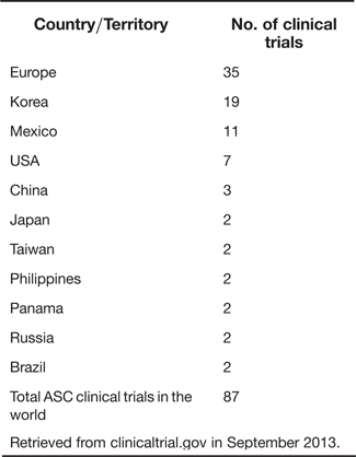
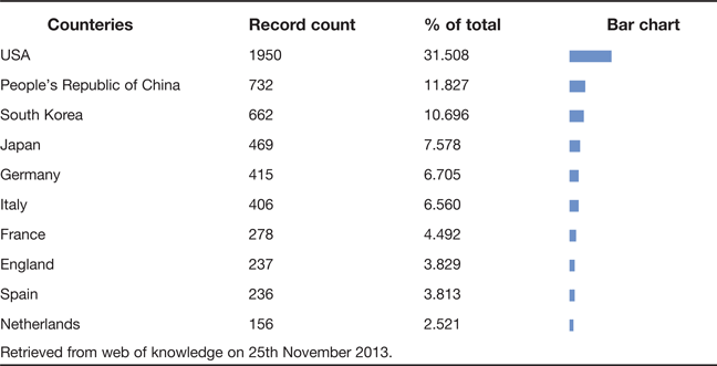
A search performed with ‘adipose stem cells’ on clinicaltrial.gov in September 2013, revealed 87 ASCs-based clinical trials performed worldwide (Table 1). EU, Korea and Mexico are the leading countries conducting the most number of ASC-based clinical trials. The USA may have the most extensive research and published journals on ASCs (Table 2), but currently it has only seven clinical trials registered (Table 1), likely attributed to the rigorous regulation of cell therapy by US FDA.
Schematic representation of cell-assisted lipotransfer (CAL). The procedure involves generation of ASC-rich fat tissue by supplementation with SVF isolated from another half of the aspirated fat sample. Thus, SVF is attached to ASC-poor fat, which serves as a scaffold in this strategy. Figure adapted and revised with permission from Yoshimura et al. (Ref. Reference Yoshimura86).

Number of ASC clinical trials by countries
Retrieved from clinicaltrial.gov in September 2013.
Top ten countries in number of journals published on ‘ASCs’
Retrieved from web of knowledge on 25th November 2013.
Most clinical studies on ASCs published thus far are not only limited in numbers, but the study designs of the ASC-based clinical trials performed till date also produced predominately lower levels of evidence according to the Oxford Centre for Evidence-Based MedicineFootnote 10. For example, the majority of these published studies are case reports, anecdotal studies or phase I/II studies performed in a relatively small sample size. Nonetheless, the reported clinical studies have covered a wide variety of specialties and indicated that ASC or SVF administration has been well tolerated (Table 3). The following subsections will categorise and summarise publications concerning the clinical use of ASCs and its outcomes in various areas of medical diseases.
Registered therapeutic clinical trials using ASCs
Retrieved from clinicaltrial.gov in September 2013.
Diseases are classified according to the International Classification of Diseases, 10th revision, Clinical Modification (ICD-10-CM).
Bone and cartilage repair
There are several case reports that examine the therapeutic potential of ASCs in healing skeletal defects in patients. One of the earliest report of using ASCs in regenerative medicine is the study done by Lendeckel et al. (Ref. Reference Lendeckel64). A 7-year-old sustained calvarial fractures after a severe head injury. A surgical procedure was conducted to replant and fix cryopreserved calvarial fragments, following craniectomy. However, osteosynthesis plates loosened and significant bone resorption resulted in an unstable skull. To rectify the situation, a surgical procedure combining autologous SVF and bone grafts was proposed. Autologous SVF was isolated and subsequently, soaked with milled bone grafts. In addition, to keep the SVF cells in place, autologous fibrin glue was applied and mechanical fixation performed with resorbable macroporous sheets. Postoperative course healing was uneventful and 3 months later, computed tomography (CT) scan showed new bone formation and complete calvarial continuity after reconstruction. Hence, the outcome from the case report suggests that autologous SVF enhances the process of bone regeneration, together with bone graft serving as an osteoconductive scaffold, although it remains to be determined how much of the effect is due to autologous SVF.
Another study on skull restoration, was performed in Finland on four patients with cranial defects due to various conditions (Ref. Reference Thesleff65). To create autologous bone for the repair of cranial defects, clinical-grade autologous ASCs cultured in a GMP facility were seeded on beta-tri-calcium phosphate (betaTCP) granules. Postoperatively, the surgical procedure was tolerated well by the patients and no adverse events reported. After 3 months to a year of follow-up, CT scans revealed satisfactory outcomes in ossification.
In the third case, Mesimaki et al. (Ref. Reference Mesimäki66) took a novel approach to repair craniofacial defects. The patient was 68 years old, with part of the hard palate in the mouth removed due to a large keratocyst. To address the issue of limited bone graft, ectopic bone was formed in a muscle-free flap region with GMP-grade ASCs combined with betaTCP and growth factor bone morphogenetic protein (BMP)-2. Subsequently, the construct was resected and transplanted to the maxillofacial defect. Four months after the surgical procedure, the implant integrated well into the bone site with normal palatal mucosa formation and provided enough structural stability for dental rehabilitation. Following the success of such a tissue-engineering triad approach, which includes ASCs with osteogenic potential, betaTCP as an osteogenic scaffold, and BMP growth factor to build a custom-made implant, more than 20 patients with craniomaxillofacial defects were similarly treated with positive results (Ref. Reference Gimble, Guilak and Bunnell67).
With approval of the KFDA, a team in Korea proceeded with ASC-based treatment of two patients with osteonecrosis of femoral heads (Ref. Reference Pak68). The procedure included autologous SVF isolated from patient's abdominal, mixed with calcium chloride-activated autologous platelet-rich plasma (PRP) and hyaluronic acid serving as the scaffold, and together this mixture was injected into the femoral head under ultrasonic guidance. Two weeks after the treatment, both patients reported significant reduction in hip pain, and post-procedure MRI showed changes that were consistent with bone regeneration. Neovascularisation at the necrotic site was speculated to support the regenerated tissues, but no bone biopsy was performed to further investigate the mechanism behind the efficacy.
The same team also reported a series of case studies that included the treatment of osteoarthritis with ASCs (Ref. Reference Pak69). The rationale was supported by previous experimental studies on animals that demonstrated the potential of ASCs in cartilage tissue engineering, by differentiating ASCs into chondrocytes in vitro or in vivo (Ref. Reference Rada, Reis and Gomes70). Two Korean women with osteoarthritis were treated with the construct of autologous SVF mixed with PRP, dexamethasone and hyaluronic acid in the medial or lateral side of the knee. Following up 3 months later, both patients exhibited significant reduction in pain, improved range of motion by the knee and significant increase in thickness of meniscus cartilage shown on MRI scans.
Ischaemic diseases
ASCs in myocardial infarction
Autologous stem cell therapy offers a novel approach to rectify cardiac functions following myocardial injury and challenges the traditional view that myocardial lesions are incapable of repair (Ref. Reference Mann71). Multiple groups have evaluated the cardioregeneration potential of ASCs in animal models of both acutely (Refs Reference Valina72, Reference Alt73, Reference Ii74, Reference Schenke-Layland75) or chronically infracted myocardium (Ref. Reference Miyahara76). These preclinical studies have shown promising results of the ASC's ability to overcome the inflammatory response and necrotic signals from apoptotic cells after acute episodes of myocardial infarction (Ref. Reference Nian77) and repair the damaged myocardium (Refs Reference Valina72, Reference Alt73, Reference Ii74, Reference Schenke-Layland75, Reference Miyahara76). With experimental evidence from preclinical studies presenting the safety and therapeutic effects of ASCs in cardioregeneration, a number of clinical trials have been carried out or are currently ongoing.
Cytori Therapeutics has completed and reported data of two clinical studies, ‘APOLLO’ (Ref. Reference Houtgraaf57) and ‘PRECISE’ (see Table 3) (Ref. Reference Sridhar56). Both studies were prospective, placebo-controlled, double-blinded Phase I studies designed to assess safety and feasibility of ASC-based therapies for acute myocardial infarction (APOLLO) and chronic myocardial ischaemia (PRECISE). ASCs (also termed ‘ADRCs’) from all trial participants were processed at point-of-care with the Celution® system, and administered to patients. Safety was measured by occurrence of major adverse cardiac or cerebral events (MACCE) and feasibility by monitoring cardiac function improvement. Results from both trials reported no adverse events directly related to the ASC therapies and suggested good efficacy consistent with their pre-clinical findings. Based on the results from APOLLO, in May 2011 Cytori initiated a multicenter, prospective, randomised, placebo-controlled phase IIb/III clinical trial that will assess the safety and efficacy of an intracoronary infusion of 2 doses of ASCs in up to 375 patients with ST-elevation myocardial infarction.
ASCs in peripheral vascular diseases (PVD)
Since several studies showed that ASCs are capable of secreting significant quantities of angiogenic (Refs Reference Nakagami78, Reference Moon79, Reference Rehman80, Reference Traktuev81) or anti-apoptotic factors (Refs Reference Rehman80, Reference Traktuev81), and transdifferentiating into endothelial cells (Refs Reference Planat-Benard82, Reference Miranville83) or into pericytes essential for vascular stabilisation (Ref. Reference Traktuev84), it is conceivable that this cell source is valuable for PVD treatment. One published pilot study enrolled 15 patients with critical limb ischaemia due to either thromboangiitis obliterans or diabetic conditions. Subsequently, the patients were treated with intramuscular injection of autologous passaged ASCs. The study reported clinical improvement in 66.7% of the enrolled patients along with significant improvement in pain scores. Furthermore, digital subtraction angiography (DSA) performed 6 months from the treatment revealed new collateral vessel formation.
Soft tissue augmentation and reconstructive surgery
ASCs as fillers in cosmetic and reconstructive surgery
Autologous fat transplantation has become a useful method for facial rejuvenation and soft tissue augmentation procedures of volume enhancement (Ref. Reference Tabit85). However, due to fat resorption, fibrosis or necrosis, outcomes of the fat transplantation are often variable or unpredictable (Ref. Reference Tabit85). To overcome the problem associated with lipotransfer, ASCs offer a novel strategy by enriching or replacing the ASC-poor fat.
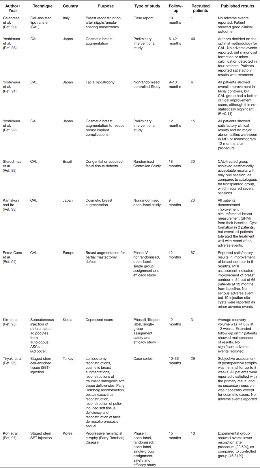
Cell-assisted lipotransfer (CAL) is one such strategy that involves the concurrent transplantation of autologous ASCs attached to aspirated fat (Ref. Reference Sterodimas87). The basic principle of CAL is addition of ASCs to aspirated fat, converting the ASC-poor fat into ASC-rich fat, thereby boosting the efficacy of autologous lipoinjection (Ref. Reference Matsumoto88) (Fig. 1). CAL used in clinical studies is generally well tolerated by the patients and tends to achieve higher aesthetically acceptable results as compared to autologous fat grafting alone in controlled studies (Refs Reference Kurita15, Reference Sterodimas89). The clinical reports of CAL are summarised in Table 4.
Clinical studies using ASCs as fillers in cosmetic and reconstructive surgery
The exact function of ASCs in CAL remains to be elucidated, although several mechanisms have been suggested. ASCs may play a role in promoting angiogenesis and graft survival, through their ability to differentiate into vascular endothelial cells and release angiogenic growth factors (e.g. vascular endothelial growth factor (VEGF)) in response to surgical injury, hypoxia or wound-healing process (Ref. Reference Matsumoto88). In addition, co-delivered ASCs may be prompted to differentiate into mature adipocytes, thereby constituting the transplanted fat (Ref. Reference Matsumoto88).
Another novel strategy that shares the same principle of CAL to enrich ASC-poor fat is staged stem cell-enriched tissue (SET) injections. While CAL combines ASCs with aspirated fat ex vivo before injecting this mixture as a single construct to patients, SET involves injection of autologous ASCs into an area that was pre-grafted with nonenriched fat just hours before. Although there is no clinical study comparing these two techniques thus far, two clinical studies using SET reported promising results with no adverse events (Refs Reference Tiryaki, Findikli and Tiryaki96, Reference Koh97).
Lastly, one study used mature adipocytes that were differentiated from autologous ASCs (ADIPOCELL) to treat 31 patients with depressed scars (Ref. Reference KIM95). The long-term follow-up (1 year) found maintenance of volume that was increased from the therapy. Therefore, it appears that mature adipocyte grafting may be a good alternative to traditional fat grafts for volume augmentation.
ASCs in wound healing
The role of ASCs in wound healing has been validated in preclinical studies done with various delivery methods (Refs Reference Altman98, Reference Kim99, Reference Lin100, Reference Maharlooei101, Reference Nie102). ASCs appear to promote wound healing through multiple mechanisms such as their direct differentiation into dermal fibroblasts and keratinocytes, and their trophic support for regenerating tissues (Refs Reference Jackson, Nesti and Tuan103, Reference Kim, Park and Sung104). However, the clinical application of ASCs in wound healing is still at its preliminary stage and mostly limited to anecdotal reports (Refs Reference Akita105, Reference Akita106, Reference Sung107). Of note, two case reports described the use of autologous SVF to treat radiation injuries in combination with dermis scaffold and growth factors. The wound was healed uneventfully with no adverse events reported (Refs Reference Akita105, Reference Akita106). On the other hand, pilot clinical studies using autologous adipose grafts on ulcerative, dystrophic or radiation wounds observed significant improvement in wound healing following lipotransfer (Refs Reference Cervelli108, Reference Rigotti109). Although further validation is required, these studies postulated that regeneration and repair of the skin were likely attributed to ASCs within the adipose matrix delivered during lipotransfer.
Immune diseases
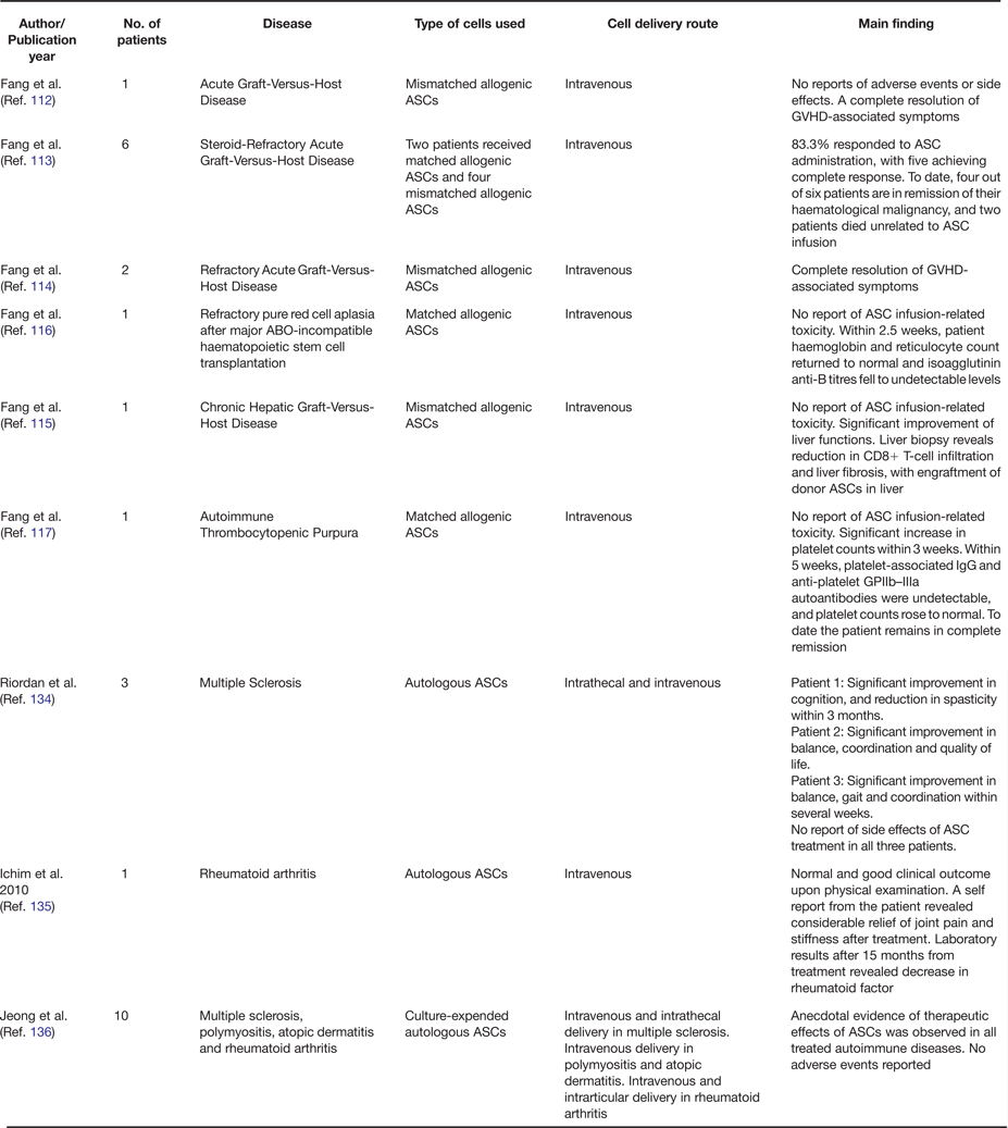
The immunomodulatory and immunoprivileged properties of ASCs are of theoretical clinical value for the application of immunologic disorders (Refs Reference Lin, Lin and Lue110, Reference Griffin, Ritter and Mahon111). From 2006 to 2009, Fang et al. reported anecdotal findings using allogenic ASCs in the treatment of graft-versus-host diseases (GVHD) (Refs Reference Fang and Song112, Reference Fang113, Reference Fang114, Reference Fang115) and haematologic disorders from ABO-incompatible transplantation (Refs Reference Fang116, Reference Fang117). Their approach was to administer intravenous infusion of allogenic ASCs isolated from healthy donors. The treatment was well tolerated by all patients and no adverse events were reported. They reported complete resolution of GVHD-associated symptoms (Refs Reference Fang and Song112, Reference Fang113, Reference Fang114, Reference Fang115) and remission of their haematological malignancy, respectively (Refs Reference Fang116, Reference Fang117). Furthermore, their findings demonstrated that matched major histocompatibility complex (MHC) was unnecessary for the immunosuppressive effects of ASCs (Ref. Reference Le Blanc118). Additional effects of ASCs were also demonstrated in autoimmune diseases with good clinical outcomes (summarised in Table 6), although they are limited to case series and case reports.
Clinical development of ASCs in treatment of Fistula
ASC case reports in autoimmune diseases
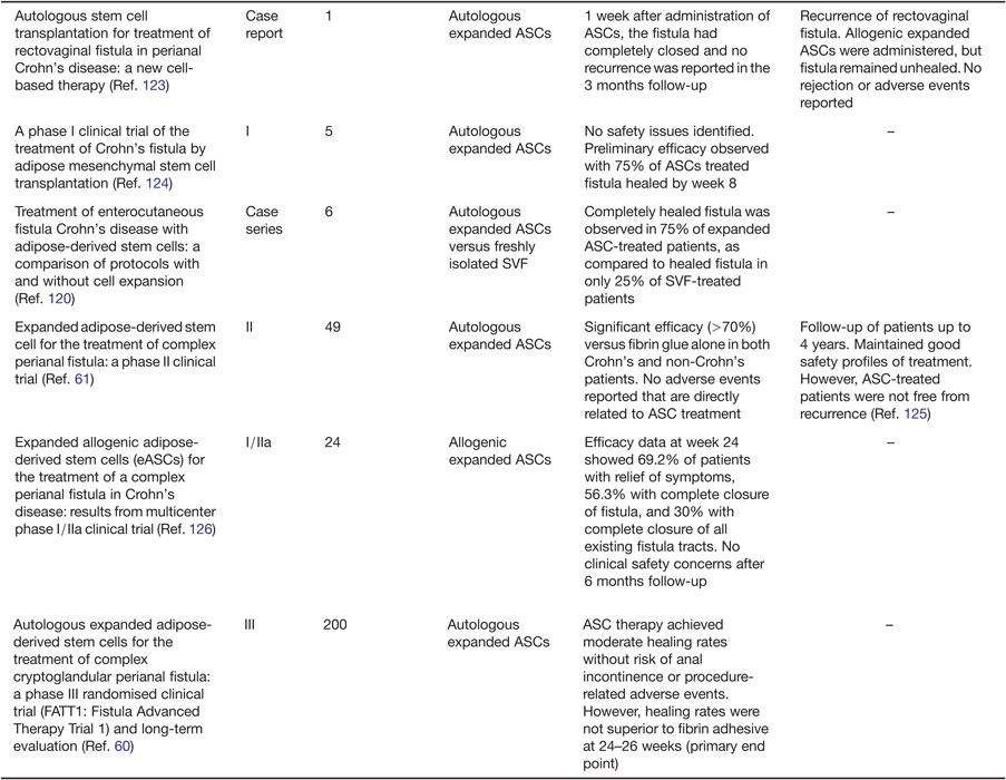
A fistula is an abnormal connection or passageway between two body cavities. Fistula management is often complicated by the high recurrence rate and its inflammatory nature, which hinders wound healing (Ref. Reference Garcia-Olmo, Garcia-Arranz and Herreros119). An independent group in Spain extensively studied and reported the therapeutic safety and effects of ASCs in fistula repair. An overview of the clinical development and published results is summarised in Table 5. Their approach was to inject ASC suspensions under the epithelium along the fistula track, and seal it with fibrin adhesive (Ref. Reference Garcia-Olmo, Garcia-Arranz and Herreros119). Additionally, an attempt to study the difference between using ASCs expanded by culture or freshly isolated SVF pointed towards better outcomes with the expanded ASCs (Ref. Reference Garcia-Olmo120). The group postulated that this may be due to the difference in immunogenic properties of ASCs and SVF (Ref. Reference Garcia-Olmo120), as shown in an in vitro study that ASCs suppressed T-cell proliferation, whereas SVF stimulated T-cell proliferation in a parallel assay (Ref. Reference McIntosh121). These findings may explain the relative success or failure of using ASCs or SVF in immunological conditions, but would require further preclinical studies for validation.
With both safety and efficacy results reported in phase I and II studies, the group had initiated and completed a phase III study, in line with an application for marketing authorisation of the ASC therapy in fistula repair. However, the study was unable to provide statistically significant results on the efficacy of ASCs when compared to the control group with fibrin glue treatment. Notably, the authors attributed the inconclusive results to the likely variability of methods and protocols introduced from the multi-centre nature of the study.
Diabetes mellitus
Vanikar et al. (Ref. Reference Vanikar122) reported improved insulin sensitivity in 11 Type 1 diabetic patients who were co-infused with insulin-secreting cells derived from ASCs and cultured bone marrow cells. This cell mixture derived from the xeno-free workflow was infused into patients via omental vein. In the 23 months follow-up, all patients were successfully treated, showing raised serum c-peptide level, less exogenous insulin requirement, decreased HbA1c level and absence of diabetic ketoacidosis events. While the exact mechanism behind the therapeutic effects remains to be elucidated, this study may present a novel potential insulin replacement therapy based on ASCs.
Perspective
Since discovery of a rich source of MSCs in adipose tissue, ASCs and SVF have made progressive advances in the clinical realm. Notwithstanding the fact that till date, the safety and efficacy evidence of ASC therapy is limited to anecdotal studies or phase I/II clinical trials, ASC therapy represents a promising therapeutic strategy to treat various diseases. However, it is crucial that any possibilities of unwanted and unexpected effects are critically assessed before embarking on a novel ASC-based therapy.
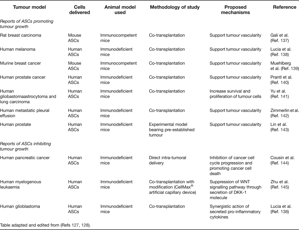
Some concerns have been raised regarding properties of ASCs to promote tumour growth. While the pro-angiogenic, anti-apoptotic and immunomodulatory properties of ASCs are attractive therapeutic aspects of ASCs, these may also act individually or together to support tumour growth (Ref. Reference Casiraghi127). Several articles have extensively reviewed the tumourigenic potential of ASCs and highlighted the striking contrast of results obtained from various experimental studies (Table 7) (Refs Reference Casiraghi127, Reference Klopp128). Many experimental models fail to faithfully recapitulate the complexity of tumour microenvironment (Refs Reference Casiraghi127, Reference Klopp128). Therefore, a definite conclusion on pro-tumourigenic effect of ASCs is still pending.
In vivo animal studies investigating the risk of ASC therapies
Table adapted and edited from (Refs Reference Casiraghi127, Reference Klopp128).
Furthermore, clinical studies with ASCs have so far not reported the speculated theoretical risk of ASCs in promoting tumour. Nevertheless, currently it may be too early to judge as the follow-up periods in many of the published clinical studies are considered relatively short to make a conclusive statement on the safety of ASCs. Therefore, having a surveillance system in place along with a rigorous follow-up or retrospective cohort studies will be crucial in identifying the long-term risk of ASCs.
On the other hand, there is concern regarding the use of culture-expanded ASCs, which may pose an additional risk of tumuorigenicity if these cells escape senescence, transform and progress to become tumuorigenic (Ref. Reference Prockop129). A group, who first reported spontaneous transformation of culture-expanded ASCs (Ref. Reference Rubio130), later retracted their findings due to cross-contamination with other cancer cell lines in their laboratory (Ref. Reference Garcia131). This highlights the importance of strict compliance with manufacturing standards in handling and preparing ASCs as well as appropriate screening of cells before transplantation.
As observed, many countries recognise the advent of the cell therapy industry and have put regulatory frameworks in place to assure safety of cell administration to human recipients. However, there is still lack of universal protocols on the isolation of ASCs or SVF (Ref. Reference Gimble, Guilak and Bunnell67). Hence, deeper understanding on the characteristics of ASCs will be useful in refining the isolation and culturing procedures to assure their clinical-grade quality. Furthermore, initiatives such as data banks that share suitable media/serum formulations will be ideal for the aim towards standardisation of optimal culturing methods (Ref. Reference Gstraunthaler18).
It is also essential that individual countries' regulatory guidelines will be designed to assure universal safety requirement of ASC-based therapy. Hence, countries considering loosening the regulatory framework for stem cell therapeutics should exercise caution as any serious adverse event in one country may potentially impede on credibility of approved cell-based therapy in all other countries. Consequently, more international collaboration would be necessary to implement global policies that safety and efficacy of ASCs are uniformly measured and shared across countries.
As ASC-based therapies are currently at the nascent stage of clinical trials, it is likely to take at least a few more years before ASCs are regarded as a standard regimen to treat some of the diseases. However, clinical trials with ASCs so far have shown promising results with notable absence of serious adverse events being reported to date. Most notably, cell therapies with ASCs in plastic and reconstructive procedures are already widely used as they offer higher aesthetically-acceptable results. Moreover, the regenerative and therapeutic secretory properties of ASCs are promising in relieving symptoms of chronic diseases, as supported by the studies previously done with bone marrow-derived MSCs (Refs Reference Vaquero and Zurita132, Reference Martin-Rendon133). Therefore, with more research efforts going into the study of ASCs in both scientific and clinical settings, ASCs may pave the way to novel clinical strategies and paradigms in treating a wide variety of diseases.
ACKNOWLEDGMENTS
We apologise those authors whose important work could not be cited due to the number limit of references. We are grateful to members of Fat Metabolism and Stem Cell Group and Laboratory of Metabolic Medicine (LMM) at Singapore Bioimaging Consortium (SBIC), who have contributed their valuable advice. S.S. is a co-founder of Adigenics/LipoStation Pte. Ltd., which has not had any financial or scientific influence on this study. Our research activities are supported by an intramural funding to LMM from Biomedical Research Council of Agency for Science, Technology and Research (A*STAR).