Introduction
Anabolic, androgenic steroids are synthetic derivatives of testosterone designed for therapeutic use, but are now predominantly self-administered to enhance performance or body image.Reference Penatti, Porter and Henderson1 The administration of anabolic, androgenic steroids disturbs the regular endogenous production of testosterone and gonadotrophins, and this disturbance may persist for months after drug withdrawal.Reference Hartgens and Kuipers2, Reference Clark, Kelton and Whitney3 Anabolic, androgenic steroid abuse by women has been associated with a number of adverse effects, including menstrual abnormalities and reproductive dysfunction. However, the physiological effects of anabolic, androgenic steroid abuse by adolescent females are largely unknown, and there is concern that some of these effects may be permanent.Reference Damrose4
Androgen therapy lowers the vocal pitch, and the effects of anabolic steroids on voice quality have been well documented. However, relatively few studies have investigated the effects of anabolic steroid use on laryngeal structure.Reference Ray, Masood, Pickles and Moumoulidis5
Epidermal growth factor receptor is the predominant member of the epidermal growth factor receptor family of cell surface receptors. It is a trans-membrane tyrosine kinase receptor. Activation of the epidermal growth factor receptor results in a cascade of phosphorylations affecting cell cycle regulation and cell differentiation, movement and survival. Epidermal growth factor receptor ligands have been found to accelerate wound healing in mice,Reference Schultz, White, Mitchell, Brown, Lynch and Twardzik6 to stimulate keratinisation and epithelial proliferation in skin and oral mucosa,Reference Steidler and Reade7 and to prompt cell cycle progression and inhibition of apoptosis.Reference Carpenter8, Reference Langner, Ratschek, Rehak, Schips and Zigeuner9 Deregulation and activation of epidermal growth factor receptor expression have been associated with benign and malignant hyper-proliferative skin diseases.Reference Jost, Kari and Rodeck10
Anti-androgens are substances that prevent androgens from expressing their activity in target tissues. Flutamide is a non-steroidal anti-androgen of known efficacy in hirsute patients. It has also been shown to restore ovulation in anovulatory polycystic ovary syndrome patients.Reference De Leo, Lanzetta, D'Antona, la Marca and Morgante11 Furthermore, its administration has been found to eliminate the effects of anabolic steroids on the day of first vaginal oestrus in rats.Reference Clark, Kelton and Whitney3
The present study aimed to investigate (1) histological changes and (2) immunohistochemical localisation of epidermal growth factor receptor (as a marker for cell differentiation), within the rat true vocal fold following the administration of anabolic, androgenic steroids. The study also aimed to evaluate the effect of anti-androgen administration on these parameters.
Materials and methods
All procedures were performed in accordance with the medical research ethics committee of Mansoura University.
Thirty-two adult female albino rats were used, each weighing 180–210 g. Each rat was housed in an individual cage (25 × 13 × 16 cm) at 22–25°C. The animals were maintained in a light-controlled environment, with cycle of 14 hours of light then 10 hours of dark, and had free access to standard rat chow and water.
The 32 rats were randomly allocated to three groups. Group I comprised control animals, subdivided into negative controls (n = 6) receiving no treatment and positive controls (n = 6) receiving intramuscular saline injections. Group II (n = 10) and group III (n = 10) rats were injected intramuscularly with the androgenic steroid nandrolone decanoate (10 mg/kg/week) for two months.Reference Takahashi, Tatsugi and Kohno12 In addition, group III rats also received concurrently the anti-androgen flutamide (10 mg/kg twice daily, as Eulexin® capsules; Sigma, St. Louis, MO, USA), dissolved in distilled water and administered via a orogastric tube.Reference Whitney and Clark13
Group II and III rats were sacrificed, via an intra-peritoneal injection of pentobarbital (50 mg/kg),Reference Chen, Brahmbhatt, Gupta and Sharma14 one week after the final injection of nandrolone decanoate.Reference Takahashi, Tatsugi and Kohno12 Group I rats were sacrificed at the same time point.
Tissue sample preparation
Following sacrifice, each rat's larynx was dissected out and the true vocal folds were fixed in 10 per cent neutral buffered formalin overnight at a temperature of 4°C. Tissue samples were then serially dehydrated in alcohol, cleared in Xylol (Jiangyin Golden Bridge Chemical Co., Ltd, China) and embedded in paraplast (Monoject Scientific Inc. Athy, Co. Kildare, Ireland).
Tissue sections of 5 µm thickness were stained for light microscopy, using haematoxylin and eosinReference Stevens, Wilson, Bancroft, Stevens and Turner15 and Mallory trichromeReference Luna16 stains. Tissue sections of 4 µm thickness were used for immunohistochemical localisation of epidermal growth factor receptor.Reference Altunbas, Ozfiliz, Yagci and Zik17
Rat kidney tissue was also obtained, and was prepared and sectioned at a thickness of 4 µm, in order to supply positive and negative controls for epidermal growth factor receptor localisation.Reference Gesualdo, Di Paolo, Calabró, Milani, Maiorano and Ranieri18
Immunohistochemical localisation of epidermal growth factor receptor
We performed immunohistochemical localisation of epidermal growth factor receptor within prepared tissue samples, using pre-prepared target retrieval solution (S1700; Dakocytomation, Denmark A/S), monoclonal anti epidermal growth factor receptor primary antibody (EGFR (1005)sc-03; Santa Cruz, Biotechnology, Inc. California, USA) and ABC Staining System Kits (sc-2018, Santa Cruz). We also used K 0673 universal detection kits (Dakocytomation), which are based on a modified labelled avidin-biotin technique in which a biotinylated secondary antibody forms a complex with peroxidase-conjugated streptavidin.
Immunostaining of epidermal growth factor receptor proceeded as follows.
Tissue sections were dewaxed in Xylol for 20 minutes (two changes) and hydrated in descending grades of alcohol, down to distilled water. They were then immersed in a target retrieval solution preheated to 95–99°C in a water bath (without boiling), for 40 minutes, removed from the bath, and allowed to cool for 20 minutes at room temperature. Sections were rinsed three times with phosphate-buffered saline. Excess liquid was tapped off the slides. Enough hydrogen peroxide was applied to cover the specimen, and left for 5 minutes. The slides were then rinsed gently with phosphate-buffered saline, and excess liquid was tapped off.
Enough primary antibody (at a 1/1000 dilution) was applied to cover each tissue section. Slides were incubated for two hours in a humidity chamber at room temperature, then rinsed in phosphate-buffered saline. Biotinylated link solution was applied to sections for 10 minutes; they were then rinsed in phosphate-buffered saline. Streptavidin Horseradish peroxidase (HRP) reagent was applied to the sections for 10 minutes, followed again by rinsing in phosphate-buffered saline. Freshly prepared 3,3-diaminobenzidine tetrahydrachloride (DAB) substrate chromogen solution (containing one drop of DAB chromogen per 1 ml of substrate buffer) was removed from 2–8°C storage and applied to the sections for 10 minutes.
The sections were rinsed gently in distilled water, immersed in haematoxylin for 30 seconds and rinsed in tap water until blue. The sections were then dehydrated in ascending grades of alcohol, cleared in Xylol, mounted using Canada Balsam (Santa Cruz Biotechnology, Inc., California) and covered with a cover slip.
Negative control slides were prepared using the same steps, except that they were incubated with antibody diluent instead of primary antibody.
Upon light microscopy, a positive immunohistochemical reaction appeared as fine brown granules.Reference Altunbas, Ozfiliz, Yagci and Zik17
Statistical analysis
The statistical data analysed included the number of cell layers observed in serial sections of epithelium. Data were expressed as the arithmetic mean ± standard deviation. The Student t-test was used to test the significance of differences in the number of epithelial cell layers in group II and III specimens, compared with the group I negative and positive controls. Statistical significance was realised at a probability level of p < 0.05. Statistical analysis of the data was performed using MedCalc medical statistics software.Reference Schoonjans, Zalata, Depuydt and Comhaire19
Results
Statistical analysis of cell layers
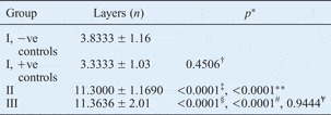
Vocal fold tissue from group II (receiving nandrolone decanoate) and group III (receiving nandrolone decanoate plus flutamide) animals revealed a significant increase in the number of epithelial cell layers, compared with the group I negative and positive controls (p < 0.05). However, there was no significant difference between the number of epithelial cell layers observed in groups II and III (p > 0.05) (Table I).
Epithelial cell layers in the 3 study groups

Layers data represent means ± standard deviations. *p < 0.05 (Student t-test) indicates statistically significant change. †Group I negative controls vs positive controls; ‡group II vs group I negative controls; **group II vs group I positive controls; §group III vs group I negative controls; #group III vs group I positive controls; ¥group II vs group III. +ve = positive; −ve = negative
Histological appearance
Group I
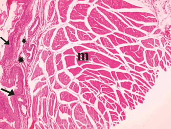
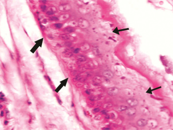
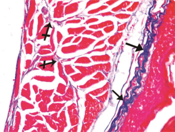
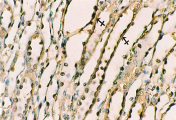
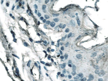
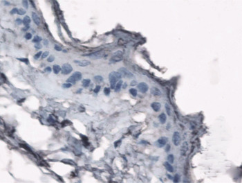
Upon light microscopy, the negative and positive control rats of group I both showed the same true vocal fold histological structure. Haematoxylin and eosin staining revealed that the true vocal folds were lined with three to four layers of low, stratified squamous epithelium (Table I) resting upon basal lamina. Subendothelial lamina propria and vocalis muscle fibres were also evident (Figures 1 and 2). Mallory trichrome staining revealed the presence of few collagen fibres in the lamina propria and between muscle fibres (Figure 3). Positive control slides prepared from rat kidney tissue showed positive immunohistochemical staining for epidermal growth factor receptor, in the form of fine brown granules within the medullary tubules (Figure 4). Negative control slides were prepared from both rat kidney and true vocal fold tissue (Figures 5 and 6, respectively). Negative immunohistochemical staining for epidermal growth factor receptor was seen in almost all the group I true vocal fold tissue examined (Figure 7).
Photomicrograph of true vocal fold tissue from a group I rat, showing a thin layer of stratified squamous epithelium (arrows), subendothelial lamina propria (stars) and muscle fibres (m). (H&E; ×100)

Photomicrograph of true vocal fold tissue from a group I rat, showing three to four layers of stratified squamous epithelial cells (thin arrows) resting on basal lamina (thick arrows). (H&E; ×1000)

Photomicrograph of true vocal fold tissue from a group I rat, showing minimal collagen fibres in the lamina propria (arrows) and between muscle fibres (crossed arrows). (Mallory trichrome; ×400)

Photomicrograph of rat kidney tissue, used as a positive control, showing a positive immunohistochemical reaction for epidermal growth factor receptor, appearing as fine brown granules within the medullary tubules (crossed arrows). (×1000)

Photomicrograph of rat kidney tissue, used as a negative control, showing a negative immunohistochemical reaction for epidermal growth factor receptor. (×1000)

Photomicrograph of true vocal fold tissue from a group I rat, used as a negative control, showing a negative immunohistochemical reaction for epidermal growth factor receptor. (×1000)

Photomicrograph of true vocal fold tissue from a group I rat, showing a negative immunohistochemical reaction for epidermal growth factor receptor. (×1000)

Group II
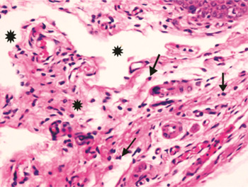
In the true vocal fold tissue of rats receiving nandrolone decanoate only, haematoxylin and eosin staining showed an increase in the number of stratified squamous epithelial cell layers, together with thick keratin and many mitotic figures. Slides also showed thick, oedematous lamina propria with many inflammatory cells, together with thick muscle fibres (Figures 8, 9 and 10). Mallory trichrome staining revealed the presence of excess collagen fibres in the lamina propria and between the muscle fibres (Figures 11 and 12). Immunohistochemical staining was positive in the superficial layers of the thickened epithelium, appearing as fine brown granules (Figure 13).
Photomicrograph of true vocal fold tissue from a group II rat, showing thickened stratified squamous epithelium (arrows), thick lamina propria (stars) and thick muscle fibres (m). (H&E; ×100)

Photomicrograph of true vocal fold tissue from a group II rat, showing many inflammatory cells (arrows) within a thick, oedematous lamina propria (stars). (H&E; ×400)

Photomicrograph of true vocal fold tissue from a group II rat, showing many layers of stratified squamous epithelium, mitotic figures (thin arrows) and a thick keratin layer (thick arrow). (H&E; ×1000)

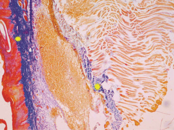
Photomicrograph of true vocal fold tissue from a group II rat, showing excess collagen fibres within the lamina propria (stars). (Mallory trichrome; ×400)

Photomicrograph of true vocal fold tissue from a group II rat, showing excess collagen fibres between muscle fibres (arrows). (Mallory trichrome; ×400)

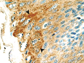
Photomicrograph of true vocal fold tissue from a group II rat, showing a positive immunohistochemical reaction for epidermal growth factor receptor within the superficial epithelial layers (arrowheads). (×1000)

Group III
In the true vocal fold tissue of rats receiving nandrolone decanoate plus flutamide, haematoxylin and eosin staining showed thickened stratified squamous epithelium and thick keratin. The lamina propria was also thick, with the presence of extravasated blood cells. The vocalis muscle fibres were thin and appeared to have degenerated (Figure 14). Mallory trichrome staining revealed the presence of excess collagen fibres within the lamina propria (Figure 15). Immunohistochemical staining was positive in the superficial layers of the thickened epithelium, appearing as fine brown granules (Figure 16).
Photomicrograph of true vocal fold tissue from a group III rat, showing thick stratified squamous epithelium (arrows) with thick keratin (arrowhead), thick lamina propria (star) with extravasated blood cells (crossed arrow), and thin muscle fibres with signs of degeneration (m). (H&E; ×100)

Photomicrograph of true vocal fold tissue from a group III rat, showing excess collagen fibres in the lamina propria (stars). (Mallory trichrome; ×400)

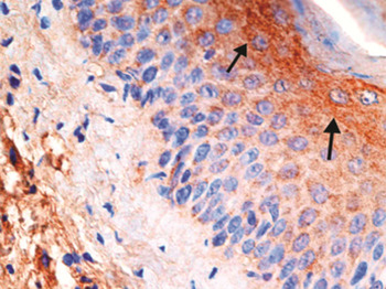
Photomicrograph of true vocal fold tissue from a group III rat, showing a positive immunohistochemical reaction for epidermal growth factor receptor within the superficial epithelial layers (arrows). (×1000)

Discussion
The vocal folds protect the airway and modulate airflow through the glottis during phonation.Reference Pedroso, Brasil, Martins, Nader and Simões Mde20 In the present study, rats were used as animal models as their vocal fold lamina propria is similar to that of humans.Reference Tateya, Sohn, Tateya and Bless21 The histological structure of the rat true vocal fold observed in the current study was similar to that described by others.Reference Lewis and Prentice22, Reference Hirano23
The use of anabolic, androgenic steroids is becoming increasingly popular among adolescent girls.Reference Clark, Kelton and Whitney3 Hormones have an important influence on the female voice.Reference Pedroso, Brasil, Martins, Nader and Simões Mde20 Androgens, encountered during development or adulthood, may cause anatomical and enzymatic sexual dimorphism in the larynx.Reference Boyd, Wissing, Heinsz and Prins24
In the current study, various structural changes were observed in the vocal folds of group II rats, which received the androgen nandrolone decanoate. Thickening was seen in the keratin and lining epithelial layers, with many mitotic figures in the latter. Similar results have been described by other authorsReference Talaat, Talaat, Kelada, Angelo, Elwany and Thabet25 (for example, Tammi and Santti found that testosterone caused a significant increase in the number of granular cells in the epidermis).Reference Tammi and Santti26 In addition, the present study observed a thickened lamina propria with many inflammatory cells, and a thick muscle layer. Other authors have found that anabolic, androgenic steroids induce an increase in muscle tissue bulk which can be attributed to both hypertrophy and new muscle fibre formation, with satellite cells playing a key role.Reference Hartgens and Kuipers2, Reference Rocha, Carmo, Roque, Hashimoto, Rossoni and Frimm27
The vibration of the vocal folds depends on their size and the constituents of the lamina propria.Reference Pedroso, Brasil, Martins, Nader and Simões Mde20 In the present study, Mallory trichrome staining showed an excess of collagen within the lamina propria and between muscle fibres, in the true vocal fold tissue of rats receiving nandrolone decanoate with or without flutamide. Anabolic steroids have also been reported to cause excessive collagen deposition within the cardiac extracellular matrix.Reference Andrade, Santos, Busato, Medeiros, Abreu and Moysés28–Reference Montisci, Cecchetto, Ruscazio, Snenghi, Portale and Viel29
In the present study, immunohistochemical staining for epidermal growth factor receptor was negative in true vocal fold tissue from the group I control rats. Other studies have found epidermal growth factor levels to be much higher in male compared with female mice,Reference Byyny, Orth and Cohen30 and have also found a general lack of expression of epidermal growth factor receptor in normal and dysplastic epithelia.
In the present study, true vocal fold tissue from group II rats (which received nandrolone decanoate only) showed positive immunohistochemical staining for epidermal growth factor receptor. Cook et al. found that keratinocytes can produce several epidermal growth factor receptor ligands which contribute to keratinocyte proliferation, both in vitro and in vivo.Reference Miyaguchi, Olofsson and Hellquist31 Roberts found that testosterone is the most important hormone in the control of epidermal growth factor accumulation in mice submandibular salivary glands.Reference Cook, Pittelkow and Shipley32,Reference Roberts33 Abnormalities in epidermal growth factor receptor expression are a common feature of hyperproliferative activity. One report found high levels of epidermal growth factor receptor expression in both simple and abnormal hyperplastic laryngeal lesions.Reference Krecicki, Jelen, Zalesska-Krecicka, Rak, Szkudlarek and Jelen-Krzeszewska34
Ray et al. have reported the case of a 47-year-old male professional body builder who presented with progressive stridor and hoarseness following abuse of anabolic steroids over a period of two years. Conservative management failed to resolve his symptoms, and a planned tracheostomy was necessary to secure the airway. This patient was subsequently treated with multiple laser resections, and was eventually decannulated.Reference Ray, Masood, Pickles and Moumoulidis5
The anti-androgen flutamide has been successfully used to treat cases of pachydermia laryngis and hirsutism.Reference Loewit, Hussl, Richter and Schwartz35 The present study investigated whether flutamide conferred protection against the effects of anabolic, androgenic steroids. The true vocal folds of group III rats (which received nandrolone decanoate plus flutamide) were observed to be thickened, with positive immunohistochemical staining for epidermal growth factor receptor, similar to group II rats (which received nandrolone decanoate alone). However, in the group III rats the vocalis muscle fibres were thin and appeared degenerate. Other authors have shown that administration of flutamide to metamorphic frogs prevented laryngeal muscle fibre hypertrophy in males receiving exogenous testosterone.Reference Sassoon and Kelley36
Extravasated blood cells were observed in the lamina propria of group III rat true vocal fold tissue. Flutamide has been reported to cause direct vasodilation by inducing endothelial nitric oxide releaseReference Iliescu, Campos, Schlegel, Morano, Baltatu and Bader37 and mesenteric vein vasculitis.Reference Wright and Cacala38
The exact mechanism of action of anabolic, androgenic steroids is unclear. They appear to act via multiple steroid signalling mechanisms. The effects of these steroids depend not only on the balance of androgen receptors versus oestrogen receptors, but also on the ability of the steroids to alter steroid metabolism and thus the endogenous steroid milieu. In addition, many anabolic, androgenic steroids may, upon aromatisation, exert physiological effects via classical oestrogen receptor pathways.Reference Penatti, Porter and Henderson1
• This study of female albino rats found that anabolic steroid administration was associated with structural changes in the true vocal folds
• Epidermal growth factor receptor was detected in the true vocal fold epithelium of rats given anabolic steroids, by immunohistochemical localisation
• Co-administration of anti-androgens did not prevent these changes
In the present study, the anti-androgenic drug flutamide could not prevent vocal fold changes produced by the androgen nandrolone decanoate. Other authors have reported that the changes produced by anabolic steroids in the female larynx are permanent and irreversible.Reference Talaat, Talaat, Kelada, Angelo, Elwany and Thabet25
Conclusion
Anabolic steroids cause various structural changes in the true vocal folds of adult female albino rats. Co-administration of anti-androgen therapy did not prevent these structural changes. This suggests that anti-androgens have a limited role in the management of anabolic-steroid-related changes in the true vocal folds of human females.