Introduction
Functional imaging studies reveal that patients with schizophrenia exhibit abnormal task-related neural activation, including both diminished and excessive regional activity (Manoach, Reference Manoach2003; Glahn et al. Reference Glahn, Ragland, Abramoff, Barrett, Laird, Bearden and Velligan2005). Fletcher et al. (Reference Fletcher, McKenna, Frith, Grasby, Friston and Dolan1998) demonstrated a curvilinear relationship between memory load and frontal lobe activation in patients with schizophrenia such that activation initially increased with increasing load but then decreased with further increase in load; by contrast, healthy controls exhibited a steepening increase in activation with increasing load. Using the n-back working memory task, Mendrek et al. (Reference Mendrek, Laurens, Kiehl, Ngan, Stip and Liddle2004) reported an attenuated increase in frontal cortex activation with memory load in patients with schizophrenia that proved to be due to excessive activity in the patients relative to controls during the 0-back condition combined with diminished activation during the more difficult 2-back condition, whereas Potkin et al. (Reference Potkin, Turner, Brown, McCarthy, Greve, Glover, Manoach, Belger, Diaz, Wible, Ford, Mathalon, Gollub, Lauriello, O'Leary, van Erp, Toga, Preda and Lim2009) found frontal hyperactivity in patients with schizophrenia during a working memory task that was most pronounced at intermediate memory loads. They concluded that their data supported neither hyper- nor hypofrontality in schizophrenia, but rather frontal lobe inefficiency that may be manifested in either direction depending on task demands.
This interpretation is supported by a meta-analysis of 41 studies of executive function in schizophrenia by Minzenberg et al. (Reference Minzenberg, Laird, Thelen, Carter and Glahn2009), which revealed diminished activation in regions implicated in top-down control, but excessive activation in what they suggest may be compensatory ventral, medial and posterior regions implicated in task maintenance functions. The authors note that inefficient cerebral recruitment in schizophrenia is a possible explanation of their findings, although they could also be explained by a disorder-specific difference in cognitive control networks topography.
Using a working memory task, Karlsgodt et al. (Reference Karlsgodt, Sanz, van Erp, Bearden, Nuechterlein and Cannon2009) found an inverted-U curvilinear relationship between activation in dorsolateral prefrontal cortex (DLPFC) and memory load in both patients and healthy controls, and also confirmed the findings of an earlier study (Karlsgodt et al. Reference Karlsgodt, Glahn, van Erp, Therman, Huttunen, Manninen, Kaprio, Cohen, Lönnqvist and Cannon2007), in which high-performing patients showed greater activation across conditions whereas low-performing patients showed reduced activation across conditions. They interpreted their finding as supporting a model in which higher activation levels are associated not only with intermediate task difficulty but also with task impairment, a conclusion borne out by another of their findings (Karlsgodt et al. Reference Karlsgodt, Glahn, van Erp, Therman, Huttunen, Manninen, Kaprio, Cohen, Lönnqvist and Cannon2007) that unaffected twins of patients with schizophrenia showed an intermediate load–activation relationship, suggesting that hyperactivation may be a compensatory mechanism in milder cases, and in those vulnerable for the disorder.
Hyperactivation in schizophrenia is not confined to brain regions normally recruited during tasks (task-positive regions). In healthy individuals, during many tasks that require attention to the external environment, deactivation is seen in a task-negative network of cerebral loci known as the default mode network (DMN) (Raichle & Snyder, Reference Raichle and Snyder2007); this deactivation tends to be greater in tasks that demand more attention to external stimuli (McKiernan et al. Reference McKiernan, Kaufman, Kucera-Thompson and Binder2003). DMN loci include the medial prefrontal cortex (MPFC); precuneus; posterior cingulate cortex (PCC); and lateral parietal, occipital and mid-temporal areas. As in studies examining hypo- and hyperactivation in schizophrenia of brain regions normally recruited by tasks, studies of DMN function in schizophrenia have also reported diverse findings, including attenuated task-induced DMN deactivation (Whitfield-Gabrieli et al. Reference Whitfield-Gabrieli, Thermenos, Milanovic, Tsuang, Faraone, McCarley, Shenton, Green, Nieto-Castanon, LaViolette, Wojcik, Gabrieli and Seidman2009; Hasenkamp et al. Reference Hasenkamp, James, Boshoven and Duncan2011), increased DMN connectivity (Whitfield-Gabrieli et al. Reference Whitfield-Gabrieli, Thermenos, Milanovic, Tsuang, Faraone, McCarley, Shenton, Green, Nieto-Castanon, LaViolette, Wojcik, Gabrieli and Seidman2009), increased attention-related DMN deactivation (Mannell et al. Reference Mannell, Franco, Calhoun, Cañive, Thoma and Mayer2010), and different abnormalities within different parts of the DMN (Garrity et al. Reference Garrity, Pearlson, McKiernan, Lloyd, Kiehl and Calhoun2007). It is plausible, then, that cerebral inefficiency in schizophrenia will be manifest not only by abnormal activation of regions required for performance of the task but also by interference from inappropriate activation in task-negative regions.
Although siblings of patients with schizophrenia have only about 9% risk of illness (Mortensen et al. Reference Mortensen, Pedersen, Westergaard, Wohlfahrt, Ewald, Mors, Andersen and Melbye1999), there is evidence for diverse dimensions of cognitive dysfunction in siblings (Egan et al. Reference Egan, Goldberg, Gscheidle, Weirich, Rawlings, Hyde, Bigelow and Weinberger2001), suggesting that some degree of abnormality may be present even in those who do not become ill. This would be consistent with the hypothesis that familial risk arises from a large number of genes (and/or non-genetic familial influences) each of small effect, but nonetheless contributing to a vulnerability for schizophrenia. Like patients with schizophrenia, siblings of patients with schizophrenia exhibit excessive regional activity in task-positive regions during the n-back working memory task compared with healthy controls (Callicott et al. Reference Callicott, Egan, Mattay, Bertolino, Bone, Verchinksi and Weinberger2003) and, similarly, non-psychotic first-degree relatives of patients with schizophrenia or schizo-affective disorder exhibit both excessive activation of frontal cortex during a working memory task (Seidman et al. Reference Seidman, Thermenos, Poldrack, Peace, Koch, Faraone and Tsuang2006; Whitfield-Gabrieli et al. Reference Whitfield-Gabrieli, Thermenos, Milanovic, Tsuang, Faraone, McCarley, Shenton, Green, Nieto-Castanon, LaViolette, Wojcik, Gabrieli and Seidman2009) and attenuated deactivation of the DMN (Whitfield-Gabrieli et al. Reference Whitfield-Gabrieli, Thermenos, Milanovic, Tsuang, Faraone, McCarley, Shenton, Green, Nieto-Castanon, LaViolette, Wojcik, Gabrieli and Seidman2009). In a meta-analysis of executive function findings in relatives of patients with schizophrenia and healthy controls by Goghari (2010), 13 studies reported hypoactivity and 14 reported hyperactivity in the right middle frontal region. Similarly, MacDonald et al. (Reference MacDonald, Thermenos, Barch and Seidman2009) found evidence of both hypo- and hyperactivation in studies of various aspects of cognitive function in the non-psychotic relatives of patients with schizophrenia.
Thus, there is evidence that people with vulnerability for schizophrenia can exhibit both hypo- and hyperactivity and that the difficulty of the task for the individual is an important factor affecting whether cerebral inefficiency is manifest as hypo- or hyperactivation. To resolve the question as to whether the mixed findings of hypo- and hyperactivation in the literature reflect different manifestations of an underlying core problem of cerebral inefficiency in those vulnerable to schizophrenia, and whether this inefficiency differs with diagnostic status, it is therefore vital to minimize confounds from individual differences in task difficulty.
We therefore designed a functional magnetic resonance imaging (fMRI) study in which adolescent and young adult participants vulnerable for schizophrenia (patients with a diagnosis of schizophrenia and siblings of patients) and healthy control participants performed a very easy auditory target-detection task. This age range was chosen to minimize the sampling of siblings who have already passed through the age of highest risk, thereby biasing the sample in favour of resilient individuals whose vulnerability has been compensated for by inherited or acquired protective factors. We matched the groups on task accuracy and compared their brain activity for the trial type expected to produce the smallest neural effects in healthy controls (non-targets). Under such conditions (easy task, matched performance, minimal expected neural response) we anticipated that cerebral inefficiency in those vulnerable for schizophrenia would be consistently manifest as hyperactivation, thus avoiding confounding by non-linearities in relationships between brain activation and task difficulty, and allowing group differences in cerebral inefficiency to be compared directly.
We predicted that, during non-target trials, subjects with vulnerability for schizophrenia (patients and healthy siblings of patients) would exhibit excessive activation regardless of diagnostic state (being ill or unaffected) relative to non-vulnerable subjects (healthy controls), in regions with net activation (across groups) by target trials. We also predicted possible attenuated deactivation at DMN sites with net deactivation (across groups) by target trials. We anticipated that unaffected siblings would differ in some respects from patients with schizophrenia, either by exhibiting attenuated hyperactivation or by additional compensatory hyperactivation.
Method
Participants
Ethical approval was granted by the Trent Multi-centre Research Ethics Committee (MREC) and by the Research and Development Department of the Nottinghamshire Healthcare National Health Service (NHS) Trust. Participants gave informed written consent if aged ⩾16 years; for those aged <16 years, written parental consent and verbal participant assent was obtained.
Participants were sampled from three populations: patients with schizophrenia; siblings of patients with schizophrenia; and healthy control participants. To maximize the likelihood that any differences in educational attainment or global cognitive function could be attributed to vulnerability to schizophrenia and not to educational opportunity or parental IQ, the groups were carefully matched for parental occupational status (ONS, 2004).
We recruited 21 patients satisfying DSM-IV criteria (APA, 1994) for schizophrenia, who were either patients from adolescent services or young adult cases within 5 years of onset of illness. The Schedules for Clinical Assessment in Neuropsychiatry (SCAN; Wing et al. Reference Wing, Babor, Brugha, Burke, Cooper, Giel, Jablenski, Regier and Sartorius1990) were administered. Clinical diagnosis was confirmed by consensus between three psychiatrists using all available information from clinical case-records and the standardized interviews. Current symptoms at the time of scanning were assessed using the Signs and Symptoms of Psychotic Illness (SSPI) scale (Liddle et al. Reference Liddle, Ngan, Duffield, Kho and Warren2002). All but one of the patients were medicated with atypical antipsychotic medication; of these, one was also on a mood stabilizer (carbamazepine) and three on an antidepressant (reboxitine, fluoxetine, citalopram). The median dose of antipsychotic medication in chlorpromazine equivalents (Woods, Reference Woods2003) was 350 (range 0–1200) mg/day.
Twenty unaffected full siblings of cases from the adolescent service were also recruited, and assessed for psychotic and prodromal symptoms using the Structured Interview for Prodromal Symptoms (SIPS; Miller et al. Reference Miller, McGlashan, Rosen, Cadenhead, Ventura, McFarlane, Perkins, Pearlson and Woods2003) and the Psychosis Screening Questionnaire (PSQ; Bebbington & Nayani, Reference Bebbington and Nayani1995). Siblings were also assessed using the Schizotypal Personality Questionnaire (SPQ; Raine, Reference Raine1991). Additionally, 41 healthy control participants with neither a current Axis I psychiatric diagnosis nor a personal or family history of psychotic illness, matched for age, sex and parental socio-economic status (SES) with the patient and sibling groups, were recruited from the general population.
To ensure that all participants in the study found the target-detection task similarly easy, any participant making more than 10% omission errors (failing to respond to targets) or more than 20% commission errors (responding to non-targets) was excluded from further analyses. As a result, three patients and two unaffected siblings were excluded. A subset of healthy control participants reaching these performance criteria was then selected, matched group-wise for age and accuracy of task performance (omission and commission errors) to the retained vulnerability group (patients plus unaffected siblings). This left a total of 62 participants in the study: 26 healthy control participants (15 male), 18 patients (12 male) and 18 unaffected siblings (seven male). Within the vulnerability groups there were eight sibling pairs. There was no significant difference in age between the healthy controls (mean = 20.3 years, s.d. = 4.9) and the vulnerability group (mean = 20.4 years, s.d. = 3.5), nor any significant differences in parental occupation. However, the patients were significantly older than the unaffected siblings [patient mean age = 22.4 years, s.d. = 3.3; unaffected siblings mean age = 18.3 years, s.d. = 2.2; t(24) = 4.447, p < 0.001]. There was no significant differences between these final groups in the distribution of parental SES codes [χ2(2) = 0.525, p = 0.773].
Task procedure
Two hundred and six stimuli were presented to the participant binaurally through insert earphones embedded within 25-dB sound-attenuating MRI-compatible headphones. Each train of stimuli comprised low-pitch non-target stimuli at 1000 Hz and high-pitch target stimuli at 1500 Hz, in fixed random order and with approximately equal probability (initial random selection resulted in 52% targets, 48% non-targets). All stimuli were presented at approximately 80 dB and all participants demonstrated that they could hear and discriminate the stimuli against background scanner noise. Stimulus duration was 200 ms with an intertrial interval (ITI: time between onsets of trial stimuli) drawn randomly from a skewed distribution (minimum = 2 s; median = 3.375 s; maximum = 19.75 s). The long ITIs were included to allow good sampling of baseline. Participants were instructed to respond with their right index finger as quickly as possible to the target stimuli only.
Imaging
Imaging was performed on a 3-T magnet (Oxford Magnet Technology, UK), custom-built head gradient set and birdcage quadrature radiofrequency coil. A T2-weighted inversion recovery image was acquired from each participant to obtain an anatomical image. A total of 312 functional (T2*-weighted) whole-brain images were collected in two scanning runs (156 scans/run), with a gradient-echo sequence [repetition time/echo time (TR/TE) 3000/40 ms, flip angle 90°, field of view (FOV) 24 × 24 cm, 64 × 64 matrix, 62.5 kHz bandwidth, 3.73 × 3.75 mm in-plane resolution, 5 mm slice thickness, 29 slices].
Analysis
Behavioral
For each participant, target detection rates (probability of responding to a target) and commission error rates (probability of responding to a non-target stimulus) were computed. These probabilities were transformed to z scores using a p to z transform with a mean of zero and a standard deviation of 1. The commission error z scores were then subtracted from the target detection z scores to give a d′ score, a measure of stimulus discriminability that is unconfounded by response bias (Green & Swets, Reference Green and Swets1966).
Image processing
Using SPM version 5 (Wellcome Trust Centre for Neuroimaging; www.fil.ion.ucl.ac.uk/spm), functional volumes were realigned and unwarped to minimize movement-by-susceptibility artifact distortion; spatially normalized to the participant's segmented, normalized anatomical image; and spatially smoothed with an 8-mm full-width at half-maximum (FWHM) Gaussian kernel.
For within-subjects analyses, stimulus onsets were modeled as delta functions and convolved with a canonical hemodynamic response and a temporal derivative. Targets and non-targets were modeled with separate regressors. Eight nuisance regressors (six sets of realignment parameters and the mean signal from voxels with >0.8 probability of being white matter or cerebrospinal fluid respectively) were included in the model. Three contrast images for each subject were computed: one representing targets minus baseline; one representing non-targets minus baseline; and one representing targets minus non-targets.
These contrast images were then entered into three random-effects between-subject analyses (one for each set of contrast images). Each model included three binary regressors representing group (healthy controls; patients; unaffected siblings) and three continuous covariates [representing age; median reaction time (RT); and error rate on the appropriate trial type (commission errors for non-targets; omission errors for targets)]. In addition, the regression model included eight dummy binary variables to model the familial shared variance within each of the eight sibling pairs.
Because of the large number of participants (n = 62) and hence the large number of contrast images for each analysis, for a substantial number of voxels at least one participant's contrast image was missing for that voxel. Therefore, to estimate between-subject β values for voxels with incomplete data across subjects, a multiple imputations procedure (Schafer & Olsen, Reference Schafer and Olsen1998) using five imputations was used to impute data for any voxel in which not more than nine (15%) participants had missing data.
Group analyses
To ascertain whether participants activated and deactivated the expected regions in response to target stimuli, the between-subjects images for responses to targets were thresholded at a voxel level of p < 0.05 after false discovery rate (FDR) correction for multiple comparisons. Next, to test the primary hypothesis of group differences in brain responses to non-targets, orthogonal contrasts between groups were computed as follows: non-vulnerable participants (healthy controls) were compared with vulnerable participants (patients plus siblings); and patients with schizophrenia were compared with unaffected siblings. We used a cluster-level significance criterion based on the spatial extent of suprathreshold voxel clusters proposed by Friston et al. (Reference Friston, Worsley, Frackowiak, Mazziotta and Evans1993): we used a voxel-level inclusion threshold of p < 0.01, uncorrected, and a cluster-level significance of p < 0.05, corrected.
To ascertain whether group differences in these clusters represented additional activation or attenuated deactivation relative to baseline, the mean voxel value in each cluster was computed for each participant's contrast image. Single-sample t tests were used to determine whether, for each cluster, the group mean of the single-subject means was significantly above or below zero. It should be noted that these one-sample tests are orthogonal to the tests of between-group differences used to identify the clusters.
To ascertain whether brain areas represented by these clusters represented areas significantly activated or deactivated by target trials across groups, mean voxel values within the same groups of voxels were computed for each participant's contrast image for target stimuli. The grand mean of these values across subjects was then tested for significant deviation from zero, again using one-sample t tests.
To determine whether subjects with vulnerability for schizophrenia and controls showed a significant difference in brain responses to targets as compared with non-targets, a test for group differences (healthy controls minus participants with vulnerability for schizophrenia) in the contrast of targets minus non-targets was performed.
Finally, to determine whether the siblings showed significant differences from controls during processing of non-targets, the contrast of activation in siblings with that in healthy controls was examined. However, it should be noted that this contrast is not orthogonal to the primary contrast of interest, namely the contrast of participants vulnerable for schizophrenia relative to controls.
Anatomical areas included in clusters were identified using automated anatomical labeling (aal; Tzourio-Mazoyer et al. Reference Tzourio-Mazoyer, Landeau, Papathanassiou, Crivello, Etard, Delcroix, Mazoyer and Joliot2002).
Results
Clinical features
The mean SSPI symptom score in the patients with schizophrenia was 15.8 (s.d. = 7.6, range 2–28), consistent with the majority of cases being in partial remission and receiving treatment in the community at the time of scanning (for two patients SSPI scores were not available for the time of scanning). The siblings all scored zero on the SIPS and the PSQ, demonstrating no clinical psychotic or prodromal features. The mean SPQ score was 14.8 (s.d. = 10.4, range 0–40), indicating some minor but non-specific psychopathology.
Behavioral performance
There was no difference between groups on mean d′ scores (F < 1) or on either commission error rates (F < 1) or omission error rates (F 2,59 = 1.213, p = 0.305). However, there was a significant difference between groups in RT (F 2,59 = 6.774, p = 0.002). Patients had significantly longer median RT to targets (mean = 526 ms, s.d. = 129 ms) than healthy control subjects (mean RT = 427 ms, s.d. = 90 ms, p = 0.006) and unaffected siblings (mean RT = 418 ms, s.d. = 78 ms, p = 0.006). There was no significant difference in RT between the two healthy (unaffected) groups.
Brain activation
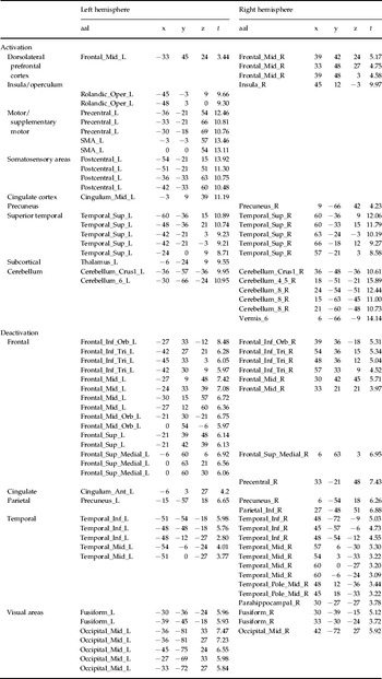
Across all participants, targets elicited robust activation relative to baseline and to non-targets in brain regions, which included those implicated in auditory processing (superior temporal cortex); bottom-up attention (insula, operculum, anterior cingulate); executive planning (right DLPFC); and motor preparation and response [motor and supplementary motor area (SMA); cerebellum]. Significant activation was also elicited in somatosensory areas (post-central cortex) and the thalamus. Targets also elicited robust deactivation relative to baseline bilaterally in brain regions of the DMN, namely the MPFC, precuneus, PCC, and lateral parietal, occipital and mid-temporal areas. In addition, there was robust suppression in regions implicated in visual processing (extra-striate regions bilaterally; fusiform gyrus bilaterally) and also in left frontal regions implicated in language processing (Broca's area; left DLPFC) (Table 1).
Target stimuli: locations (MNI coordinates) and t values of local maxima (>4 mm apart), thresholded at a voxel level of p <0.05 (FDR corrected), representing event-related activation (above implicit baseline) and deactivation (below implicit baseline)

MNI, Montreal Neurological Institute; FDR, false discovery rate; aal, automated anatomical labelling; SMA, supplementary motor area; R, right; L, left.
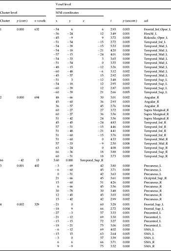
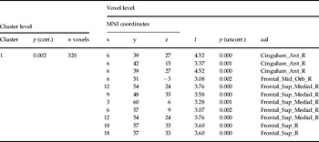
For non-targets, as predicted, participants with vulnerability for schizophrenia (unaffected siblings and patients) showed significant clusters of voxels exhibiting greater activation than in healthy controls (Table 2). These included three clusters in regions activated by targets across subjects (temporal cortex bilaterally and SMA) and one cluster in a region deactivated by targets (precuneus). When compared with patients, siblings showed one significant cluster of greater activation in the MPFC (Table 3). There were no significant clusters of voxels in which activity was greater in controls than in participants with vulnerability to schizophrenia.
Non-targets: locations and t values of local maxima (>4 mm apart), within significant clusters (p <0.05 corrected) thresholded at a voxel level of p <0.01 (uncorrected), representing brain regions in which greater activation was found in participants vulnerable for schizophrenia than in healthy controls

MNI, Montreal Neurological Institute; corr., corrected; uncorr., uncorrected; SMA, supplementary motor area; R, right; L, left.
Non-targets: locations and t values of local maxima (>4 mm apart), within significant clusters (p <0.05 corrected) thresholded at a voxel level of p <0.01 (uncorrected), representing brain regions in which greater activation was found in healthy siblings than in patients

MNI, Montreal Neurological Institute; corr., corrected; uncorr., uncorrected; aal, automated anatomical labeling; R, right; L, left.
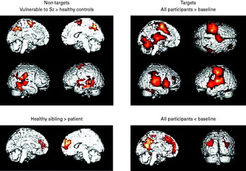
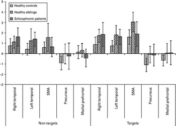
One-sample t tests (Fig. 1) conducted on mean voxel values within these clusters for each group indicated that, for non-targets, in both temporal clusters, siblings and patients showed significant activation relative to baseline. Siblings also showed significant activation relative to baseline in the SMA cluster. For the precuneus cluster, for non-targets, healthy controls showed significant deactivation relative to baseline.
Significant voxels and clusters for each contrast shown projected onto the cortical surface. Non-targets (left): clusters (voxel inclusion threshold: p < 0.05, uncorrected; cluster size threshold p < 0.01, corrected), where (top) patients and siblings of patients showed significantly greater activation than healthy controls and (bottom) where siblings of patients showed greater activation than patients. Targets (right): voxels [voxel inclusion threshold: p < 0.05, false discovery rate (FDR) corrected], where all participants showed event-related activation significantly greater than baseline.

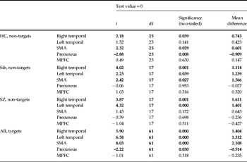
Examination of the activation elicited by targets within the clusters in which the activation elicited by non-targets (Fig. 2) differed between participants with vulnerability for schizophrenia and controls revealed that the mean intensity elicited by targets in the two temporal clusters and the SMA cluster (averaged across voxels and participants) was significantly greater than zero, indicating net activation. By contrast, the mean intensity in the precuneus cluster was significantly below zero, indicating net deactivation, although post-hoc tests conducted on the groups separately indicated that this only reached significance in the healthy control group [t(25) = 3.348, p = 0.003]. For the MPFC cluster that showed significantly greater activation for non-targets in siblings than in patients, the activation elicited by targets was not significantly different from zero, although it should be noted that a post-hoc t test showed significant deactivation for the healthy controls [t(25) = 2.524, p = 0.018] (Table 4).
Mean values in each of the non-target clusters in which group differences were found, for both target and non-target trials. SMA, supplementary motor area.

Results of single-sample t tests for the mean difference from zero of intensity values for target stimuli within the five statistically significant clusters of voxels that differentiated groups for non-target stimuli. The right and left temporal clusters and the SMA cluster all covered regions that were significantly activated, across groups, by target stimuli; the precuneus cluster was significantly deactivated, across groups, by target stimuli. The MPFC cluster, which showed greater activation by non-targets in healthy siblings than in patients, was not significantly activated or deactivated across groups by target stimuli, although it was significantly deactivated by target stimuli in healthy controls

HC, Healthy controls; Sib, siblings; SZ, schizophrenia patients; MPFC, medial prefrontal cortex; SMA, supplementary motor area; df, degrees of freedom.
Differences significant at p < 0.05 are shown in bold.
The two-way contrast (healthy controls minus participants with vulnerability for schizophrenia, targets minus non-targets), when thresholded at a voxel level of p < 0.05 (uncorrected), yielded one large, significant cluster of 1181 voxels (cluster-level significance p = 0.004 corrected). This included the fusiform and lingual gyrus, hippocampus and parahippocampus, cerebellum and thalamus. For the opposite contrast (participants with vulnerability for schizophrenia minus healthy controls), there were no significant clusters.
Finally, activation elicited by non-targets in each of the vulnerable groups was compared separately with that in controls, thresholded at a voxel level of p < 0.05 (uncorrected). These comparisons yielded large significant clusters (FDR corrected) of hyperactivation for both groups [siblings: one large cluster of 7114 (p < 0.001) voxels; patients: two large clusters of 1882 (p < 0.001) and 932 voxels (p = 0.004], and no significant clusters of hypoactivation.
Discussion
Although the groups were matched for task accuracy, both sibling and patient groups showed greater brain activation in response to non-target stimuli compared with healthy controls. Moreover, each group showed large clusters of hyperactivation compared with the control group, and no significant hypoactivation. Areas of hyperactivation included temporal and motor preparation regions significantly activated by target stimuli across groups, along with attenuated deactivation in a medial posterior cluster of voxels that included the precuneus and PCC, a region that forms part of the DMN, and significantly deactivated by target stimuli across groups. These results therefore support the study hypothesis that inefficient cerebral recruitment during a subjectively easy task, as reflected in both hyperactivation of task-relevant areas and attenuated suppression of areas normally deactivated by task stimuli, is a marker of vulnerability for schizophrenia and is observed in patients with overt disorder and in well siblings.
Potkin et al. (Reference Potkin, Turner, Brown, McCarthy, Greve, Glover, Manoach, Belger, Diaz, Wible, Ford, Mathalon, Gollub, Lauriello, O'Leary, van Erp, Toga, Preda and Lim2009) concluded from their study of working memory that frontal lobe inefficiency might be manifest as either hyperfrontality or hypofrontality depending on task demands. Our finding of hyperactivation in task-relevant brain regions outside of the frontal cortex suggests that inefficient recruitment also affects brain areas involved in relatively low-level cognitive processing of the stimuli, whereas our finding of attenuated suppression of a posterior medial region of the DMN (precuneus) suggests that this inefficiency may also be reflected in failure to switch off regions that may interfere with the recruitment of task-relevant circuits.
The participants with vulnerability for schizophrenia exhibited excessive activation during non-target trials in regions that were activated by targets in the entire sample. This suggests that the inefficient cerebral recruitment in participants with vulnerability for schizophrenia might arise, at least in part, from inappropriate allocation of behavioral salience to non-target stimuli. This would be consistent with the hypothesis of a deficit in assignment of proximal salience (Palaniyappan & Liddle, Reference Palaniyappan and Liddle2012), based on a substantial body of evidence indicating abnormal function of the salience network delineated by Seeley et al. (Reference Seeley, Menon, Schatzberg, Keller, Glover, Kenna, Reiss and Greicius2007) embracing bilateral insula and anterior cingulate cortex, leading to abnormal recruitment or suppression of other distributed networks, including the DMN, during simple information processing tasks (White et al. 2010 a,b). It would also be consistent with the ‘aberrant salience’ (Kapur, Reference Kapur2003) or ‘incentive salience’ (Heinz & Schlagenhauf, Reference Heinz and Schlagenhauf2010) hypothesis of schizophrenia. In addition, the attenuated deactivation of the DMN in participants with vulnerability for schizophrenia might indicate diminished ability to shift attention from internal stimuli to external stimuli, and would suggest that salience processing abnormalities do not merely involve an inappropriate assignment of salience to non-salient stimuli but an abnormal salience response in which neural activity in task-positive regions is increased while activity in task-negative regions is inadequately suppressed.
Siblings differed from patients in two respects. First, their median RTs were significantly shorter than those of patients but were not significantly different to those of controls. Second, they showed additional frontal activation in a region of MPFC relative to patients. This region showed significant deactivation in response to target stimuli in healthy control participants, and forms part of the DMN. We had no prior hypotheses concerning differences between siblings and patients, so any interpretation of this finding must remain speculative. With that proviso, it is worth noting that one role of this region is in monitoring erroneous responses (Ridderinkhof et al. Reference Ridderinkhof, Ullsperger, Crone and Nieuwenhuis2004), suggesting that a possible interpretation of the finding is that, although in response to non-targets, patients and unaffected siblings hyperactivated regions normally engaged in response to targets, this aberrant activity led to activation of a response–error monitoring system in the siblings that was not available to the patients. It is noteworthy therefore that, although the groups were matched for accuracy, the level of accuracy in the patients was only achieved at lower response speeds than in the healthy participants (unaffected siblings and healthy controls).
Patients with schizophrenia whose first contact with mental health services is early are likely to have greater familial loading (Byrne et al. Reference Byrne, Agerbo and Mortensen2002). Our sibling sample was recruited from families in which the member with schizophrenia was young; moreover, the sibling sample itself was young, and therefore had not yet passed through the period of high risk. It therefore represents a population in whom the risk of subsequently developing schizophrenia is relatively high. However, being free of psychiatric history or symptoms at the time of the study, they were also unmedicated. As they shared the same patterns of attenuated deactivation and increased activation to non-target stimuli as did the medicated patients, it is unlikely that our patient findings are artifacts of medication.
In a review of the possible role of genes involved in dopaminergic and glutamatergic neurotransmission in the dysfunctional and perhaps compensatory prefrontal cortical systems in schizophrenia, Tan et al. (Reference Tan, Callicott and Weinberger2007) speculated that the neurobiology of schizophrenia involves dynamic changes to cortical systems as they adapt to adverse neurobiological circumstances through the additional engagement of related cortical networks – possibly those usually engaged by processes lower in the cognitive processing hierarchy. Our study provides evidence that excessive recruitment of brain regions engaged in relatively low-level cognitive processing during a relatively easy target-detection task might be a marker for predisposition to schizophrenia. The observed attenuated DMN suppression in the precuneus indicates that there is also a failure to switch off task-irrelevant processing, raising the possibility that the abnormality of cerebral recruitment might entail a defect in the mechanism for switching efficiently between circuits according to current task demands.
Conclusions
These findings clarify the conflicting evidence from previous functional neuroimaging studies regarding hyper- and hypoactivation in patients with schizophrenia and their relatives, and show that when a task is sufficiently easy for all participants, significant hyperactivation, but not hypoactivation, is observed in both patients and healthy siblings of patients. Moreover, our results indicate that this hyperactivation is found not merely in the frontal cortex but is also widespread in other brain regions, including not only regions normally activated by salient stimuli but also regions normally suppressed in response to salient stimuli. This may indicate that vulnerability to schizophrenia is marked by inefficient cerebral processing that may reflect inadequate suppression of intrinsic activity in addition to aberrant hyperactivation to non-task-relevant stimuli.
Acknowledgments
This study was funded by the Public–Private Partnership (PPP) Foundation and partly by the Medical Research Council (UK), Grant no. 79708. We are grateful for technical assistance from K. Head and for J. Bell's extensive work on data preprocessing.
Declaration of Interest
P.F.L. has received speaker fees from Bristol Myers Squibb, Janssen Pharmaceuticals, Eli Lilly and AstraZeneca. D.D. has received sponsorship and hospitality to attend conferences from AstraZeneca, Janssen Pharmaceuticals and Eli Lilly. C.H. has received speaker fees from Janssen Pharmaceuticals, Eli Lilly and Shire. M.J.G. has received speaker fees from Eli Lilly and sponsorship to attend conferences from Shire.