I. INTRODUCTION
ZnSn(OH)6 (ZHS) is a kind of typical perovskite-structured hydroxide materials. Reference Jena, Kutty and Kutty1 In the compound, the metal atoms are octahedrally coordinated with oxygen atoms to form Sn(OH)6 and Zn(OH)6 polyhedra, which share their “O” corners to build the structural framework. Reference Christensen, Broch, Heidenstam and Nilsson2 ZHS has shown extraordinary performance as flame-, smoke-, and toxic gas-retardant. Reference Chiang, Wager, Hoffman, Jeong and Keszler3 It is mainly used as a kind of novel, nontoxic flame-retardant and environment-friendly polymeric materials. Reference Xu, Zhang, Qu and Tian4–Reference Hornsby, Winter and Cusack6 Recently, it is found that ZHS exhibits good photocatalytic activity in dye degradation. Reference Wang, Zhu and Yang7–Reference Wang, Ma, Liang, Wu and Wu9 Several methods have been developed to prepare ZHS, including solid-state metathesis approaches, solvothermal reactions, hydrothermal reactions, and wet sonochemical methods. Reference Xu, Liu and Niu10,Reference Lu and Tang11 The ZHS prepared via varying methods exhibited different photocatalytic activities. Wang et al. synthesized single-crystalline ZnSn(OH)6 hollow cubes via self-templated synthesis at room temperature, which exhibited certain photocatalytic activity for the degradation of phenol under UV light irradiation. Reference Wang, Tang, Liu, Wang, Sheng and Cheng12
In our previous study, we found that various morphologies of ZHS particles show different photocatalytic performance. Hollow cubes ZHS have a larger surface area, larger adsorption capacity, and higher crystallinity than the particles with other morphologies. Hence, hollow ZHS exhibited better photoactivity than other samples. However, ZHS, as a ternary wide band gap semiconductor (3.2–4.2 eV), can only be excited to produce electron–hole pairs under UV light irradiation, Reference Chen, Li, He, Hu, Ruan, Lin, Hu, Zheng and Shao13 which is only 3–5% of solar irradiation. This greatly limited the usage of ZHS. To extend the optical response of photocatalyst to the visible light region, various approaches have been used to modify it, including noble metal deposition, coupling with a semiconductor, ion doping, photosensitization, and surface reduction treatment.
In recent years, noble metals such as gold and silver nanoparticles (NPs) have attracted much attention because of their unique optical properties. The localized surface plasmon resonance (SPR) effect in gold and silver NPs results in strong surface plasmon absorption bands, usually in the visible region, and has been exploited to develop visible light plasmonic photocatalysts. Among these photocatalysts, silver and silver/silver halide coupled systems have been widely investigated. For example, Wang et al. Reference Wang, Huang, Zhang, Zhang, Qin, Dai, Zhan, Yu, Liu and Lou14 prepared a plasmonic photocatalyst of Ag/AgCl by treating Ag2MoO4 with HCl. They proposed that Ag NPs were photoexcited by the SPR effect under visible light irradiation. The photoexcited electrons were then trapped by the Ag NPs, which reduced the probability of electron–hole recombination. At the same time, the holes migrated to the AgCl surface and reacted with Cl ions to form Cl0 radicals. These plasmonic photocatalysts have been further loaded onto TiO2 to extend the optical response of TiO2 and reduce the electron–hole recombination efficiency. Hu et al. prepared a visible light photocatalyst of Ag/AgBr/TiO2 using the deposition-precipitation method, with cetylmethylammonium bromide (CTAB), Degussa P25, and AgNO3 as raw materials. Under alkaline conditions, CTAB can be adsorbed on the surface of TiO2 to limit the number of nucleation sites for AgBr, resulting in a homogeneous dispersion of AgBr. Reference Hu, Lan, Qu, Hu and Wang15
In this paper, Ag/AgCl/ZHS photocatalyst was prepared and investigated in Rhodamine B (RhB) degradation. The synthesized composites present much higher photoactivity than pure ZHS particles under visible light and UV light.
II. EXPERIMENTAL
A. Materials and reagents
Tin(IV) chloride pentahydrate (SnCl4⋅5H2O, ≥99.9%), zinc(II) chloride (ZnCl2, 100%), silver nitrate (AgNO3, ≥99.9%), citric acid monohydrate (C6H8O7⋅H2O, ≥99.5%), and sodium hydroxide (NaOH, ≥98.0%) were purchased from Aladdin Chemistry Co. Ltd. All reagents were of analytical grade and used without further purification.
B. Preparation
A typical synthesis of hollow ZHS cubes by co-precipitation method is as follows: 0.137 g ZnCl2 and 0.21 g C6H8O7⋅H2O were first dissolved in 10 mL of distilled water. Simultaneously, 0.351 g SnCl4⋅5H2O was dissolved in 5 mL of anhydrous ethanol. The Sn4+ solution was dropped into the Zn2+ solution to obtain a mixed solution. Then, NaOH solid was directly added to obtain a white precipitate. Reference Kim, Jeong, Song, Park, Cao and Moon16 After stirring at room temperature for 10 min, 25 mL of distilled water was also added. Then 20 mL of 2 M NaOH solution was added by dropwise into the precursor solution. The whole process was conducted in a magnetic stirring apparatus. The obtained white precipitate was centrifuged and rinsed thoroughly with distilled water and anhydrous ethanol several times, and finally dried at 60 °C for 10 h.
The synthesized hollow ZHS cubes (1 g) were dispersed in distilled water, ultrasonicated for 15 min at room temperature, and then mixed with 10 mL of 0.05, 0.1, and 0.2 M AgNO3 solution. After stirring for 10 min at room temperature, 10 mL of 0.05, 0.1, and 0.2 M HCl aqueous solution was added. The mole ratios of Ag to ZHS are 1/2, 1/1, and 2/1, respectively. The mixture was ultrasonicated for 10 min and stirred for 20 min at room temperature. The filtered powder was washed with distilled water and dried at 80 °C for 12 h. Then the dried powder was irradiated with a 500 W halogen tungsten lamp for 30 min to reduce part of the Ag+ ions to Ag0 species. The synthesized plasmonic photocatalyst was denoted as Ag/AgCl/ZHS. The experimental conditions for the preparation of samples are listed in Table I.
Experimental conditions for the preparation of samples.

C. Characterization
X-ray diffraction (XRD) patterns of the catalysts were obtained on a Philips x-ray diffractometer (PW 3040/60, Amsterdam, Netherlands) using Cu Kα radiation. The accelerating voltage and the current were 40 kV and 40 mA, respectively. The scanning electron microscope (SEM) images were taken on a field emission scanning electron microscope (Hitachi S-4800, Tokyo, Japan). UV-vis diffuse reflectance spectra were recorded in the range of 200–800 nm on a UV-vis spectrometer (Nicolet, evolution 500, Thermo, MA), where BaSO4 was used as a reference sample. Pore structure parameters of the samples, including Brauner–Emmett–Teller (BET) surface areas and density-functional-theory pore size distribution, were determined according to the nitrogen adsorption–desorption isotherms measured at 77 K using a physisorption apparatus (Quantachrome Autosorb-1, Boynton Beach, FL).
D. Catalytic performances
The catalytic activity was measured in a self-made photoreaction apparatus. The photocatalytic activity of catalyst was evaluated by the degradation of RhB. A 500 W Hg lamp was used as a source of UV light, the dominant wave length was 284 nm. The initial concentration of RhB is 1.0 × 10⁻⁴ mol L−1. A 250 W Xe lamp equipped with a UV cutoff filter (>420 nm) was used as a source of visible light. The initial concentration of RhB is 1.0 × 10⁻⁵ mol L−1, and the concentration of catalyst is 0.5 g L−1. To disperse the catalyst powder and reach the adsorption–desorption equilibrium on the photocatalyst surface, the suspensions were stirred for 1 h in the dark before irradiation. During the RhB photodecomposition, the samples were withdrawn at regular intervals and centrifuged by a low speed centrifuge. The absorption of filtrate was measured in the range of 450–600 nm by using a UV-vis spectrophotometer (TU-1810).
The examination experiment process of reactive species was similar to the photodegradation experiment. A quantity of scavengers were introduced into the RhB solution prior to addition of the catalyst. The concentration of scavengers was controlled to be 0.2 mol L−1 according to the previous studies. Reference Li, Wong, Zhang, Hu, Yu, Chan and Wong17,Reference Cao, Xu, Luo, Lin and Chen18 These quenchers included isopropanol (IPA, a quencher of •OH), benzoquinone (BQ, a quencher of •O2 −), and ammonium oxalate (AO, a quencher of h+).
III. RESULTS AND DISCUSSION
A. Characterization of photocatalysts
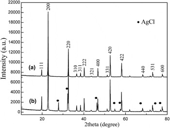
The phase purity of the as-prepared samples was obtained by XRD and the results are shown in Fig. 1. The XRD peaks of ZHS match SnZn(OH)6 (JCPDS No. 20-1455). No additional peaks of other impurities were observed, indicating that the as-prepared sample (S1) is pure ZHS. In addition, the average particle size of particles was calculated using the Debye–Scherrer formula,
 ${D_{{\rm{hkl}}}} = {{K{\rm{\lambda }}} \mathord{\left/ \kern-\nulldelimiterspace} {{\rm{\beta cos\theta }}}}$
, where λ is the wave length of the Cu Kα used, β is the full width at half maximum of the diffraction angle considered, K is a shape factor (0.94), and θ is the angle of diffraction.
Reference Wang, He, Li, Cai, Luo and Zhao19
These particles have an average size of 44 nm. Figure 1(b) shows the XRD of Ag/AgCl/ZHS (S3). No other crystalline phase was observed except cubic ZHS and AgCl. The two phases all exhibit sharp and strong peaks, which might be ascribed to their high crystallinity.
Reference Fu, Leung, Wang, Xue and Fu20
Furthermore, as shown in the XRD patterns of the Ag/AgCl/ZHS (S3), the diffraction peaks of ZHS became stronger, indicating that the crystallinity of ZHS particle was increased. This change can be due to a series of processes during the loading of Ag/AgCl particles. Besides the cubic ZHS phase, other diffraction peaks were indexed to a cubic phase of AgCl (JCPDS No. 00-31-1238). Nevertheless, the diffraction peaks of Ag NPs were not observed, suggesting that the amount or size of the Ag NPs was small in the Ag/AgCl/ZHS (S3) catalysts.
XRD pattern of the as-prepared S1 (a) and S3 (b).

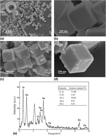
Figures 2(a) and 2(b) show the SEM images of as-prepared ZHS samples. It can be observed that the morphology and size of ZHS (S1) particles are uniform. They have an average edge length of 500–600 nm. The hollow cubes contain sharp corners and some cubes exhibit holes in the corners [Fig. 2(a) inset], indicating that the particles have a hollow interior. The thickness of the cell wall is about 50–100 nm. This hollow structure means a large surface area of the sample, which benefits the adsorption of small organic and inorganic NPs (Ag/AgCl NPs) inside the hollow cubes. In addition, this structure facilitates the transport of photogenerated carriers during a photocatalytic reaction. Reference Wen and Ding21 After loading of Ag/AgCl NPs, the morphology of ZHS shows no obvious change, as shown in Figs. 2(c) and 2(d). Some NPs are attached on the surface of the cubes which might be Ag or AgCl NPs.
SEM images of S1 (a, b), S3 (c, d), and EDS spectrum of S3 (e).

Figure 2(e) shows the EDS analysis of as-prepared Ag/AgCl/ZHS (S3) particles. Approximately 9.36 at.% Sn element is present in the catalyst. The atomic ratio of zinc:tin:oxygen in the cubes is close to 1:1:6, consistent with the composition of ZnSn(OH)6 determined by XRD. Furthermore, approximately 4.6 at.% Ag and 3.42 at.% Cl are present in the catalyst, which also agrees well with the XRD result.
B. Photoabsorption property
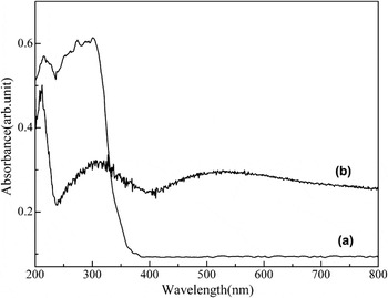
The photoabsorption ability of materials can affect their photocatalytic activities greatly. Figure 3 shows the UV-vis absorption spectra of these as-prepared samples. The absorption edge of the pure ZHS (S1) was located at 270 nm, indicating a strong photoabsorption property in the UV light region. Compared with ZHS hollow cubes (S1), Ag/AgCl/ZHS (S3) exhibited an absorption edge near 370 nm corresponding to the interband transition of ZHS and a broad absorption band between 350 and 800 nm. The latter band is attributed to the SPR absorption of Ag NPs, which further verified that the Ag/AgCl/ZHS (S3) contained silver NPs as a result of the photochemical decomposition or photocatalytic reduction of AgCl. Clearly, the loading of Ag/AgCl NPs extended the absorption of the ZHS hollow cubes to the visible region. This indicates that the synthesized Ag/AgCl/ZHS composite might be active under visible light irradiation.
The UV-vis absorption spectra of S1 (a) and S3 (b).

C. Surface areas and pore structure
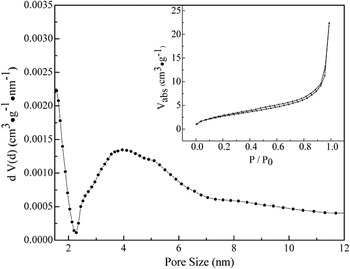
The BET specific surface area of the as-prepared ZHS (S1) sample, calculated from N2 isotherms at 77 K, was 54.4 m2 g−1. The adsorption–desorption isotherm (Fig. 4) of the hollow ZHS sample (S1) shows the type IV isotherm, indicating its mesoporous structure. The average pore size of the hollow sample was 4.0 nm with 3.0 nm range distribution of pore size. The BET specific surface area of Ag/AgCl/ZHS (S3) was 51.7 m2 g−1.
The nitrogen adsorption–desorption isotherm (inset) and the pore size distribution plot for ZHS.

D. Photodegradation of RhB under visible and UV light irradiation
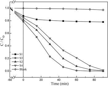
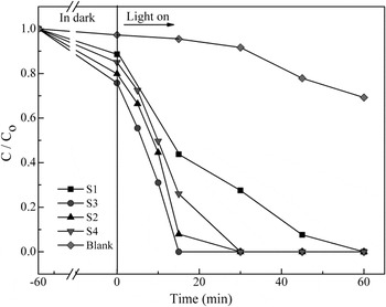
The photocatalytic decolorization of RhB dye was used to evaluate the photocatalytic performance of the photocatalysts under visible and UV light irradiation. ZHS (S1) was chosen as the reference photocatalyst. The results are shown in Fig. 5, where c 0 is the initial dye concentration and c is the dye concentration at time t. To disperse the catalyst powder and reach the adsorption–desorption equilibrium on the photocatalyst surface, the suspensions were stirred for 1 h in the dark before irradiation. Both pure ZHS cubes and Ag/AgCl/ZHS exhibited strong adsorptive capacities for RhB in the dark. The dye concentration decreased to 15% of its initial value by the adsorption on pure ZHS hollow cubes (S1). For Ag/AgCl/ZHS (S3), the value is 25%. This result suggests that the adsorptive capacity of the ZHS hollow cubes increased after the loading of Ag/AgCl NPs. Under visible light irradiation, nearly no dye molecules were degraded in the presence of ZHS. However, when Ag/AgCl was loaded on ZHS, the photocatalytic activity was improved greatly. After visible light irradiation for 60 min, RhB was completely degraded in the presence of S3 sample (Fig. 5). The degradation rate is calculated to be 0.00101 min−1, which is higher than that of S2 (0.00044 min−1) and S4 (0.00029 min−1) [see Fig. S2]. Under UV light, the RhB degradation process was further accelerated (Fig. 6). The stronger photoactivity can be mainly attributed to the increased ability of catalyst in light adsorption and electron–hole separation, which results from the plasmonic effect of Ag/AgCl particles. Reference Zhang, Chen, Liu and Tsai22 However, S3 exhibited the highest activity, indicating that the concentration of Ag/AgCl has great effect on the photoactivity. For the sample with a low concentration of Ag/AgCl, the promotion effect in light adsorption or electron–hole separation might be limited. On the contrary, when the content of Ag/AgCl is too high, the loaded Ag/AgCl might cover ZHS. Considering that the photogenerated electron gathers on the ZHS, the covering by Ag/AgCl would suppress the photocatalytic reaction. That is why there has an optimal concentration of Ag/AgCl (S3 sample) in the paper, just as the previous literature reported. Reference Dong, He, Li, Cai, Hu, Wang, Lin, Luo, Yi, Zhao, Weng and Wan23–Reference Li, Hua, Yue and Li25 Clearly, the loading of optimal Ag/AgCl on ZHS created a new photocatalyst with high activity under visible light. Reference Kawahara, Suzuki, Ohko and Tatsuma26,Reference Wang, Huang, Qin, Zhang, Dai, Wei and Whangbo27
Concentration percent of photocatalytic degradation of RhB as a function of irradiation time under visible light.

Concentration percent of photocatalytic degradation of RhB as a function of irradiation time under UV light.

E. Photocatalytic mechanism
In general, all photocatalytic activity is mainly influenced by three factors: (i) the adsorption ability of the reactants, (ii) the photoabsorption ability in the available light energy region, (iii) the separation and transporting rate of the photogenerated electrons and holes in catalyst. Reference Chang, Rao, Kuo, Cai and Wong28 The hollow structure might improve the utilization of light since the incident light can be reflected in the hole. At the same time of light irradiation, hollow structure can absorb much more of light and generate more electron–hole pairs, which is beneficial for the photocatalytic reaction.
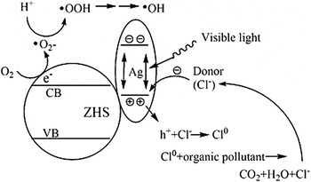
The photocatalytic activity of prepared Ag/AgCl/ZHS can be understood by the following suggested mechanism (as shown in Fig. 7), which is similar to the mechanism of plasmon-induced charge separation by Tatsuma et al. and the mechanism of Ag@AgCl plasmonic photocatalysis by Huang et al.
Reference Wang, Huang, Qin, Zhang, Dai, Wei and Whangbo27
Under visible light irradiation, photogenerated electron–hole pairs are formed in Ag NPs due to SPR. The photoexcited electrons at the silver NPs are injected into the ZHS conduction band, as shown in Fig. 7. The conduction band potential of ZHS was calculated to be −0.19 eV (see Table SI and Fig. S1) which is more negative than the potential of
 ${{\rm{O}}_2}/ \bullet {\rm{O}}_2^ - \left( {{E_{{{\rm{O}}_2}/ \bullet {\rm{O}}_2^ - }}} \right) = - 0.046\;{\rm{V}}$
.
Reference Fujishima, Rao and Tryk29–Reference Sawyer and Valentine31
Hence, the injected electrons on the conduction band of ZHS can reduce molecular oxygen to form the superoxide radical anions (•O2
−) and the subsequent active species •OOH and •OH radicals.
Reference Tian and Tatsuma32
These active species will result in the degradation and mineralization of RhB. Meanwhile, the holes transfer to the surface of the AgCl particles and most likely are terminated by Cl− ions because the surface of AgCl particles is negatively charged due to the Xe lamp induced reduction of partial Ag+ ions. The transferred holes will cause the oxidation of Cl− ions to Cl0 atoms. As chlorine atoms are also reactive radical species, they are able to oxidize RhB and be reduced to chloride ions again,
Reference Wu, Liu and Zhao33
so Ag NPs can be rapidly regenerated and the Ag/AgCl/ZHS system remains stable. The major reaction steps in this plasmonic photocatalytic mechanism under visible light irradiation are summarized in Eqs. (1)–(8).
 $${\rm{Ag - NPs}} + h\nu \to {\rm{Ag - NP}}{{\rm{s}}^*}\quad{\rm{,}}$$
 $${\rm{Ag - NP}}{{\rm{s}}^ * } + {\rm{ZHS}} \to {\rm{Ag - NP}}{{\rm{s}}^{ + \bullet }} + {\rm{ZHS(e)\quad,}}$$
 $${\rm{ZHS(e)}} + {{\rm{O}}_2} \to {\rm{ZHS}} + \bullet {\rm{O}}_2^ -\quad {\rm{,}}$$
 $$\bullet {\rm{O}}_2^ - + {{\rm{H}}^ + } \to \bullet {\rm{OOH}}\quad,$$
 $$\bullet {\rm{OOH}} + {\rm{ZHS(e)}} + {{\rm{H}}^ + } \to {{\rm{H}}_2}{{\rm{O}}_2} + {\rm{ZHS\quad,}}$$
 $${{\rm{H}}_2}{{\rm{O}}_2} + {\rm{ZHS(e)}} \to \bullet {\rm{OH}} + {\rm{O}}{{\rm{H}}^ - } + {\rm{ZHS\quad,}}$$
 $${\rm{Ag - NP}}{{\rm{s}}^{ + \bullet }} + {\rm{C}}{{\rm{l}}^ - } \to {\rm{Ag - NPs}} + {\rm{C}}{{\rm{l}}^0}\quad{\rm{,}}$$
 $$\eqalign{ & {\rm{Organic}}\;{\rm{pollutant}} + {\rm{C}}{{\rm{l}}^0}({\rm{or}}\; \bullet {\rm{OH}}\;{\rm{or}} \bullet {{\rm{O}}_2}\;{\rm{or}}\;{{\rm{H}}_2}{{\rm{O}}_2}) \to \to \cr & {\rm{Degraded}}\;{\rm{or}}\;{\rm{mineralized}}\;{\rm{products}}{\rm{.}} \cr}$$
Schematic diagram for the charge separation in a visible light irradiated Ag/AgCl/ZHS system.

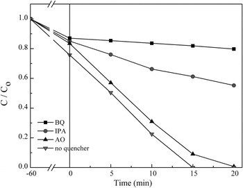
The suggested mechanism was partly verified by the reactive species trapping experiment. As shown in Fig. 8, AO (h+ scavenger) shows a little effect on the photocatalytic activity. Meanwhile, the addition of BQ (•O2 − scavenger) and IPA (•OH quencher) significantly decreased the photocatalytic activity of photocatalyst. Reference He, Cai, Li, Wu, Lin, Zhao and Luo34,Reference He, Cai, Li, Wu, Yi, Zhao and Luo35 The result in Fig. 8 indicated that •O2 − and •OH species were the main active species, which is consistent with the aforementioned mechanism.
Photocatalytic activity of as-prepared catalyst with different quenchers under UV light.

IV. CONCLUSIONS
In summary, ZHS particles (S1) with hollow structure were synthesized by an alkali-assisted dissolution process at room temperature. Ag/AgCl/ZHS composite, which is a plasmonic photocatalyst, was prepared by the photodepositing method. The as-prepared Ag/AgCl/ZHS exhibited good photocatalytic decolorization activity for RhB under visible light, which can be attributed to a synergistic effect between SPR effect and large adsorption capacity of the catalyst.
Supplementary Material
To view supplementary material for this article, please visit http://dx.doi.org/jmr.2014.1022.
ACKNOWLEDGMENTS
This work was supported by grants from the Nature Science Foundation of China (Nos. 21373188 and 21243010), Nature Science Foundation of Zhejiang Province (LY12B03001), and the National Natural Science Foundation of China (21003109).