Coarctation of the aorta is a very common congenital heart malformation occurring in ~6–8% of all congenital heart diseases.Reference Anderson, Baker, Macartney, Rigby, Shinebourne and Tynan 1
It is frequently associated with other abnormalities, for example tubular hypoplasia of the aortic arch (63%), left ventricular outflow tract obstruction (40%), bicuspid aortic valve (40%), ventricular septal defect (28%) and atrial septal defect (12%).Reference Becker, Becker and Edwards 2 , Reference Teo, Cannell, Babu-Narayan, Hughes and Mohiaddin 3
Echocardiography is the diagnostic modality for congenital heart diseases in most of the children including those with coarctation of the aorta.Reference Lai, Geva and Shirali 4 , Reference Mertens, Ganame and Eyskens 5
Coarctation of the aorta usually manifests as a discrete constriction of the aortic isthmus with a posterior shelf. However, it is more likely to represent a spectrum of aortic narrowing from discrete constriction to tubular hypoplasia. Colour Doppler may reveal the presence of turbulent flow and abnormal diastolic flow in most of the cases. There is increased velocity of the blood flow at the narrowed segment with diastolic run-off detected by pulsed and continuous wave Doppler.Reference Kenny and Hijazi 6
In neonates and young infants, coarctation of the aorta may be missed or underestimated by echocardiography, especially in the presence of a large patent ductus arteriosus or severe concurrent illness, for example sepsis or shock. This may be due to the presence of a patent ductus arteriosus without flow acceleration at the isthmus, poor image quality, lack of coplanarity of the aortic arch, ductus, and descending aorta, and the location of the coarctation further downstream in the descending aorta.
Allowing the patent ductus arteriosus to close under observation to allow the coarctation to “declare itself” can be done, but this may take the form of acute development of aortic obstruction with potential consequences including distal hypoperfusion and metabolic acidosis, renal injury, left ventricular dysfunction, pulmonary oedema and pulmonary hypertension.Reference Fesseha, Eidem and Dibardino 7
The carotid-subclavian artery index, defined as the ratio of the distal transverse arch diameter to the distance between the left common carotid and left subclavian arteries, was proposed because of the frequent difficulty in confidently establishing the diagnosis of coarctation of the aorta, particularly in the smallest and youngest patients with a reported excellent sensitivity and specificity.Reference Dodge-Khatami, Ott, Di Bernardo and Berger 8
The association of coarctation with displacement of the left subclavian artery and hypoplasia of the transverse aortic arch is well known.Reference Glancy, Morrow, Simon and Roberts 9 , Reference Morrow, Huhta, Murphy and McNamara 10
The isthmus/descending aorta ratio was also proposed as a supplementary tool for the detection of coarctation of the aorta using two-dimensional echocardiography.Reference Lu, Wang and Chang 11
The objectives of our study were to evaluate in greater detail the validity of such indexes and to look for other two-dimensional echocardiographic predictors of coarctation of the aorta. The study was approved by the centre’s research committee.
Methods
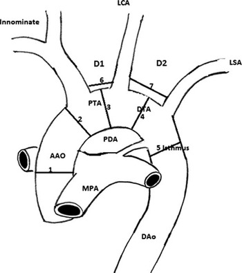
From July, 2010 to December, 2011, echocardiographic evaluation was reviewed for consecutive patients with coarctation of the aorta, as well as the control group. All echocardiograms were done by Vivid 7 Echo machines. Echo was reviewed by The Echo PAC Dimension clinical workstation (GE Healthcare, United States of America). End-systolic measurements were obtained from different sites of the aortic arch (Fig 1). We included neonates and infants 3 months or less of age at the time of echocardiography. All patients diagnosed to have coarctation of the aorta had surgery, which confirmed the diagnosis. Children with anomalies of the head and neck vessels, for example common brachiocephalic trunk or a vertebral branch arising from the aortic arch, or interrupted aortic arch, were excluded. Variables assessed include demographic data, such as gender, age, weight. Echocardiographic measurements include ascending aorta diameter proximal to the first aortic arch branch, proximal transverse arch diameter (Between the brachiocephalic and left common carotid arteries), distal transverse arch diameter (between the left common carotid and the left subclavian arteries), aortic isthmus diameter (just beyond the left subclavian artery), the distance between the brachiocephalic and left common carotid arteries (named distance 1), and the distance between the left common carotid and left subclavian arteries (named distance 2). Measurements were recorded according to the published guidelines.Reference Lai, Geva and Shirali 4 , Reference Lopez, Colan and Frommelt 13 Distances 1 and 2 were measured just above the origin of the head and neck vessels from the aortic arch. We used the Echo PAC Dimension clinical workstation (GE Healthcare), and hence there was no need for calibration for off-line measurements of the cardiac dimensions.
Echocardiographic measurements performed. AAO=ascending aorta; D1=distance 1 – distance between innominate (brachiocephalic) and left common carotid arteries; D2=distance 2 – distance between left common carotid and left subclavian arteries; DAo=descending aorta; DTA=distal transverse arch; Innominate=Innominate or brachiocephalic trunk; LCA=left common carotid artery; LSA=left subclavian artery; MPA=main pulmonary artery; PTA=proximal transverse arch.

We also reviewed the aortic valve anatomy – unicuspid, bicuspid or tricuspid – as well as the associated findings. The surgical diagnosis was reviewed from the surgical reports. We indexed the following: distal transverse arch/ascending aorta ratio, carotid-subclavian artery index (distal transverse arch/distance 2 ratio), isthmus/ascending aorta ratio, as well as the ratio between distance 2 and the ascending aorta.
Data analysis
Data were analysed using the SPSS 16.0 for Windows (by IBM) software program. Continuous variables were expressed as mean±standard deviation. Categorical variables were expressed as frequencies and percentages. Univariate analyses were used to compare between those with and without coarctation. The unpaired independent sample t-test was used for comparing continuous variables, and the Pearson χ2 test was used for comparing categorical variables. Receiver Operating Characteristic curve was applied to detect the significant predictors for the presence of coarctation and the area under the curve was estimated. The sensitivity, specificity, predictive values and likelihood ratios were calculated. Statistical significance was considered at a p-value≤0.05.
Results
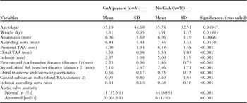
During the study period, we included 31 patients with coarctation of the aorta and 50 patients with no coarctation. All patients underwent echocardiography at an age of 3 months or less. A comparison between the two groups is presented in Table 1. The mean age±standard deviation for those with coarctation was 35±44 days compared with 35±32 days for the control group (p-value 0.95). The mean weight±standard deviation for those with coarctation was 3.3±0.9 kg compared with 3.9±1.3 kg for the control group (p-value 0.034).
Comparison between those with and without coarctation of the aorta.

CoA=coarctation of the aorta; SD=standard deviation; TAA=thoracic aortic aneurysm
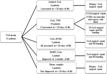
Among the 31 patients with coarctation of the aorta, 6 had isolated coarctation and 25 had associated defect – ventricular septal defect in 19, atrioventricular septal defect in three, double-outlet right ventricle in one, and Shone complex in two. All had patent ductus arteriosus. Twenty two patients were diagnosed to have coarctation before the 28th day of life, and nine were diagnosed beyond the neonatal period (29 days to 3 months of age). Among all 8 patients presented with shock and the others presented with shortness of breath and/or the detection of cardiac murmur on cardiovascular examination (Fig 2). All patients were admitted and given prostaglandin infusion once coarctation was diagnosed or suspected. All were referred for cardiac surgery. Those with isolated coarctation, small muscular ventricular septal defects and Shone complex (11 patients) had coarctation surgical repair only. Those with associated large ventricular septal defects, double-outlet right ventricle and atrioventricular septal defect had coarctation repair and pulmonary artery banding (20 patients) (Fig 2).
Different diagnosis and time of presentation, as well as the surgical procedure(s) for those with coarctation of the aorta.

Among the 50 patients in the control group with no coarctation – 27 neonates and 23 infants <3 months of age – patent ductus arteriosus was present in six, isolated ventricular septal defect was present in eight, one had secundum atrial septal defect, and one had secundum atrial septal defect and ventricular septal defect.
Abnormal aortic valve was present in 20 patients (65%) with coarctation – unicuspid in two and bicuspid in 18 patients – compared with six (12%) in the control group (p<0.001).
There were no significant differences in the diameter of the ascending aorta between the two groups. The diameters of the proximal transverse arch, the distal transverse arch, and the isthmus were smaller in the group with coarctation of the aorta compared with the control group. The distance between the aortic arch branches (distance 1 and 2) was longer in the coarctation group. Regarding distance 1, most cases with coarctation as well as in the control group had a distance <3 mm. Only three neonates in the coarctation group had a distance more than 3 mm (Table 1).
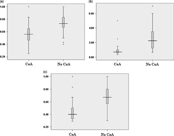
There was some overlap between those with coarctation and the control group regarding the diameter of the distal transverse arch and distance 2. A longer distance 2 was present more in those with coarctation of the aorta (Fig 3).
Stem and leaf diagrams of the carotid subclavian index (carotid-subclavian artery index), distal transverse arch/ascending aorta ratio (DTA/AAo), and the isthmus/ascending aorta ratio (I/AAo index) for those with CoA and the control group. The boxes represent the interquartile range, the horizontal line inside the box represents the median, and the ‘I’ bars represent the 10th and 90th percentiles of the values. The black dots represent values above the 90th percentile or below the 10th percentile. ( a ) Distal transverse arch/ascending aorta ratio (DTA/AAo ratio). ( b ) Carotid subclavian artery index in those with and without coarctation – distal transverse aortic arch diameter divided by the distance between the left common carotid artery and the left subclavian artery. ( c ) Aortic isthmus/ascending aortic arch ratio.

Results of receiver operating characteristic curves
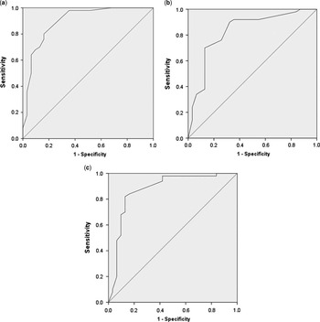
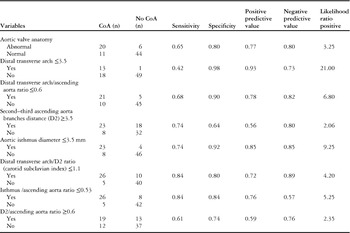
Receiver operating characteristic curves were created for all variables including the distal transverse arch, isthmus, distance 2, carotid-subclavian index, distal transverse arch/ascending aorta ratio, isthmus/ascending aorta ratio, and for distance 2/ascending aorta ratio. (Fig 4). The area under the curve was significant for all of them. The sensitivity, specificity, predictive value and likelihood ratio were calculated (Table 2).
ROC curves. ( a ) ROC curve, carotid subclavian index – distal transverse arch/D2 ratio. Area under the curve was 0.9. The smallest cut value was 1.1, at which the sensitivity and specificity for the presence of CoA were 84% and 80%, respectively. ( b ) ROC curve, distal transverse arch/ascending aorta ratio. The area under the curve was 0.83. The smallest cut value was 0.6, at which the sensitivity and specificity for the presence of coarctation of the aorta were 68% and 90%, respectively. ( c ) ROC curve, isthmus/ascending aorta ratio. The area under the curve was 0.87. The smallest cut value was 0.53, at which the sensitivity and specificity for the presence of coarctation of the aorta were 84% and 84%, respectively. ROC=receiver operating characteristic.

Frequency with sensitivity and specificity for the different predictors of coarctation of the aorta.

CoA=coarctation of the aorta
If the distal transverse aortic arch diameter is <3.5 mm, the sensitivity and specificity to have coarctation of the aorta were 42% and 98%, respectively. The low sensitivity was because a significant number of patients with coarctation of the aorta have a distal transverse arch diameter of more than3.5 mm.
With regard to the distance between the left common carotid and left subclavian arteries (distance 2), there was some overlap between those with coarctation and the control group. A longer distance was present to a greater extent in those with coarctation. If distance 2 is more than 3.5 mm, the sensitivity and specificity to have coarctation were 74% and 64%, respectively. The low sensitivity was because of the presence of neonates with coarctation and a distance <3.5 mm (eight patients). There were 18 normal neonates with a distance more than 3.5 mm, leading to a specificity of 64% (Table 2).
To eliminate the effect of the weight and body size in the comparison between those with coarctation and the control group, the ratios – carotid-subclavian artery index, distal transverse arch/ascending aorta ratio, the isthmus/ascending aorta ratio, and distance 2/ascending aorta ratio – were calculated. Instead of calculating the isthmus/descending aorta ratio, it may be easier to calculate the isthmus/ascending aorta ratio to avoid the problem of lack of good-quality image of the descending aorta. All except distance 2/ascending aorta ratio were smaller in those with coarctation compared with the control group (Table 1). For a carotid-subclavian artery index equal or <1.1, the sensitivity and specificity to have coarctation of the aorta were 84% and 80%, respectively.
For a distal transverse arch/ascending aorta ratio of 0.6 or less, the sensitivity and specificity to have coarctation were 68% and 90%, respectively. This was because of the presence of a significant number of neonates with coarctation with a distal transverse arch/ascending aorta ratio of >0.6 (10 patients).
For an isthmus/ascending aorta ratio of 0.53 or less, the sensitivity and specificity to have coarctation were 84% and 84%, respectively.
Discussion
Echocardiography is the diagnostic modality for congenital heart diseases in most of the children including those with coarctation of the aorta.Reference Lai, Geva and Shirali 4 , Reference Mertens, Ganame and Eyskens 5 Two-dimensional echocardiography is highly specific in diagnosing aortic arch obstruction. It is less sensitive for the diagnosis of isthmic hypoplasia in the neonatal period.Reference Petros, Spilios, Ralph, Katherine and Rodney 14 The association of coarctation of the aorta with displacement of the left subclavian artery and hypoplasia of the transverse aortic arch is well known.Reference Glancy, Morrow, Simon and Roberts 9 , Reference Morrow, Huhta, Murphy and McNamara 10 The carotid-subclavian index and the isthmus-descending aorta diameter ratio were proposed by Dodge Khatami et al and Wa et al, respectively, as supplementary diagnostic tools for the detection of coarctation of the aorta by two-dimensional echocardiography.Reference Dodge-Khatami, Ott, Di Bernardo and Berger 8 , Reference Lu, Wang and Chang 11 The carotid-subclavian artery index, defined as the ratio of the distal transverse arch diameter to the distance between the left common carotid and the left subclavian arteries, suggests coarctation when it is below 1.5. Despite the reported excellent sensitivity and specificity of this index, it is important that it is not to be applied in isolation because there are occasional neonates with transverse aortic arch hypoplasia and a large patent ductus arteriosus who does not develop coarctation of the aorta, as well as neonates with a long distance between the left common carotid and left subclavian arteries with no coarctation of the aorta.Reference Jaquiss and Frommelt 15
It was reported that the isthmus/descending aorta ratio, defined as the ratio of the isthmus to the descending aorta, suggests coarctation when it is below 0.64.Reference Lu, Wang and Chang 11 A carotid-subclavian artery index <1.5 was reported to be highly suggestive of coarctation independent of age and of the presence of other cardiac defects. The isthmus/descending ratio alone is less specific than the carotid-subclavian artery index alone at any age and for any associated cardiac lesion. The association of both indexes improves sensitivity and permits diagnosis of coarctation in all patients based solely on a bedside echocardiographic measurement.Reference Mivelax, Di Bernardo, Meijboom and Sekarski 12
In our study, a significant percentage of patients with coarctation of the aorta have small transverse aortic arch, as well as a long distance between the aortic arch branches, especially the distance between the left common carotid and left subclavian arteries (distance 2). In the current study, we recruited more patients in the control group (50) than what was previously done by other investigators – 23 in the study by Dodge Khatami; 24 in the study by Yvan M.; and 19 in the study by Lu C.W.Reference Dodge-Khatami, Ott, Di Bernardo and Berger 8 , Reference Lu, Wang and Chang 11 , Reference Mivelax, Di Bernardo, Meijboom and Sekarski 12 In the current study, we found that normal infants usually have a small distance between the aortic arch branches. There are also patients with coarctation of the aorta with a small distance, as well as neonates with no coarctation of the aorta and a long distance between the aortic arch branches, especially distance 2 (Table 2).
In the current article, there was some overlap between those with and without coarctation regarding distance 2. A longer distance was present more in those with coarctation.
To eliminate the effect of the weight and body size, the ratios – carotid-subclavian artery index, distal transverse arch/ascending aorta ratio, the isthmus/ascending aorta ratio, and distance 2/ascending aorta ratio – were calculated. Instead of calculating the isthmus/descending aorta ratio, it may be easier to calculate the isthmus/ascending aorta ratio to avoid the problem of lack of good-quality image of the descending aorta. All except distance 2/ascending aorta were smaller in the coarctation of the aorta group than in the control group (Table 1). For a carotid-subclavian artery index equal or <1.1, the sensitivity and specificity to have coarctation were 84% and 80%, respectively. Long distance 2, small distal transverse arch, small distal transverse arch/ascending aorta ratio, small isthmus/ascending aorta ratio, and small carotid-subclavian artery index should lead the cardiologist to look for the presence of coarctation.
In our cohort, there are some patients with coarctation of the aorta and a distal transverse arch diameter of more than 4 mm, as well normal infants with distance 2 of more than 3.5 mm. This leads to the lower sensitivity and specificity than what was reported in previous studies.
It should be noted that during echocardiographic evaluation of the aortic arch we should combine the two-dimensional echocardiographic findings with the colour Doppler and pulsed/continuous Doppler findings. We should also look for other indirect echo findings, which suggest the presence of coarctation, for example damped flow in the abdominal aorta, pulmonary hypertension and depressed cardiac function.Reference Houston, Simpson, Pollock, Jamieson, Doig and Coleman 16 In our study, we concentrated mainly on the two-dimensional echocardiographic findings. Not mentioning the Doppler findings in our patient could be a limitation of our study.
During interpretation of the echocardiographic cardiovascular structures measurements, it is important to relate these measurements to the body size (body surface area) and indexing the size of structures to body surface area. An increasingly popular approach in paediatric cardiology has been the use of z-scores.Reference Lopez, Colan and Frommelt 13 In our study, we did not used the z-score but used ratios between structures/measurements in the same individual. Not reporting the z-scores for the different crude measurements could also be a limitation of our study.
Conclusion
The carotid-subclavian artery index, isthmus/ascending aorta ratio, and the distal transverse arch/ascending aorta ratio are simple screening echocardiographic parameters, easily measured using the suprasternal view. On the basis of the previous and this study, the presence of abnormal aortic valve, a carotid-subclavian artery index <1.1, distal transverse arch/ascending aorta ratio <0.6, and isthmus/ascending aorta ratio <0.53 suggest the presence of coarctation of the aorta. Neonates with these findings need close observation until the patent ductus arteriosus closes. If the assessment of the isthmus is difficult by two-dimensional echocardiography, the patient may need further imaging to rule out coarctation of the aorta.
Acknowledgements
The authors acknowledge Professor Mustafa Abolfotouh, Consultant Family Medicine, KSUH, NGHA; Riyadh, Saudi Arabia, for helping us in the statistical analysis.
Financial Support
This research received no specific grant from any funding agency, commercial or not-for-profit sectors.
Conflicts of Interest
None.