Introduction
Bipolar disorder (BD) is a chronic, severe, and fluctuating psychiatric disease, characterized by vacillations in mood from the lows of depression to the highs of mania (BD I)/hypomania (BD II) with the relatively normal mood in between (Prieto et al., Reference Prieto, Cuellar-Barboza, Bobo, Roger, Bellivier, Leboyer and Frye2014). However, depressive episodes are the most common mood manifestation of the illness duration in BD, which leads to a high rate of morbidity and risk of suicide (Azorin et al., Reference Azorin, Kaladjian, Besnier, Adida, Hantouche, Lancrenon and Akiskal2010; Gonda et al., Reference Gonda, Pompili, Serafini, Montebovi, Campi, Dome and Rihmer2012; Tarai et al., Reference Tarai, Mukherjee, Gupta, Rizvanov, Palotas, Chandrasekhar Pammi and Bit2019a). It is difficult to distinguish depressed BD from major depressive disorder (MDD) in terms of clinical symptoms, leading to inadequate treatment, higher health care costs, and poor clinical outcomes (Sasayama et al., Reference Sasayama, Hori, Teraishi, Hattori, Ota, Matsuo and Kunugi2011). Accumulating evidence suggests widespread structural and functional brain abnormalities in patients with MDD and BD (Kempton et al., Reference Kempton, Salvador, Munafo, Geddes, Simmons, Frangou and Williams2011; Rive et al., Reference Rive, Mocking, Koeter, van Wingen, de Wit, van den Heuvel and Schene2015). However, the underlying differences in pathophysiology between BD and MDD given the paucity of direct comparisons are still unclear.
A growing number of neuroimaging studies have demonstrated structural (Konarski et al., Reference Konarski, McIntyre, Kennedy, Rafi-Tari, Soczynska and Ketter2008; MacMaster, Carrey, Langevin, Jaworska, & Crawford, Reference MacMaster, Carrey, Langevin, Jaworska and Crawford2014) and functional (Felger et al., Reference Felger, Li, Haroon, Woolwine, Jung, Hu and Miller2016; Han et al., Reference Han, He, Duan, Tang, Chen, Yang and Chen2018; Jiang et al., Reference Jiang, Dai, Kale Edmiston, Zhou, Xu, Zhou and Tang2017; Pan et al., Reference Pan, Sato, Salum, Rohde, Gadelha, Zugman and Stringaris2017) abnormalities in the striatum in both BD and MDD. Thus, the striatum has been proposed to play an important role in mood disorders (Ng, Alloy, & Smith, Reference Ng, Alloy and Smith2019). The striatum is involved in a variety of motor-related functions and cognitive and affective functions as a component of cortico-striato-thalamo-cortical loops and anatomically receives innervations from multiple cortical regions and midbrain (Alexander & Crutcher, Reference Alexander and Crutcher1990; Di Martino et al., Reference Di Martino, Scheres, Margulies, Kelly, Uddin, Shehzad and Milham2008). The ventral striatum inferior (VSi) and superior (VSs) parts are involved in the affective limbic system, which receives projections mainly from the prefrontal cortex (PFC), anterior cingulate cortex (ACC), temporal lobe, and limbic structures (Hu, Salmeron, Gu, Stein, & Yang, Reference Hu, Salmeron, Gu, Stein and Yang2015). The dorsal-caudal putamen (DCP) and dorsal-rostral putamen (DRP), which receive projections mainly from precentral/posterior central gyrus, are believed to play important roles in the sensorimotor system (Guo et al., Reference Guo, Liu, Xu, Hou, Chen, Zhang and Chen2018). The ventral-rostral putamen (VRP) and dorsal caudate (DC) receive projections mainly from the association cortex (mainly the dorsal lateral PFC), which are involved in executive function (Di Martino et al., Reference Di Martino, Scheres, Margulies, Kelly, Uddin, Shehzad and Milham2008; Hu et al., Reference Hu, Salmeron, Gu, Stein and Yang2015; Lin et al., Reference Lin, Wang, Zhang, Kirkpatrick, Ongur, Levitt and Wang2018). Several resting-state functional magnetic resonance imaging (rs-fMRI) studies found aberrant functional connectivity (FC) between the striatum and the precuneus (He et al., Reference He, Sheng, Lu, Long, Han, Pang and Chen2019), and the inferior parietal lobule (IPL) (Marchand et al., Reference Marchand, Lee, Garn, Thatcher, Gale, Kreitschitz and Wood2011) in patient with BD. Altered FC of the striatum was also reported in patients with MDD, mainly with the medial PFC (mPFC), ACC, and middle/superior temporal cortex (Gabbay et al., Reference Gabbay, Ely, Li, Bangaru, Panzer, Alonso and Milham2013; Liu et al., Reference Liu, Zhao, Lu, Zhu, Chen, Wang and Lv2018). Only one recent rs-fMRI study found commonly increased FC between the striatum and the dorsolateral PFC in BD and MDD, and altered FC of the striatum with precuneus/cuneus was observed only in BD (He et al., Reference He, Sheng, Lu, Long, Han, Pang and Chen2019). Their findings suggested that the striato-precuneus FC could be considered as a marker to differentiate BD from MDD. However, the sample size of this study was relatively small, and the majority of patients were treated with medications.
Until recently, most FC studies on BD and MDD implicitly assumed that FC was stationary throughout the entire resting scan period. It has been shown that human brain connectivity is dynamic and associated with ongoing rhythmic activity over time rather than stationarity (Allen et al., Reference Allen, Damaraju, Plis, Erhardt, Eichele and Calhoun2014; Reinen et al., Reference Reinen, Chen, Hutchison, Yeo, Anderson, Sabuncu and Holmes2018). An emerging approach is dynamic FC (dFC) analysis, which can measure the variability in the spatial dynamic organization of the FC and estimate time-varying characteristics between brain regions during the entire scan period (Hutchison, Womelsdorf, Gati, Everling, & Menon, Reference Hutchison, Womelsdorf, Gati, Everling and Menon2013; Liao et al., Reference Liao, Chen, Li, Ji, Wu, Long and Biswal2019; Qiu et al., Reference Qiu, Xia, Cheng, Yuan, Kuang, Bi and Gong2018). Recently, several researchers have successfully applied dFC analysis in neuropsychiatric diseases, such as BD (Rashid, Damaraju, Pearlson, & Calhoun, Reference Rashid, Damaraju, Pearlson and Calhoun2014), MDD (Qiu et al., Reference Qiu, Xia, Cheng, Yuan, Kuang, Bi and Gong2018), schizophrenia (Rashid et al., Reference Rashid, Damaraju, Pearlson and Calhoun2014), and Alzheimer's disease (Cordova-Palomera et al., Reference Cordova-Palomera, Kaufmann, Persson, Alnaes, Doan, Moberget and Westlye2017), providing a novel understanding of their pathology. In addition, dFC analysis provides a potential tool to capture sensitive changes that occur in psychiatric disorders (Dong et al., Reference Dong, Duan, Wang, Zhang, Jia, Li and Luo2018; Keilholz, Reference Keilholz2014). For example, a rs-fMRI study found some robust dFC features rather than traditional static FC to discriminate between schizophrenia and BD (Rashid et al., Reference Rashid, Arbabshirani, Damaraju, Cetin, Miller, Pearlson and Calhoun2016). However, no dFC study has explored the use of striato-cortical circuitry to sensitively measure different rhythmic activities over time between BD and MDD.
In addition, patients with BD I and BD II exhibited different symptoms and severity, the cognitive (Schenkel, Chamberlain, & Towne, Reference Schenkel, Chamberlain and Towne2014), genetic (Lee et al., Reference Lee, Chen, Chang, Chen, Chu, Huang and Lu2010, Reference Lee, Chen, Chen, Huang, Tzeng, Chang and Lu2011), and metabolic (Nikolaus, Muller, & Hautzel, Reference Nikolaus, Muller and Hautzel2017) studies indicated that there are different pathophysiological and neurobiological mechanisms between BD I and BD II. Patients with BD II are characterized by more recurrent episodes of depression and longer time spent in a state of depression than patients with BD I (Judd et al., Reference Judd, Akiskal, Schettler, Coryell, Maser, Rice and Keller2003). Neuroimaging studies have indicated the differences in functional (Caseras et al., Reference Caseras, Murphy, Lawrence, Fuentes-Claramonte, Watts, Jones and Phillips2015; Dell'Osso et al., Reference Dell'Osso, Cinnante, Di Giorgio, Cremaschi, Palazzo, Cristoffanini and Altamura2015) and structural brain changes (Abe et al., Reference Abe, Ekman, Sellgren, Petrovic, Ingvar and Landen2016; Maller, Thaveenthiran, Thomson, McQueen, & Fitzgerald, Reference Maller, Thaveenthiran, Thomson, McQueen and Fitzgerald2014) between BD I and BD II. However, most previous studies focused on patients with BD I or mixed samples with BD I and BD II. Few studies directly compared the brain abnormalities between BD II and MDD patients.
In this study, rs-fMRI data were collected from a large sample of patients with unmedicated BD II and MDD during a depressive episode. Then, the whole-brain dFC of each striatum subdivision was calculated using the sliding-window method. It was hypothesized that this study could confirm the shared and specific dynamic functional pathway abnormalities of the striato-cortical circuitry in both disorders, including the affective limbic, sensorimotor, and executive circuitries.
Methods
Participants
A total of 132 right-handed, currently depressed patients diagnosed with BD II and 143 right-handed, currently depressed patients with MDD were recruited from the psychiatry department, First Affiliated Hospital of Jinan University, Guangzhou, China. The patients were aged from 18 to 55 years. All patients met the Diagnostic and Statistical Manual of Mental Disorders, Fifth Edition (known as DSM-V) criteria for BD II and MDD according to the diagnostic assessment using the Structured Clinical Interview for DSM-V Patient Edition (SCID-P) by two experienced psychiatrists (YJ and SZ, with 20 years and 5 years of experience in clinical psychiatry, respectively). The clinical state was assessed using the 24-item Hamilton Depression Rating Scale (HDRS) and the Young Mania Rating Scale (YMRS) during the 3-days period prior to the imaging session. The inclusion criterion was a total HDRS-24 score of >21 and a total YMRS score of <7 for the depressed patients with BD II whereas a total HDRS-24 score of >21 for the patients with MDD. The exclusion criteria were patients with other Axis-I psychiatric disorders, a history of electroconvulsive therapy, neurological disorders, any history of organic brain disorder, mental retardation, pregnancy, alcohol/substance abuse, cardiovascular diseases, or presence of a concurrent and major physical illness. At the time of testing, all patients were neither medicated naïve nor medicated for at least 6 months. In addition, 135 right-handed healthy controls (HCs) were recruited via local advertisements. They were carefully screened through a diagnostic interview, the Structured Clinical Interview for DSM-V Nonpatient Edition (SCID-NP), to rule out the presence of current or past history of any psychiatric illness. Further exclusion criteria for HCs were any history of psychiatric illness in first-degree relatives and current or past significant medical or neurological illness.
This study was approved by the Ethics Committee of First Affiliated Hospital of Jinan University, Guangzhou, China. All participants were right-handed and signed a written informed consent form after a full-written and verbal explanation of the study. Two senior clinical psychiatrists confirmed that all participants had the ability to consent to participate in the examination.
MRI data acquisition and preprocessing
All MRI data were obtained on a GE Discovery MR750 3.0T System with an eight-channel phased-array head coil. The participants were scanned in a supine, head-first position with symmetrically placed cushions on both sides of the head to decrease motion. During the scanning, the participants were instructed to relax with their eyes closed without falling asleep. After the experiment, each participant confirmed not having fallen asleep.
The rs-fMRI data were acquired using a gradient-echo echo-planar imaging sequence with the following parameters: time repetition (TR)/time echo (TE) = 2000/25 ms; flip angle = 90°; voxel size = 3.75 × 3.75 × 3 mm3; field of view (FOV) = 240 × 240 mm2; matrix = 64 × 64; slice thickness/gap = 3.0/1.0 mm; 35 axial slices covering the whole brain; and 210 volumes acquired in 7 min. In addition, a three-dimensional brain volume imaging (3D-BRAVO) sequence covering the whole brain was used for structural data acquisition with the following parameters: TR/TE = 8.2/3.2 ms; flip angle = 12°; bandwidth = 31.25 Hz; slice thickness/gap = 1.0/0 mm; matrix = 256 × 256; FOV = 240 × 240 mm2; NEX = 1; and acquisition time = 3 min 45 s. Routine MRI examination images were also collected for excluding any anatomic abnormality. All participants were found by two experienced neuroradiologists (YM and YS, with 8 and 3 years of experience in neuroimaging, respectively) to confirm the absence of any brain structural abnormalities.
Functional image preprocessing
The preprocessing was carried out using Data Processing Assistant for Resting-State fMRI (DPABI_V2.3, http://restfmri.net/forum/DPABI) (Yan, Wang, Zuo, & Zang, Reference Yan, Wang, Zuo and Zang2016), which is based on Statistical Parametric Mapping (SPM12, http://www.fil.ion.ucl.ac.uk/spm/). The detailed functional image preprocessing process could be found in Supplementary Material.
Dynamic functional connectivity variability analysis
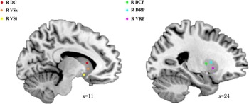
Following previous work (Di Martino et al., Reference Di Martino, Kelly, Grzadzinski, Zuo, Mennes, Mairena and Milham2011; Fornito et al., Reference Fornito, Harrison, Goodby, Dean, Ooi, Nathan and Bullmore2013; Hu et al., Reference Hu, Salmeron, Gu, Stein and Yang2015), seed-based dFC analyses were performed by placing bilateral, 6-mm-radius, spherical regions of interest (ROIs) within 6 a priori defined subdivisions of the striatum representing the affective limbic, sensorimotor, and executive loops. The affective limbic loop ROIs were the VSi (±9, 9, −8) and the VSs (±10, 15, 0). The sensorimotor loop ROIs were the DCP (±28, 1, 3) and the DRP (±25, 8, 6). The executive loop ROIs were the DC (±13, 15, 9) and the VRP (±20, 12, −3) (Di Martino et al., Reference Di Martino, Scheres, Margulies, Kelly, Uddin, Shehzad and Milham2008; Lin et al., Reference Lin, Wang, Zhang, Kirkpatrick, Ongur, Levitt and Wang2018) (Fig. 1). The dFC variability characteristics of the striato-cortical circuitry were calculated using the sliding-window method based on the Temporal Dynamic Analysis (TDA) toolkits integrated in the DPABI software (http://rfmri.org/DPABI). The Hamming sliding window was selected for the whole-brain blood oxygenation level-dependent (BOLD) signal time series; 50 TRs window length and step width of 1 TRs were selected for dFC analysis. The minimum window length should be no less than 1/f min (1/0.01 s = 100 s) according to previous studies (Leonardi & Van De Ville, Reference Leonardi and Van De Ville2015; Li, Duan, Cui, Chen, & Liao, Reference Li, Duan, Cui, Chen and Liao2019); the f min was defined as the minimum frequency of time series. Shorter window lengths might increase the risk of introducing spurious fluctuations in the observed dynamic FC. The window length of 50 TRs (100 s) was selected to compute the temporal variability of FC because a longer window length might hinder the description of the temporal variability dynamics. Also, other window lengths (30 TRs and 70 TRs) and shifting step (1 TRs) were tried to further examine their possible effects on dFC results (Liao et al., Reference Liao, Wu, Xu, Ji, Zhang, Zang and Lu2014). In total, 151 sliding windows of dFC were obtained. For each sliding window, correlation maps were produced by computing the temporal correlation coefficient between the truncated time series of the striatum seeds and all the other voxels. Consequently, 151 sliding-window correlation maps were obtained for each individual. To improve the normality of the correlation distribution, each correlation map was converted into z-value maps using Fisher's r-to-z transformation. Then, the dFC maps were computed by calculating the standard deviation of 151 sliding-window z-value maps. Then, z-standardization was applied for the dFC maps. Finally, all the dFC maps were smoothed using a 4 mm full width at half maximum Gaussian kernel.
Six seeds of the striatum in the right hemisphere. DC, dorsal caudate; VSs, ventral striatum superior; VSi, ventral striatum inferior; DCP, dorsal-caudal putamen; DRP, dorsal-rostral putamen; VRP, ventral-rostral putamen.

Statistical analysis
One-way analysis of variance (ANOVA) and post-hoc analysis were performed to compare the demographic and clinical data among groups using SPSS 19.0 software (SPSS, IL, USA). A chi-squared test was used to compare the gender differences among the three groups. All tests were two-tailed, and a p value less than 0.05 was considered statistically significant (Bonferroni corrected for post-hoc analysis). The one-sample t test was performed to demonstrate the within-group dFC variability distribution of each striatum seed in patients with BD II, patients with MDD, and HCs. The significant level was set at a p value less than 0.05 (uncorrected). To further examine the difference in dFC variability patterns among the three groups, one-way ANOVA was performed on the standard deviation in the z value at each voxel within the union mask of one-sample t test results of the three groups. Age, gender, years of education, and the mean frame-wise displacement (FD) were included as nuisance covariates in the comparisons. The Gaussian random field (GRF) theory was used for the cluster-level multiple comparison correction (minimum z > 2.3; cluster significant: p < 0.05, corrected). The brain regions showing significant differences based on the results of one-way ANOVA were defined as ROIs for further post-hoc analysis for the comparison of each of the two groups (Bonferroni corrected, p < 0.05).
Once the brain regions showed significant group differences in dFC variability for each seed of the striatum, the Pearson correlation coefficients were calculated between the dFC variability values and the clinical variables in patients with BD II and patients in MDD. The clinical variables included the number of episodes, onset age of illness, 24-item HDRS scores, YMRS scores, and the duration of illness. The Bonferroni correction was used for multiple comparisons.
Validation analysis
Another 2 supplementary window lengths (30 TRs and 70 TRs) were applied to validate the main results of dFC with the window length of 50 TRs.
Results
Demographic data and clinical comparisons
Table 1 shows the demographic and clinical data of all study participants. Four patients with BD II, three patients with MDD, and three HCs were excluded from further analyses because of excessive head motion during image acquisition. Finally, the analyzed participants included 128 patients with BD II depression, 140 patients with MDD, and 132 HCs. The three groups had no significant differences in terms of sex and age. In addition, no significant differences were found in HDRS-24 scores, YMRS scores, and duration of illness between the two groups. However, significant differences were observed in education among the three groups, besides significant differences in the age of onset and the number of episodes between the two groups.
Demographic and clinical data and (standard deviations) by group

BD, bipolar disorder; MDD, major depressive disorder; HCs, healthy controls; HDRS, Hamilton Depression Rating Scale; YMRS, Young Mania Rating Scale.
Means (with standard deviations in parentheses) are reported unless otherwise noted.
* The p values were obtained by one-way ANOVA.
† The p value for gender distribution was obtained by chi-square test.
a The education level showed significant differences between BD II and HCs, between MDD and HCs.
b The p values were obtained by two-sample t test between BD II and MDD.
Dynamic functional connectivity variability of the striatum seeds
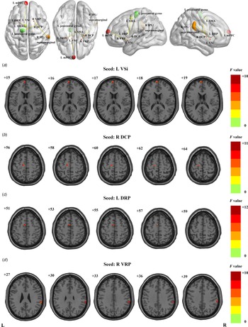
The one-sample t test revealed the dFC variability patterns for each striatum seed in the three groups (online Supplementary Fig. S1, p < 0.05, uncorrected for visual inspection). The one-way ANOVA demonstrated the significant differences in dFC variability among the three groups for each striatum seed (Fig. 2 and Table 2, minimum z > 2.3; cluster significance: p < 0.05, GRF corrected). Specifically, significant differences in dFC variability were found between the left VSi and the left mPFC, the right DCP and the left precentral gyrus, the left DRP and the left supplementary motor area (SMA), and the right VRP and the right IPL. Post-hoc analysis was performed for the significantly different regions among the three groups (Fig. 3 and Table 2; p < 0.05, Bonferroni correction). Briefly, both patients with BD II and patients with MDD exhibited increased dFC variability between the left DRP and the left SMA, and between the right VRP and the right IPL, compared with HCs. The patients with BD II had increased dFC variability between the right DCP and the left precentral gyrus compared with patients with MDD and HCs. The patients with MDD had increased dFC variability between the left VSi and the left mPFC compared with patients with BD II and HC.
The significant dFC variability differences among the three groups for striatum seed, respectively (minimum z > 2.3; cluster significance: p < 0.05, GRF corrected). The color bar indicates the F value from One-Way ANOVA analysis. dFC, dynamic functional connectivity; GRF, Gaussian random field; VSi, ventral striatum inferior; DCP, dorsal-caudal putamen; DRP, dorsal-rostral putamen; VRP, ventral-rostral putamen; L (R), left (right) hemisphere.

The bar graph for the post-hoc analysis of the significant dFC variability differences among the three groups for striatum seed, respectively (Bonferroni corrected, p < 0.05). dFC, dynamic functional connectivity; VSi, ventral striatum inferior; mPFC, medial prefrontal cortex; DCP, dorsal-caudal putamen; DRP, dorsal-rostral putamen; SMA, supplementary motor area; VRP, ventral-rostral putamen; IPL, inferior parietal lobule; L (R), left (right) hemisphere. *, denotes p < 0.05 Bonferroni correction; BD, bipolar disorder; MDD, major depressive disorder; HC, healthy control; L (R), left (right) hemisphere.

The significant dynamic FC variability differences among three groups

FC, functional connectivity; GRF, Gaussian random field; BA, Brodmann Area; MNI, Montreal Neurological coordinate; VSi, ventral striatum inferior; mPFC, medial prefrontal cortex; MDD, major depressive disorder; BD, bipolar disorder; DCP, dorsal-caudal putamen; DRP, dorsal-rostral putamen; SMA, supplementary motor area; VRP, ventral-rostral putamen; IPL, inferior parietal lobule; L (R), left (right) hemisphere.
No significant correlations were noted between the significantly different dFC variability of striatum seeds and any clinical variable (including onset age of illness, number of episodes, duration of illness, HDRS-24 scores, and YMRS scores) in patients with BD II and patients with MDD (p > 0.05, two-tailed).
Validation results
The main results of 30 TRs and 70 TRs sliding-window length validated the main results (50 TRs) (online Supplementary Fig. S2 and S3 in the Supplementary Material).
Discussion
This study was novel in comparing the temporal dynamics of the striato-cortical connectivity in patients with unmedicated BD II depression, patients with MDD, HCs, using a seed-based dFC approach. Compared with HCs, patients with BD II and patients with MDD showed increased dFC variability between the right VRP and the right IPL and between the left DRP and the left SMA. Moreover, patients with BD II showed increased dFC variability between the right DCP and the left precentral gyrus, while patients with MDD showed increased dFC variability between the left VSi and the left mPFC. These findings provided new evidence for shared and specific neuropathological mechanisms underlying MDD and BD from the perspective of the dFC pattern.
The IPL was thought to be an important node capable of accessing the brain network associated with motor intention and awareness (Desmurget et al., Reference Desmurget, Reilly, Richard, Szathmari, Mottolese and Sirigu2009; Desmurget & Sirigu, Reference Desmurget and Sirigu2012). It was primarily involved in execution, somesthesis processing, action inhibition, social cognition, and spatial cognition (Morita et al., Reference Morita, Saito, Ban, Shimada, Okamoto, Kosaka and Naito2018; Vickery & Jiang, Reference Vickery and Jiang2009; Wang et al., Reference Wang, Zhang, Rong, Wei, Zheng, Fox and Jiang2016). In this study, patients with BD II and patients with MDD showed increased dFC variability between the right VRP and the right IPL/supramarginal gyri, suggesting excessive variability in the ventral striatal-cognitive circuitry in BD and MDD. A previous rs-fMRI study found increased static FC between the right striatum and the right IPL in patients with BD II depression (Marchand et al., Reference Marchand, Lee, Garn, Thatcher, Gale, Kreitschitz and Wood2011). Several structural MRI studies found abnormal cortical thickness (Lan et al., Reference Lan, Chhetry, Oquendo, Sublette, Sullivan, Mann and Parsey2014), gray matter volumes (Rubin-Falcone et al., Reference Rubin-Falcone, Zanderigo, Thapa-Chhetry, Lan, Miller, Sublette and Mann2018) and white matter volumes (Yuan et al., Reference Yuan, Zhang, Bai, Yu, You, Shi and Jiang2009) of the IPL in BD and MDD. Rs-fMRI studies on BD and MDD showed abnormal functional activities, such as Regional Homogeneity (ReHo) (Liang et al., Reference Liang, Zhou, Yang, Yang, Fang, Chen and Huang2013), fractional amplitude of low-frequency fluctuations (fALFF) (Qiu et al., Reference Qiu, Xia, Cheng, Yuan, Kuang, Bi and Gong2018), FC strength (Wang et al., Reference Wang, Zhang, Rong, Wei, Zheng, Fox and Jiang2016), and connection (Liu et al., Reference Liu, Wu, Zhang, Guo, Long and Yao2015) in the IPL and supramarginal gyri. A task-based fMRI study reported increased activation in the IPL/supramarginal gyri during working memory tasks in BD (Monks et al., Reference Monks, Thompson, Bullmore, Suckling, Brammer, Williams and Curtis2004) and MDD (Hugdahl et al., Reference Hugdahl, Specht, Biringer, Weis, Elliott, Hammar and Lund2007). Patients with MDD underwent fMRI during the performance of a self-paced letter/digit task-switching paradigm, demonstrating decreased activity in the right IPL and suggesting subtle abnormalities of cognitive flexibility (Remijnse et al., Reference Remijnse, van den Heuvel, Nielen, Vriend, Hendriks, Hoogendijk and Veltman2013). Therefore, these findings of increased dFC variability between the VRP and IPL in BD and MDD might contribute to the similar cognition deficits in both disorders.
In this study, patients with BD II depression and patients with MDD showed increased dFC variability between the left DRP and the left SMA compared with HCs. Additionally, patients with BD II showed increased dFC variability between the right DCP and the left precentral gyrus compared with patients with MDD and HCs. These findings suggested excessive variability in the dorsal striatal-sensorimotor circuitry in the two disorders, specifically in BD II. The SMA and the precentral gyrus consist of the primary motor cortex and play an important role in complex motor learning, planning, and decision components in movements (Exner, Reference Exner2002; Nachev, Kennard, & Husain, Reference Nachev, Kennard and Husain2008; Nitsche et al., Reference Nitsche, Schauenburg, Lang, Liebetanz, Exner, Paulus and Tergau2003). Structural MRI studies on patients with BD and MDD showed abnormal gray matter and white matter volumes in the sensorimotor regions, including the SMA and the precentral gyrus (Adler, Levine, DelBello, & Strakowski, Reference Adler, Levine, DelBello and Strakowski2005; Bracht et al., Reference Bracht, Federspiel, Schnell, Horn, Hofle, Wiest and Walther2012; Cheng et al., Reference Cheng, Xu, Chai, Li, Luo, Yang and Xu2010; Fung et al., Reference Fung, Deng, Zhao, Li, Qu, Li and Chan2015; Jorgensen et al., Reference Jorgensen, Nerland, Norbom, Doan, Nesvag, Morch-Johnsen and Agartz2016). Functional MRI studies showed abnormal local activity and FC in the SMA in BD and MDD (Liu et al., Reference Liu, Hu, Wang, Guo, Zhao, Li and Chen2012; Wei et al., Reference Wei, Chang, Womer, Zhou, Yin, Wei and Wang2018). Felger et al. (Reference Felger, Li, Haroon, Woolwine, Jung, Hu and Miller2016) using rs-fMRI, reported decreased connectivity between the dorsal striatum and SMA in depression, which correlated with increased psychomotor slowing. A task-based fMRI study found abnormal activations in the SMA and putamen in patients with BD depression and motor retardation during finger tapping (Liberg et al., Reference Liberg, Adler, Jonsson, Landen, Rahm, Wahlund and Wahlund2013). One rs-fMRI study reported greater connectivity strength between the sensorimotor network and the left precentral gyrus in young adults with BD, which was significantly related to depression and anxiety symptoms (Thomas et al., Reference Thomas, Christensen, Schettini, Saletin, Ruggieri, MacPherson and Dickstein2019). Moreover, several studies found abnormal structural (Frazier et al., Reference Frazier, Breeze, Papadimitriou, Kennedy, Hodge, Moore and Makris2007; Paillere Martinot et al., Reference Paillere Martinot, Lemaitre, Artiges, Miranda, Goodman, Penttila and Consortium2014) and functional (Doucet, Bassett, Yao, Glahn, & Frangou, Reference Doucet, Bassett, Yao, Glahn and Frangou2017) connectivity of sensorimotor regions in patients with BD and their first-degree relatives. Notably, a recent neuroimaging meta-analysis of affective experiences reported that sensorimotor regions might be involved in affective-related activity in adult healthy participants (Satpute et al., Reference Satpute, Kang, Bickart, Yardley, Wager and Barrett2015). An event-related fMRI study showed diminished activation in the precentral gyrus of patients with BD in response to positive words, which might reflect motor activity and emotion-related processing (Malhi et al., Reference Malhi, Lagopoulos, Owen, Ivanovski, Shnier and Sachdev2007). Taken together, the findings of excessive temporal variability of the dorsal striatal-sensorimotor FC in BD and MDD in this study might be related to the psychomotor symptoms and dysfunctional emotion processing in affective disorders, especially in BD.
The mPFC is heavily connected with emotional-limbic structures, such as the ventral striatum and the hypothalamus, which has been thought to be an important region integrating emotional and affective processes (Haber, Kunishio, Mizobuchi, & Lynd-Balta, Reference Haber, Kunishio, Mizobuchi and Lynd-Balta1995; Raichle et al., Reference Raichle, MacLeod, Snyder, Powers, Gusnard and Shulman2001; Tarai, Mukherjee, Qurratul, Singh, & Bit, Reference Tarai, Mukherjee, Qurratul, Singh and Bit2019b; Zhi et al., Reference Zhi, Hou, We, Zhang, Li and Yuan2018). This study showed increased dFC variability between the left VSi and the left mPFC in patients with MDD but not in patients with BD II, which suggest excessive variability in the ventral striatal-affective circuitry. The results indicated that might be an MDD-specific phenomenon. Several researches also reported that disrupted mood-related nerve circuitries in depression, including abnormal volume of the PFC and striatum (Tarai et al., Reference Tarai, Mukherjee, Gupta, Rizvanov, Palotas, Chandrasekhar Pammi and Bit2019a). A previous rs-fMRI study showed attenuated static FC between the ventral striatum and the mPFC and subgenual ACC in MDD (Furman, Hamilton, & Gotlib, Reference Furman, Hamilton and Gotlib2011). Felger et al. (Reference Felger, Li, Haroon, Woolwine, Jung, Hu and Miller2016) also observed that reduced connectivity between the ventral striatum and mPFC was in turn associated with symptoms of anhedonia. A rs-fMRI study using ICA observed increased FC in the mPFC, which correlated positively with the rumination score in MDD (Zhu et al., Reference Zhu, Wang, Xiao, Liao, Zhong, Wang and Yao2012). Additionally, Kaiser et al. (Reference Kaiser, Whitfield-Gabrieli, Dillon, Goer, Beltzer, Minkel and Pizzagalli2016) found the excessive dFC variability of the mPFC and the insula, which might signify increased sensitivity to salient emotional or self-referential information that triggered rumination in MDD. As described earlier, it was speculated that excessive dFC variability between the VSi and the mPFC in patients with MDD might suggest that these patients displayed typical depressed emotions, such as anhedonia and rumination, which might be a specific phenomenon not reported in patients with BD and HCs.
Limitations
The present study had some limitations. First, it lacked longitudinal data. The patients with MDD had no family history of BD. Moreover, we had tracked the MDD patients after we acquired the data and found that no MDD patients had switched to BD by the time of the submission of this paper. However, whether some patients would turn to BD in the future could not be predicted. Second, 50 TRs window length dFC analysis and fresh window lengths (30 TRs and 70 TRs) were used to further examine their possible effects on dFC results, but the results were not exactly the same. Few studies are available on this topic, and hence further large-sample studies are required. Third, we fail to find the correlation between abnormal dFC variability of the striatum and clinical variable in patients with BD II or MDD. In addition, we did not record the numbers of depressive episodes and hypomanic episodes separately in BD II group that may affect the correlation analysis between the dFC and the number of episodes. Therefore, further longitudinal studies are needed to clarify the relationship between the abnormal dFC and clinical information such as number of mood episodes, severity or symptoms, etc. Finally, head motion remained the confounding factor for the resting-state FC. A series of procedures, such as realignment correction and motion regression, were used in the models. Even so, the head motion effect was not fully eliminated.
Conclusions
The findings revealed that patients with BD and MDD shared common excessive variability in the dorsal striatal-sensorimotor circuitry and ventral striatal-cognitive circuitry. Excessive variability in the ventral striatal-affective circuitry was found specific to patients with MDD. These findings suggested the important roles of dysfunctional striato-cortical loops in the neuropathological mechanisms of BD and MDD.
Supplementary material
The supplementary material for this article can be found at https://doi.org/10.1017/S0033291720002378.
Acknowledgements
The study was supported by grants from the National Natural Science Foundation of China (81671670, 81501456, and 81971597); Project in Basic Research and Applied Basic Research in General Colleges and Universities of Guangdong, China (018KZDXM009). The funding organizations play no further role in study design, data collection, analysis and interpretation, and paper writing.
Author contributions
Ying Wang designed the study; Guanmao Chen, Ying Wang contributed to data sources and study selection; Guanmao Chen, Pan Chen, Jiaying Gong, Yanbin Jia, Shuming Zhong, Feng Chen, Jurong Wang, Zhenye Luo, Zhangzhang Qi contributed to data acquisition; Guanmao Chen contributed to data analysis; Guanmao Chen, Pan Chen wrote the manuscript; Guanmao Chen, Pan Chen, Jiaying Gong, Li Huang, Ying Wang revised the manuscript. All authors contributed and approved the final manuscript.