Introduction
Inflammatory pseudotumour is an uncommon, benign, non-specific granulation consisting of inflammatory cells, histiocytes and fibroblasts.Reference Huang and Dai1 A very early report described two cases of inflammatory pseudotumour of the lung, and numerous cases in various organs have subsequently been reported.Reference Ushio, Takeuchi, Kikuchi and Kaga2, Reference Brunn3 However, the pathogenesis and biological characteristics of inflammatory pseudotumour are still under debate. Inflammatory pseudotumour is frequently observed in the lungs and orbits, but also occasionally in the head and neck region, including the sinonasal tract.Reference Huang and Dai1, Reference Maruya, Kurotaki, Hashimoto, Ohta, Shinkawa and Yagihashi4–Reference Hanna, Blenke, Sharma and Knight6
Although a generally accepted therapeutic strategy for inflammatory pseudotumour does exist, it is not well established. Standard options include corticosteroids, surgical excision and radiotherapy.Reference Matsubara, Tan-Liu, Kenney and Mark7–Reference Ribeiro, Joshi, Funkhouser and Mukherji10 Because of anatomical limitations, wide surgical resection of inflammatory pseudotumour arising in the head and neck region is often difficult, especially in children. To the best of our knowledge, no previous study has reported the effect of arterial embolisation of inflammatory pseudotumour in the head and neck region.
In this case report, we present a two-year-old girl with a massive inflammatory pseudotumour of the maxillary sinus, who was successfully managed with arterial embolisation.
Case report
A two-year-old girl had initially presented to the dental clinic with a short history of discomfort over the right maxillary bone. The patient's medical history had been unremarkable.
She had undergone extraction of a right upper fifth deciduous tooth. After the extraction, a gingival mass in the right mandible had appeared. The mass had been biopsied, and histological examination had revealed respiratory epithelium with chronic inflammation and fibrous tissue.
The patient was admitted to our hospital.
Physical examination identified a reddish, granular, mass lesion with aggressive enlargement and frequent haemorrhaging.
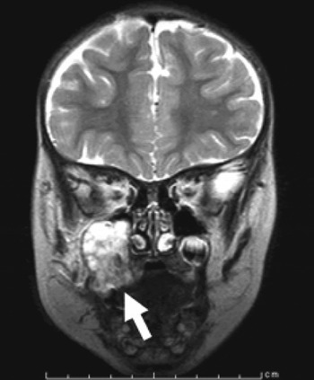
Computed tomography scanning of the paranasal region showed a diffuse soft tissue density in the right maxillary sinus, with severe destruction of the maxillary bone. Magnetic resonance imaging (MRI) of the paranasal sinuses demonstrated an abnormal, enhancing soft tissue mass (Figure 1). A plain radiograph of the chest was normal.
Coronal magnetic resonance imaging scan showing a massive, non-homogeneous lesion in the right maxillary sinus (arrow).

Laboratory blood analysis showed an increased white blood cell count (11 300/μl), platelet count (58.7 × 10⁴/μl), serum alkaline phosphatase activity (541 IU/l), serum calcium level (10.5 mg/dl) and C-reactive protein level (0.8 mg/dl). A decreased red blood cell count (3.61 × 10⁶/μl) and haemoglobin concentration (9.9 g/dl) were also observed.
A malignant tumour was highly suspected.
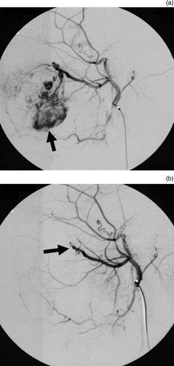
As the patient suffered frequent, substantial bleeding, she underwent embolisation of the major feeding vessels prior to re-biopsy. Angiography under general anaesthesia revealed that a branch of the maxillary artery was the main feeding vessel of the mass, with a branch of the facial artery also supplying a small amount of the circulation (Figure 2a). Embolisation was achieved by injecting polyvinyl alcohol via a catheter into the branch of the maxillary artery; this resulted in almost complete disappearance of the previously visualised angiography enhancement (Figure 2b). After embolisation, another biopsy was taken from the lesion.
Lateral view of transcatheter angiography, (a) before and (b) after arterial embolisation. (a) A branch of the maxillary artery is the main feeding vessel of the lesion (arrow). (b) Contrast enhancement disappears after arterial embolisation (arrow).

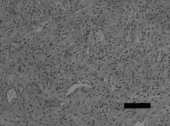
Histological examination revealed reactive granulation. The regeneration of stratified squamous epithelium was observed. There was fibrosis and a mild myxoid change in the interstitium. Infiltration of inflammatory cells including neutrophils was observed. The number of vessels was increased, and vascular endothelial cells were enlarged. There was no evidence of malignancy or angiosarcoma. The pathological diagnosis was inflammatory pseudotumour (Figure 3).
Photomicrograph showing inflammatory pseudotumour (H&E; bar = 100 µm)

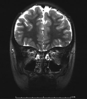
After arterial embolisation, bleeding stopped and the mass gradually reduced. The patient was followed up using MRI scans. The mass completely disappeared without any additional treatment, and no recurrence was observed in the five years after embolisation (Figure 4). The patient's right upper permanent teeth begin to appear. Facial development was not affected, and the patient had no subsequent clinical symptoms.
Follow-up coronal magnetic resonance imaging scan taken after arterial embolisation, showing complete remission of the inflammatory pseudotumour.

Discussion
Inflammatory pseudotumours are composed of a proliferation of myofibroblasts against a background of chronic inflammatory cells, such as plasma cells, lymphocytes and eosinophils. Because of this condition's pathologically heterogeneous composition, a variety of names have been applied, including plasma cell granuloma, pseudosarcomatous myofibroblastic proliferation, xanthomatous pseudotumour, inflammatory myofibroblastic tumour and atypical fibromyxoid nodule.Reference Van Weert, Manni and Driessen11
Several classification systems have been reported.Reference Travis, Colby, Koss, Rosado de Christenson, Muller, King and King12 For example, inflammatory pseudotumour has been sub-categorised histologically into the following: (1) lymphoid subset (mostly lymphocytes with minimal fibrosis); (2) granulomatous subset (diffuse infiltration of lymphocytes, plasma cells, eosinophils and histiocytes, with a variable degree of fibrosis); and (3) sclerosing subtype (increased fibrosis with limited inflammatory infiltrate).Reference Fujii, Fujisada, Kondo, Takahashi and Okada13 Newlin et al. reported that early lesions tended to be predominantly lymphoid, and that advanced lesions contained a greater component of fibrosis.Reference Newlin, Werning and Mendenhall14
There have recently been several reported cases of inflammatory pseudotumour of the lung or liver with extensive immunoglobulin (Ig) G4 positive plasma cell infiltration.Reference Yamamoto, Yamaguchi, Aishima, Oda, Kohashi and Oshiro15, Reference Zen, Kitagawa, Minato, Kurumaya, Katayanagi and Masuda16 Microscopic examination of the latter case revealed a slight plasma cell infiltration, together with fibrosis and the infiltration of other inflammatory cells, including neutrophils; IgG4-positive cells represented less than 1 per cent of the total cell population.
Previous studies have reported effective treatment of inflammatory pseudotumour using high-dose corticosteroids, surgical excision and radiotherapy. Surgical resection has generally been proposed, with radiation therapy for unresectable or recurrent cases.Reference Matsubara, Tan-Liu, Kenney and Mark7, Reference Weisman and Osguthorpe9 Maruya et al. reported that high-dose corticosteroid treatment should be carefully applied prior to consideration of resection or radiotherapy.Reference Maruya, Kurotaki, Hashimoto, Ohta, Shinkawa and Yagihashi4 A literature review indicates that radiotherapy has been proposed for lymphoid-predominant lesions, while granulomatous lesions may respond better to high-dose corticosteroids. Sclerosing lesions with a large fibrotic component exhibit more aggressive features, and show little response to high-dose corticosteroids or radiotherapy.Reference Weisman and Osguthorpe9, Reference Newlin, Werning and Mendenhall14, Reference Batsakis, el-Naggar, Luna and Goepfert17–Reference Hadley, Coady and Milford19
• The pathogenesis and characteristics of inflammatory pseudotumour are controversial
• Surgical excision, high-dose corticosteroids and radiation therapy have been used to treat this condition
• A case of inflammatory pseudotumour in the maxillary antrum is presented, in which arterial embolisation was successful, suggesting an additional therapeutic option for this condition
Ahuja et al. reported successful combination therapy of a proximal haemophilic pseudotumour arising in a child with mild haemophilia, using arterial embolisation prior to surgery. However, no previous report has presented the utility of arterial embolisation as the sole treatment for inflammatory pseudotumour.Reference Ahuja, Sidonio, Raj, Bertolone, Silverman and Antekeier20
The precise effect of embolisation on tumour cells remains largely unknown.Reference Liapi and Geschwind21 The ischaemia and hypoxia generated by arterial embolisation are possible mechanisms for the observed reduction in inflammatory pseudotumour mass.
Conclusion
Wide surgical resection of inflammatory pseudotumour is difficult for head and neck lesions, especially in children. Furthermore, radiotherapy may have late-onset side effects in children. Hence, we propose that arterial embolisation may be a therapeutic option for inflammatory pseudotumour presenting in children.
Acknowledgement
We thank Dr Mitsuhiro Okano for his help during the preparation of this manuscript.