The monotypic genus Lepidocollema was described by Vainio (Reference Vainio1890) based on a single collection of the species L. carassense Vain. from Brazil, a species which has never been collected since. The status of the genus was recently changed dramatically by the revisions in Pannariaceae made by Ekman et al. (Reference Ekman, Wedin, Lindblom and Jørgensen2014). Here, a large group of 22 tropical Parmeliella species with distinct ring-like thalline excipuli, were transferred to Lepidocollema, now with a much wider concept. One of these species is Lepidocollema polyphyllinum (P. M. Jørg.) P. M. Jørg. It was described as Parmeliella polyphyllina P. M. Jørg. by Jørgensen (Reference Jørgensen2001), based on a holotype from Queensland, Australia, with three additional samples cited; another one from Queensland, and one each from Indonesia and Papua New Guinea. This species was described as being squamulose with radiating peripheral lobes 2–3 mm wide, and central parts with numerous, erect and fragile lobules. Jørgensen & Sipman (Reference Jørgensen and Sipman2006) added 11 localities from Papua New Guinea, one from Malaysia and one from the Philippines.
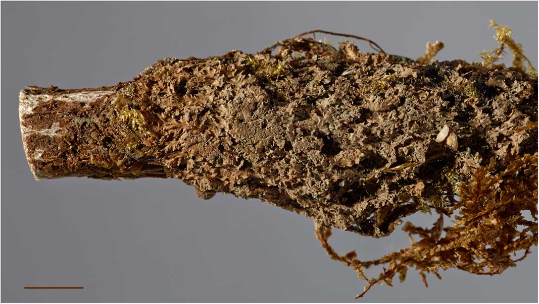
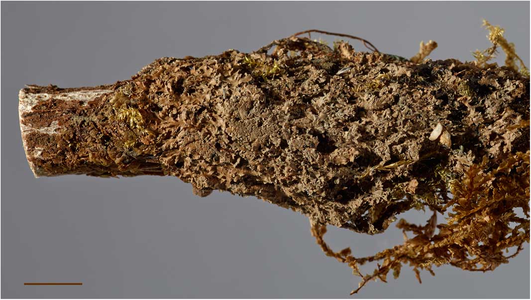
Two collections at BM from elevations between 1900–2200 m on Mount Popomansiu, Guadalcanal Island, the major island of the Solomon Island archipelago, have now been studied by the present author, and the species is reported here as new to the Solomon Islands. The specimens are basically small-foliose, with radiating lobes 0·3–0·6 mm wide, 80–100 µm thick, resting on a thin, blackish prothallus (Fig. 1). Central parts of the thalli are covered by very different lobules, semi-erect, 0·1–0·2 mm wide, 40–60 µm thick, up to 3 mm long and subdichotomously branched, almost forming a mat. The colour of the latter is more greyish on old herbarium specimens, compared with the more olivaceous brown colour of the primary lobes. The secondary lobules look rather like the large cephalodia found in some tripartite austral Pannaria and Psoroma species or in the new genus Gibbosporina (Elvebakk et al. Reference Elvebakk, Hong, Park, Robertsen and Jørgensen2016). However, L. polyphyllinum is a bipartite species.
Lepidocollema polyphyllinum from the Solomon Islands (Jackson Hill 9603). Scale=5 mm. In colour online.
On closer inspection, a difference between the Nostoc symbiont of the primary thallus versus that of the central lobules was found to be consistent. In the primary thallus, Nostoc cells are located in glomeruli below a 30 µm thick, plectenchymatous upper cortex. The glomeruli are c. 20 µm diam., each with 10–15 Nostoc cells, 3–4×4–6 µm, with no visible chain structures present. The colour of the Nostoc cells is a conspicuous dark sky blue. In the secondary lobules, the Nostoc cells are also in small (c. 15 µm) glomeruli without chain structures, but they are larger (5–7×6–8 µm) and are dark violet, a more common colour in Nostoc symbionts in Pannariaceae.
This structure appears to be distinct from, but parallel to, a cephalodium, when the latter is defined as a cyanobiont structure on a chlorolichen, as commonly done, for example, in Malcolm & Galloway (Reference Malcolm and Galloway1997). However, it can actually be called a cephalodium following the definition by Smith et al. (Reference Smith, Aptroot, Coppins, Fletcher, Gilbert, James and Wolseley2009): “a delimited region within or a warty, squamulose, or shrubby structure on the surface of a lichen thallus containing a photobiont (cyanobacterium) different from that characteristic of the rest of the thallus.” The brittle lobules apparently function as vegetative dispersal units, like those of proper, coralloid cephalodia (Jørgensen & Wedin Reference Jørgensen and Wedin1999). This would involve establishing lobule fragments to incorporate the deviating Nostoc strain to reinstate a primary thallus. Alternatively, this is a bipartite photosymbiodemic species, where both morphs can occur independently.
These problems in defining the symbiotic nature of Lepidocollema polyphyllinum illustrate the increasing photobiont diversity discovered in Pannariaceae. A striking example is the genus Fuscopannaria, previously comprising heteromerous bipartite cyanolichens, and now also containing tripartite species (Fryday Reference Fryday2004) and homoiomeric cyanolichens transferred to Fuscopannaria from Kroswia (Magain & Sérusiaux Reference Magain and Sérusiaux2015).
The apothecia of Lepidocollema polyphyllinum are uncommon, and a rare one studied on the specimen Jackson Hill 9485 corresponds to the description by Jørgensen (Reference Jørgensen2001), which in general is also the case with the ascospores. It can be added here that the perispores are smooth, 0·5–1·0 µm thick, with even walls, so that the perispores have the same shape as the proper spores.
Ideally, the observed cyanobiont differences should have been confirmed by molecular studies. However, the cited samples are old, and our specific project on cyanobionts in Pannariaceae was terminated by the study of Elvebakk et al. (Reference Elvebakk, Papaefthimiou, Robertsen and Liaimer2008).
Specimens examined. Solomon Islands: Guadalcanal Island: central part, Mt. Popomansiu, on ridge SE of Sutakiki River, montane rainforest, 5800–6600 ft, 1965, D. Jackson Hill 9485 (BM000731913); ‘top camp’, c. 5700 ft, 1965, D. Jackson Hill 9603 (BM000731915).
Curator Holger Thüs at the Natural History Museum, London (BM), is acknowledged for making available the samples studied and Mari Karlstad, University of Tromsø – Arctic University of Norway, Tromsø University Museum, for photographing one specimen.