Introduction
Streptococcus suis infections have been considered a major problem worldwide in the swine industry, particularly during the past 15 years. The natural habitat of S. suis is the upper respiratory tract of pigs, particularly the tonsils and nasal cavities, as well as the genital and alimentary tracts (Higgins and Gottschalk, Reference Higgins, Gottschalk, Straw, D'Allaire, Mengeling and Taylor2005). The most important clinical feature associated with S. suis infection in pigs is meningitis. However, other pathologies have also been described, such as arthritis, endocarditis, pneumonia, and septicemia with sudden death (Higgins and Gottschalk, Reference Higgins, Gottschalk, Straw, D'Allaire, Mengeling and Taylor2005). Although S. suis is considered a major swine pathogen, it has been isolated increasingly from a wide range of mammalian species (including humans) and from birds. These findings suggest new epidemiological patterns of the infection, since other animal species might also be a source of infection of swine (Devriese et al., Reference Devriese, Sustronck, Maenhout and Haesebrouck1990, Reference Devriese, Hommez, Pot and Haesebrouck1994; Devriese and Haesebrouck, Reference Devriese and Haesebrouck1992).
Infections in humans were considered sporadic infections in people working with pigs or pork-derived products (Arends and Zanen, Reference Arends and Zanen1988). However, an important outbreak that occurred during the summer of 2005 in China and affected more than 200 people with a mortality rate of nearly 20% has changed the perspective of the threat posed by this pathogen to human health. Here, we review the most recent available data on S. suis infection in humans. Although we discuss some general aspects of the infection in pigs, a complete review on infection caused by this pathogen in animals can be found in a previously published book chapter (Higgins and Gottschalk, Reference Higgins, Gottschalk, Straw, D'Allaire, Mengeling and Taylor2005).
S. suis: general characteristics of the microorganism
S. suis is an encapsulated Gram-positive coccus that possesses cell wall antigenic determinants somewhat related to Lancefield group D. There are 35 serotypes described to date and the composition of the capsule defines the serotype (Higgins and Gottschalk, Reference Higgins, Gottschalk, Straw, D'Allaire, Mengeling and Taylor2005). There is some confusion regarding the early terminology of Lancefield groups R, S and T, and the relationship of these groups with group D streptococci and the different S. suis serotypes (also referred to in the literature as capsular types or serovars), especially in papers describing human infections (Tarradas et al., Reference Tarradas, Luque, de Andres, Abdel-Aziz, Pons, Gonzalez, Borge and Perea2001b; Facklam, Reference Facklam2002; de la Hoz Adame et al., Reference de la Hoz, de la Rubia, Dominguez and Garcia2005). Various α-hemolytic streptococci were ascribed to Lancefield groups R, S, RS and T in 1963 by de Moor (de Moor, Reference de Moor1963). Years later, British researchers, working with encapsulated streptococci similar to de Moor's groups S and R, discovered that de Moor had erroneously worked with antigens extracted from the capsular material rather than from the cell wall, and clearly demonstrated that the lipoteichoic acid present in the cell wall of these strains reacts with group D antiserum (Elliott et al., Reference Elliott, McCarty and Lancefield1977). That this antigen is difficult to extract may account for the negative reaction with the existing antisera obtained in 1963 by de Moor. In fact, the conventional method for extracting streptococcal group antigens from other group D streptococci by heating in HCl at 100°C and pH 2 yields sufficient free teichoic acid to precipitate with potent group D antisera, but such extracts from S. suis may give weak or equivocal results (Elliott et al., Reference Elliott, McCarty and Lancefield1977). The ‘Lancefield groups R and S’ were later identified as S. suis (Lancefield group D), and reclassified as capsular types 1 (formerly group S) and 2 (formerly group R) (Elliott and Tai, Reference Elliott and Tai1978). Some years later, groups RS and T were also reclassified as capsular types 1/2 and 15, respectively (Perch et al., Reference Perch, Pedersen and Henrichsen1983; Gottschalk et al., Reference Gottschalk, Higgins, Jacques, Mittal and Henrichsen1989). In conclusion, the Lancefield groups R, S, RS and T do not exist and, to avoid confusion, this terminology should not be used.
As noted above, S. suis cell wall antigen shares epitopes with Lancefield group D; however, negative reactions are often observed with some strains (Chau et al., Reference Chau, Huang and Kay1983; Ho et al., Reference Ho, Woo, Tse and French1990; Gottschalk et al., Reference Gottschalk, Higgins, Jacques, Beaudoin and Henrichsen1991; Tarradas et al., Reference Tarradas, Luque, de Andres, Abdel-Aziz, Pons, Gonzalez, Borge and Perea2001b) due to either technical problems or use of low antisera with titers of antibodies. In addition, S. suis is not closely related genetically to other group D streptococci (Kilpper-Bälz and Schleifer, Reference Kilpper-Bälz and Schleifer1987). Using 16S rRNA gene sequence analysis, Chatellier et al. described a major, well-defined group of S. suis reference strains which are very closely interrelated but separated from other streptococci and enterococci, with the exception of serotypes 32, 33 and 34 that showed a higher divergence (Chatellier et al., Reference Chatellier, Gottschalk, Higgins, Brousseau and Harel1999). The analysis of chaperonin 60 gene sequences also revealed that serotypes 32 and 34 may belong to another species (Brousseau et al., Reference Brousseau, Hill, Prefontaine, Goh, Harel and Hemmingsen2001). More recently, DNA sequence data and biochemical profiles clearly indicate that these two serotypes cluster with Streptococcus orisratti, a species usually isolated from the teeth of rats (Hill et al., Reference Hill, Gottschalk, Brousseau, Harel, Hemmingsen and Goh2005).
General aspects of S. suis infections in pigs
Although the pig carrier rate is near 100%, the incidence of disease varies over time and is generally less than 5% (Clifton-Hadley et al., Reference Clifton-Hadley, Alexander and Enright1986). However, in the absence of treatment, mortality rates can reach 20% (Cloutier et al., Reference Cloutier, D'Allaire, Martinez, Surprenant, Lacouture and Gottschalk2003). In peracute cases, pigs may be found dead with no premonitory signs. Meningitis is the most striking feature and often the basis of a presumptive diagnosis. Other manifestations of S. suis infection are arthritis, endocarditis, pneumonia, rhinitis, abortion and vaginitis (Sanford and Tilker, Reference Sanford and Tilker1982; Sihvonen et al., Reference Sihvonen, Kurl and Henrichsen1988). In North America, S. suis is, by far, the infectious agent most frequently isolated from cases of endocarditis in pigs. Affected pigs may die suddenly or show various levels of dyspnea, cyanosis and wasting (Higgins and Gottschalk, Reference Higgins, Gottschalk, Straw, D'Allaire, Mengeling and Taylor2005).
Most S. suis organisms isolated from diseased pigs belong to a limited number of serotypes, often between 1 and 8 (Galina et al., Reference Galina, Collins and Pijoan1992; Kataoka et al., Reference Kataoka, Sugimoto, Nakazawa, Morozumi and Kashiwazaki1993; Reams et al., Reference Reams, Harrington, Glickman, Thacker and Bowersock1996; Higgins and Gottschalk, Reference Higgins and Gottschalk2001). Although serotype 2 isolates predominate in most countries, the distribution may differ depending on the geographical location. For example, the prevalence of serotype 2 strains recovered from diseased animals in Canada remains relatively low (below 25%) (Higgins and Gottschalk, Reference Higgins and Gottschalk2001). In some European and Asian countries, however, serotype 2 is isolated more frequently (Wisselink et al., Reference Wisselink, Smith, Stockhofe-Zurwieden, Peperkamp and Vecht2000). In this regard, it may be hypothesized that Euroasian and North American serotype 2 strains of S. suis possess a different virulence potential (Gottschalk and Segura, Reference Gottschalk and Segura2000) (see below). Some strains belonging to less common serotypes, such as serotypes 9, 14, 7 and 5, have been associated with severe infections (Heath et al., Reference Heath, Hunt, Duff and Wilkinson1996; Cloutier et al., Reference Cloutier, D'Allaire, Martinez, Surprenant, Lacouture and Gottschalk2003; Tian et al., Reference Tian, Aarestrup and Lu2004; Higgins and Gottschalk, Reference Higgins, Gottschalk, Straw, D'Allaire, Mengeling and Taylor2005). Despite these differences, almost all studies on virulence factors, pathogenesis of the infection and mechanisms of protection have been conducted with serotype 2 strains. Indeed, animal models have been almost exclusively standardized using this serotype (Kataoka et al., Reference Kataoka, Haritani, Mori, Kishima, Sugimoto, Nakazawa and Yamamoto1991; Beaudoin et al., Reference Beaudoin, Higgins, Harel and Gottschalk1992; Quessy et al., Reference Quessy, Dubreuil and Higgins1994; Busque et al., Reference Busque, Higgins, Caya and Quessy1997; Charland et al., Reference Charland, Jacques, Lacouture and Gottschalk1997; Vecht et al., Reference Vecht, Stockhofe-Zurwieden, Tetenburg, Wisselink and Smith1997; Allen et al., Reference Allen, Bolitho, Lindsay, Khan, Bryant, Norton, Ward, Leigh, Morgan, Riches, Eastty and Maskell2001).
Presumptive diagnosis of S. suis infection in pigs is based on clinical signs and macroscopic lesions. Confirmation of the infection must be achieved by the isolation of the infectious agent and the detection of microscopic lesions in tissue (Staats et al., Reference Staats, Feder, Okwumabua and Chengappa1997). Veterinary diagnostic laboratories easily identify S. suis isolates through use of a minimum of biochemical tests, especially when the samples are recovered from diseased pigs. As proposed by Devriese et al., an α-hemolytic Streptococcus recovered from diseased pigs that produces amylase but not acetoin can be considered as S. suis (Devriese et al., Reference Devriese, Ceyssens, Hommez, Kilpper-Balz and Schleifer1991). Serotyping remains an important part of the routine diagnostic procedure. Some laboratories propose the use of multi-tests, such as the API Strep System test (BioMérieux, France), the BBL Crystal Gram-positive ID kit (Becton-Dickinson), the Vitek GPI Card (BioMérieux), and the Phoenix System PID (Becton-Dickinson); however, some strains (and/or the serotype) of S. suis can be misidentified using these commercial kits (Gottschalk et al., Reference Gottschalk, Higgins, Jacques, Beaudoin and Henrichsen1991; Michaud et al., Reference Michaud, Duperval and Higgins1996; Heidt et al., Reference Heidt, Mohamed, Hain, Vogt, Chakraborty and Domann2005; Tsai et al., Reference Tsai, Lee, Wann, Huang, Chen and Liu2005; Ip et al., Reference Ip, Fung, Chi, Cheuk, Chau, Wong, Lui, Hui, Lai and Chan2007).
Since the majority of typeable isolates belong to capsular types 1–8, it is advisable for diagnostic laboratories to use only antisera corresponding to those serotypes and to send untypeable isolates to a reference laboratory (Higgins and Gottschalk, Reference Higgins, Gottschalk, Straw, D'Allaire, Mengeling and Taylor2005). Isolation and identification of S. suis isolates from carrier animals is much more complicated. In fact, most animals harbor S. suis in their tonsils and multiple serotypes, and untypeable strains may be present in a single herd and even within the same animal (Martinez et al., Reference Martinez, Harel, Lacouture and Gottschalk2002; Cloutier et al., Reference Cloutier, D'Allaire, Martinez, Surprenant, Lacouture and Gottschalk2003; Marois et al., Reference Marois, Le Devendec, Gottschalk and Kobisch2007). In these cases, final identification of untypeable strains must be carried out with the use of S. suis species-specific PCR tests (Okwumabua et al., Reference Okwumabua, O'Connor and Shull2003). Finally, no reliable serological test has been described so far (del Campo Sepulveda et al., Reference del Campo, Altman, Kobisch, D'Allaire and Gottschalk1996; Higgins and Gottschalk, Reference Higgins, Gottschalk, Straw, D'Allaire, Mengeling and Taylor2005).
Virulence factors and pathogenesis of the infection
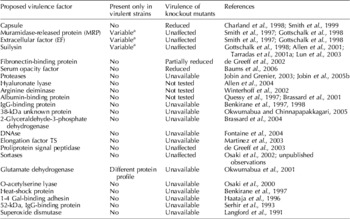
Most studies on S. suis virulence factors have been performed using serotype 2 strains. One of the problems in identifying virulence factors of S. suis is the lack of a clear definition of virulence for this pathogen, mainly as a result of the different parameters used to define whether a strain is virulent or avirulent (Gottschalk et al., Reference Gottschalk, Higgins and Quessy1999). Table 1 summarizes the most important virulence factors of S. suis proposed so far. As of 2006, the single most critical virulence factor described is the capsular polysaccharide (CPS) (Charland et al., Reference Charland, Harel, Kobisch, Lacasse and Gottschalk1998; Smith et al., Reference Smith, Damman, van der Velde, Wagenaar, Wisselink, Stockhofe-Zurwieden and Smits1999). However, most avirulent strains are encapsulated, indicating that other virulence factors are also essential (Gottschalk and Segura, Reference Gottschalk and Segura2000). With the exception of the fibronectin/fibrinogen-binding protein described by de Greeff et al. (Reference de Greeff, Buys, Verhaar, Dijkstra, van Alphen and Smith2002), which seems to be only partially involved in virulence, other factors are not essential for virulence, are found in both virulent and avirulent strains, or could not be properly studied due to the unavailability of knockout mutants. Knockout mutants of other putative virulence candidates have been obtained, but their virulence has not been tested. Recently, a serum opacity factor has been described with structural homology to members of the MSCRAMM (microbial surface components recognizing adhesive matrix molecules) family and determined to be critical for the virulence of S. suis (Baums et al., Reference Baums, Kaim, Fulde, Ramachandran, Goethe and Valentin-Weigand2006). However, like CPS, this factor is present in both virulent and avirulent S. suis strains (unpublished observations). Thus, the role in the pathogenesis of S. suis infections of most putative virulence factors (other than CPS) described so far remains to be confirmed. Using a novel signature-tagged mutagenesis system, Wilson et al. identified 22 mutants as attenuated in either pig or mouse model of infection. A variety of genes were thus identified, including previously known genes essential to the virulence of other organisms (Wilson et al., Reference Wilson, Jeffers, Rapp-Gabrielson, Martin, Klein, Lowery and Fuller2007).
Proposed virulence factors for S. suis serotype 2

a Results vary depending on the geographical origin of the strains studied.
The literature is also conflicting regarding the differentiation between virulence factors and virulence markers. Despite the lack of evidence that some of these putative virulence factors play a critical role in virulence, they nonetheless may serve as virulence markers. This seems to be the case for MRP and EF proteins (Vecht et al., Reference Vecht, Wisselink, Jellema and Smith1991), as well as suilysin, a thiol-activated hemolysin produced by some strains of S. suis (Jacobs et al., Reference Jacobs, Loeffen, van den Berg and Storm1994; Gottschalk et al., Reference Gottschalk, Lacouture and Dubreuil1995). The role of MRP and EF proteins has not been clearly defined; however, suilysin has been identified as a toxic factor for various cell types (see below). Although isogenic mutants lacking either MRP and EF proteins or suilysin were shown to be as virulent to pigs as the respective parent strain (Smith et al., Reference Smith, Vecht, Wisselink, Stockhofe-Zurwieden, Biermann and Smits1996; Allen et al., Reference Allen, Bolitho, Lindsay, Khan, Bryant, Norton, Ward, Leigh, Morgan, Riches, Eastty and Maskell2001; Lun et al., Reference Lun, Perez-Casal, Connor and Willson2003), there is a positive association between the presence of these proteins and virulence in Euroasian strains. In addition, avirulent strains possessing MRP, EF and suilysin factors have not been described so far. On the other hand, the absence of one or both of these proteins is not necessarily associated with a lack of virulence. Indeed, some European and most North American virulent isolates do not produce these factors (Quessy et al., Reference Quessy, Dubreuil and Higgins1994; Galina et al., Reference Galina, Vecht, Wisselink and Pijoan1996; Gottschalk et al., Reference Gottschalk, Lebrun, Wisselink, Dubreuil, Smith and Vecht1998; Segers et al., Reference Segers, Kenter, de Haan and Jacobs1998; Berthelot-Herault et al., Reference Berthelot-Herault, Morvan, Keribin, Gottschalk and Kobisch2000). As mentioned, it has been hypothesized that Euroasian virulent serotype 2 strains, especially those expressing MRP, EF and suilysin factors, are more virulent than North American virulent strains (Gottschalk and Segura, Reference Gottschalk and Segura2000). This might explain the higher prevalence of this serotype in single infections in European and Asian countries as well as the higher association of S. suis cases with the immunosuppressive infection caused by the porcine reproductive and respiratory syndrome virus (PRRSV) and, possibly, the higher number of human cases in those countries (see below). Indeed, higher virulence of a virulent serotype 2 European strain compared to a virulent Canadian strain was observed using a standardized experimental infection method (Berthelot-Herault et al., Reference Berthelot-Herault, Gottschalk, Morvan and Kobisch2005).
The mechanisms that enable S. suis to disseminate throughout the animal are not well understood. The bacterium is able to spread systemically from the nasopharynx, occasionally resulting in septicemia and death (Gottschalk and Segura, Reference Gottschalk and Segura2000; Madsen et al., Reference Madsen, Svensmark, Elvestad, Aalbaek and Jensen2002). The palatine and pharyngeal tonsils are both potential portals of entry for S. suis, leading to subsequent hematogenous or lymphogenous dissemination (Madsen et al., Reference Madsen, Svensmark, Elvestad, Aalbaek and Jensen2002). It is not known how S. suis, despite its low quantities on mucosal surfaces, is able to traverse the first line of host defense to initiate disease. To accomplish this, bacteria must be able to breach mucosal epithelia in the upper respiratory tract. Very few studies have investigated the interactions between S. suis and epithelial cells, and many of these have produced contradictory results. For example, some studies have reported limited invasion (Norton et al., Reference Norton, Rolph, Ward, Bentley and Leigh1999), no invasion at all (Lalonde et al., Reference Lalonde, Segura, Lacouture and Gottschalk2000), or invasion only with non-encapsulated (probably avirulent) strains (Benga et al., Reference Benga, Goethe, Beilage and Valentin-Weigand2004; Valentin-Weigand, Reference Valentin-Weigand2004).
Survival of the organism in the bloodstream may be facilitated by the CPS, which efficiently hampers phagocytosis. Although an early theory, called the Trojan horse theory, suggested that the bacteria are taken up by monocytes (in the absence of specific antibodies), survive intracellularly and then invade the central nervous system (CNS) (Williams, Reference Williams1990; Williams and Blakemore, Reference Williams and Blakemore1990), studies conducted by various laboratories over the past decade indicate that bacteria travel extracellularly, not intracellularly, either free in circulation or attached to the surface of monocytes (Segura and Gottschalk, Reference Segura and Gottschalk2002). Importantly, neutrophils and monocytes are unable to kill S. suis in the absence of specific antibodies (Chabot-Roy et al., Reference Chabot-Roy, Willson, Segura, Lacouture and Gottschalk2006). In addition, it has been shown that suilysin is toxic to not only epithelial and endothelial cells but also monocytes and neutrophils, thus suggesting a role in immune evasion. Moreover, suilysin appears to affect complement-dependent killing by decreasing the opsonization of S. suis and the bactericidal capacity of neutrophils (Charland et al., Reference Charland, Nizet, Rubens, Kim, Lacouture and Gottschalk2000; Gottschalk and Segura, Reference Gottschalk and Segura2000; Lalonde et al., Reference Lalonde, Segura, Lacouture and Gottschalk2000; Segura and Gottschalk, Reference Segura and Gottschalk2002; Chabot-Roy et al., Reference Chabot-Roy, Willson, Segura, Lacouture and Gottschalk2006; Segura et al., Reference Segura, Vanier, Al-Numani, Lacouture, Olivier and Gottschalk2006).
In the event that S. suis fails to cause acute fatal septicemia, bacteria are able to reach the CNS via mechanisms that are only partially elucidated, such as adhesion to, with or without toxicity, and invasion of brain microvascular endothelial cells (BMEC) (Charland et al., Reference Charland, Nizet, Rubens, Kim, Lacouture and Gottschalk2000; Vanier et al., Reference Vanier, Segura, Friedl, Lacouture and Gottschalk2004; Benga et al., Reference Benga, Friedl and Valentin-Weigand2005). BMEC, together with the choroid plexus epithelial cells (CPEC), constitute the structural basis of the blood–brain barrier (BBB). It has recently been shown that S. suis also affects porcine CPEC barrier function and integrity. Although apoptosis may be involved in the process of CPEC cell death, necrosis seems to be the predominant mechanism. These and probably other mechanisms facilitate S. suis invasion of the CNS (Tenenbaum et al., Reference Tenenbaum, Adam, Eggelnpohler, Matalon, Seibt, Novotny, Galla and Schroten2005, Reference Tenenbaum, Essmann, Adam, Seibt, Janicke, Novotny, Galla and Schroten2006).
Inflammation as a major player in S. suis pathology?
Clinical presentation of S. suis infection in swine and humans varies from asymptomatic bacteremia to fulminant systemic disease similar to Gram-negative sepsis. The increased severity of S. suis infections in humans observed in cases of septic shock and toxic shock-like syndrome, such as a shorter incubation time, more rapid disease progression and a higher rate of mortality, is indicative of a massive inflammation process and underscores the critical need to better understand the interactions between S. suis and cells of the host immune system. In addition, clinical signs of meningitis in swine have also been associated with inflammation in the CNS, and treatment with anti-inflammatory drugs contributes to the recovery of the affected animal (Higgins and Gottschalk, Reference Higgins, Gottschalk, Straw, D'Allaire, Mengeling and Taylor2005).
Multiple lines of evidence suggest that the immune system may play an important role not only in the development of protection against disease but also in the pathology caused by the invading pathogen. Indeed, several inflammatory and infectious diseases are associated with the overproduction of pro-inflammatory cytokines and chemokines, and the recruitment and activation of different leukocyte populations are hallmarks of acute inflammation. Given that these cytokines have been detected in the blood and cerebrospinal fluid (CSF) during septic shock and invasive meningeal infections, the ability of S. suis to induce cytokine production may have considerable biological relevance (Verhoef and Mattsson, Reference Verhoef and Mattsson1995). We previously demonstrated that S. suis is able to induce the production of pro-inflammatory cytokines by murine and human cells (Segura et al., Reference Segura, Stankova and Gottschalk1999, Reference Segura, Vadeboncoeur and Gottschalk2002). More recently, we confirmed the inflammatory properties of S. suis in a swine whole blood culture system (Segura et al., Reference Segura, Vanier, Al-Numani, Lacouture, Olivier and Gottschalk2006). Live bacteria induce high levels of tumor necrosis factor α (TNF-α), interleukin-1β (IL-1β) and IL-6 and intermediate levels of IL-8 and monocyte chemotactic protein (MCP)-1 in this system. The bacterial cell wall was observed to be the major cytokine-inducing component, whereas capsule expression was important for MCP-1 activation. The presence of specific antibodies that suppress bacterial growth also causes a significant reduction in levels of cytokine production. Thus, antibody-mediated bacterial phagocytosis combined with suppressed inflammation may be beneficial for infection control strategies (Segura et al., Reference Segura, Vanier, Al-Numani, Lacouture, Olivier and Gottschalk2006).
We further studied in vitro the receptors involved in cell activation by S. suis. We demonstrated that stimulation of human monocytes by whole encapsulated S. suis or its purified cell wall components influences the relative expression of Toll-like receptor (TLR) 2 and CD14 mRNA. Moreover, this stimulation triggers the release of cytokines (TNF-α, IL-1β and IL-6) and chemokines (IL-8 and MCP-1), which is significantly reduced by antibody-mediated neutralization of TLR2 but not TLR4. Mouse macrophages deficient in TLR2 also show reduced IL-6 and MCP-1 release in response to whole encapsulated bacteria. Given that this response is completely abrogated in MyD88-deficient macrophages, other TLRs might be involved in MyD88-dependent cytokine production induced by encapsulated S. suis. Furthermore, we demonstrated that the presence of CPS modulates S. suis interactions with TLRs. In the absence of CPS, uncovered cell wall components induce cytokine and chemokine production via TLR2-dependent as well as -independent pathways. However, CPS contributes to MCP-1 production in a MyD88-independent manner (Graveline et al., Reference Graveline, Segura, Radzioch and Gottschalk2007).
A typical type 1 inflammatory response is observed following S. suis infection. However, the failure to control this inflammatory cascade may contribute to clinical manifestations and poor outcome (Dominguez et al., Reference Dominguez-Punaro, Segura, Plante, Lacouture, Rivest and Gottschalk2007). Since most of the aforementioned cytokines are believed to mediate responses associated with clinical deterioration, multi-organ system failure and death, we developed an in vivo mouse model to study the septic shock induced by S. suis. Using the Luminex multiplex system, we quantitatively measured the kinetics of several cytokines simultaneously in a small volume of plasma. High levels of the pro-inflammatory cytokines TNF-α, IL-6, IL-12 and IFN-γ and the chemoattractants MCP-1, KC and RANTES were observed in vivo within 24 h post-infection and might be responsible in part for the sudden death of 20% of animals. On the other hand, the anti-inflammatory cytokine IL-10 was up-regulated mainly at 24 h post-infection, following the onset of most pro-inflammatory cytokines. This may indicate a negative feedback mechanism to control the extent of the inflammatory response. Increased rate of septic shock was observed in S. suis-infected mice that were treated with neutralizing antibodies against IL-10 (unpublished observations).
Meningitis is also one of the most striking features of S. suis infection and, as previously mentioned, the inflammatory reaction induced by this pathogen at the CNS may play an important role in pathology. Indeed, fibrin, edema and cellular infiltrates in the meninges are typically observed during S. suis meningitis (Madsen et al., Reference Madsen, Svensmark, Elvestad, Aalbaek and Jensen2002). S. suis can induce the release of arachidonic acid by BMEC, a mechanism suggested to facilitate the ability of bacteria to penetrate the CNS and to modulate local inflammation (Jobin et al., Reference Jobin, Fortin, Willson, Gottschalk and Grenier2005a). In addition, bacterial CPS induces human macrophages to secrete PGE2 and matrix metalloproteinase 9, which also may be involved in disruption of the BBB (Jobin et al., Reference Jobin, Gottschalk and Grenier2006). Finally, other studies from our laboratory have shown that S. suis is able to induce the release of pro-inflammatory cytokines and chemokines by human BMEC and to up-regulate the expression of adhesion molecules on human monocytes with consequent increased adherence of S. suis-activated monocytes to endothelial cells (Al-Numani et al., Reference Al-Numani, Segura, Dore and Gottschalk2003; Vadeboncoeur et al., Reference Vadeboncoeur, Segura, Al-Numani, Vanier and Gottschalk2003).
In vivo experiments showed that infected mice that survive the septicemic phase can develop serious signs of inflammation at the CNS, with suppurative and necrotizing lesions at the meninges and brain parenchyma, especially in the cortex, hippocampus, thalamus, hypothalamus and corpus callosum. Bacterial antigens were detected in association with microglia residing only in the affected zones. In situ hybridization combined with immunocytochemistry showed transcriptional activation of TLR2 and TLR3 as well as of CD14, NF-κB, IL-1β, MCP-1 and TNF-α at the aforementioned brain structures. Microglia/macrophage cells were probably the main cellular sources of cytokine induction in the brain. Early transcriptional activation of TLR2, CD14 and inflammatory cytokines in the choroid plexus and brain endothelial cells suggests these structures as possible bacterial portals of entry into the CNS (Dominguez et al., Reference Dominguez-Punaro, Segura, Plante, Lacouture, Rivest and Gottschalk2007) (Gottschalk and Segura, Reference Gottschalk and Segura2000; Vanier et al., Reference Vanier, Segura, Friedl, Lacouture and Gottschalk2004; Tenenbaum et al., Reference Tenenbaum, Adam, Eggelnpohler, Matalon, Seibt, Novotny, Galla and Schroten2005, Reference Tenenbaum, Essmann, Adam, Seibt, Janicke, Novotny, Galla and Schroten2006). These data confirm the important role of the inflammatory response in the pathogenesis of S. suis infection in mice.
S. suis infection in humans
Since the first description in Denmark in 1968 (Perch et al., Reference Perch, Kristjansen and Skadhauge1968), nearly 250 human cases of S. suis infection have been reported. Although S. suis disease has been considered in the past as a rare event in humans, it was identified more recently as the third most common culture-confirmed cause of community-acquired bacterial meningitis in Hong Kong, following Streptococcus pneumoniae and Mycobacterium tuberculosis as the most common agents (Hui et al., Reference Hui, Ng, Tong, Mok, Chow, Wu and Wong2005). In addition to Denmark, cases have been reported in The Netherlands, Italy, Spain, the United Kingdom, Belgium, Croatia, Austria, New Zealand, Sweden, Hong Kong, Singapore, Taiwan, Japan, China, Thailand, Germany, Ireland, Hungary, France, Greece, Australia, Ireland, Hungary and Argentina (Christensen and Kronvall, Reference Christensen and Kronvall1985; Colaert et al., Reference Colaert, Allewaert, Magerman, Vandeven and Vandepitte1985; Dickie et al., Reference Dickie, Bremner, Wong, North and Robertson1987; Arends and Zanen, Reference Arends and Zanen1988; Dupas et al., Reference Dupas, Vignon and Geraut1992; Hampson et al., Reference Hampson, Trott, Clarke, Mwaniki and Robertson1993; Yen et al., Reference Yen, Liu, Wang, Chen, Wang and Cheng1994; Perseghin et al., Reference Perseghin, Bezzi, Troupioti and Gallina1995; Spiss et al., Reference Spiss, Kofler, Hausdorfer, Pfausler and Schmutzhard1999; Fongcom et al., Reference Fongcom, Pruksakorn, Mongkol, Tharavichitkul and Yoonim2001; Tarradas et al., Reference Tarradas, Luque, de Andres, Abdel-Aziz, Pons, Gonzalez, Borge and Perea2001b; Watkins et al., Reference Watkins, Brooksby, Schweiger and Enright2001; Chan et al., Reference Chan, Wilder-Smith, Ong, Kumarasinghe and Wilder-Smith2002; Kopic et al., Reference Kopic, Paradzik and Pandak2002; Rosenkranz et al., Reference Rosenkranz, Elsner, Sturenburg, Weiller, Rother and Sobottka2003; Huang et al., Reference Huang, Teng, Ho and Hsueh2005; Lopreto et al., Reference Lopreto, Lopardo, Bardi and Gottschalk2005; Chang et al., Reference Chang, Wada, Ikebe, Ohnishi, Mita, Endo, Matsuo, Asatsuma, Kuramoto, Sekiguchi, Yamazaki, Yoshikawa, Watabe, Yamada, Kurita, Imai and Watanabe2006; Voutsadakis, Reference Voutsadakis2006). Mysteriously, only two cases have been reported in Canada (Trottier et al., Reference Trottier, Higgins, Brochu and Gottschalk1991; Michaud et al., Reference Michaud, Duperval and Higgins1996) and only one, very recently, in the USA (Willenburg et al., Reference Willenburg, Sentochnik and Zadoks2006).
Most cases of S. suis infection have been attributed to serotype 2 strains, generally on the basis of biochemical analysis obtained using commercial kits (Colaert et al., Reference Colaert, Allewaert, Magerman, Vandeven and Vandepitte1985; Peetermans et al., Reference Peetermans, Moffie and Thompson1989; Meecham and Worth, Reference Meecham and Worth1992; Yen et al., Reference Yen, Liu, Wang, Chen, Wang and Cheng1994; Perseghin et al., Reference Perseghin, Bezzi, Troupioti and Gallina1995; Caumont et al., Reference Caumont, Gerard, Depernet, Brasme, Eschard and Etienne1996; Asensi et al., Reference Asensi, Asensi, Arias, Moreno, Perez and Navarro2001; Alonso-Socas et al., Reference Alonso-Socas, Aleman-Valls, Roldan-Delgado and Gomez-Sirvent2006). While many of these kits claim to clearly differentiate serotype 2 from serotype 1 strains, there is no evidence of a correlation between a specific serotype and its biochemical properties (Michaud et al., Reference Michaud, Duperval and Higgins1996). In addition to serotype 2 strains, cases due to serotype 4 (Arends and Zanen, Reference Arends and Zanen1988), serotype 14 (Gottschalk et al., Reference Gottschalk, Higgins, Jacques, Mittal and Henrichsen1989; Watkins et al., Reference Watkins, Brooksby, Schweiger and Enright2001) and serotype 16 (one case in Vietnam; C. Schultz, personal communication, 2006) strains have also been reported. Two human cases associated with S. suis serotype 1 could not be confirmed because the serotype of these strains was established using only biochemical criteria and was not confirmed with a serologic reaction using specific sera (Kopic et al., Reference Kopic, Paradzik and Pandak2002). Unfortunately, these isolates are not longer viable to allow confirmation of the serotype (J. Kopic, personal communication, 2004).
In humans, S. suis usually produces a purulent or non-purulent meningitis (Arends and Zanen, Reference Arends and Zanen1988). In addition, endocarditis, cellulitis, peritonitis, rhabdomyolysis, arthritis, spondylodiscitis, pneumonia, uveitis and endopthalmitis have been reported (Walsh et al., Reference Walsh, Williams and Satsangi1992; Huang et al., Reference Huang, Teng, Ho and Hsueh2005). Arthritis affects various joints including hips, elbows, wrists, sacroiliac, spine and thumbs (Walsh et al., Reference Walsh, Williams and Satsangi1992). In most cases, arthritis reflects generalized septicemia caused by S. suis. Recently, an association between S. suis infection and colon carcinoma, as reported for Streptococcus bovis infection, has been suggested (Voutsadakis, Reference Voutsadakis2006). Finally, cases of peracute infections with shock and a high rate of mortality have been described (see below). In general, most patients with systemic S. suis infections exhibit leukocytosis and neutrophilia (Suankratay et al., Reference Suankratay, Intalapaporn, Nunthapisud, Arunyingmongkol and Wilde2004), and patients with meningitis have CSF with high numbers of leukocytes, a high percentage of neutrophils, and low sugar and high protein levels (Suankratay et al., Reference Suankratay, Intalapaporn, Nunthapisud, Arunyingmongkol and Wilde2004).
One of the most striking features of the infection is the consequence of deafness and/or vestibular dysfunction following S. suis meningitis (Lutticken et al., Reference Lutticken, Temme, Hahn and Bartelheimer1986; Huang et al., Reference Huang, Teng, Ho and Hsueh2005). Indeed, the recorded incidence of deafness following infection caused by this pathogen is consistently higher than that reported for other meningitis-causing bacteria, such as S. pneumoniae, Neisseria meniningitidis and Haemophilus influenzae, and can reach 50 and 65% in Europe and Asia, respectively (Walsh et al., Reference Walsh, Williams and Satsangi1992). The reason for these observations is unknown. The deafness (unilateral or bilateral) has been mainly high tone, and is frequently associated with vertigo. Studies have shown that the accumulation of inflammatory cells observed around the vestibular-cochlear (eighth cranial) nerve in S. suis meningitis is insufficient to impede normal neural conduction. Alternatively, cochlear sepsis, resulting from passage of the organism from the sub-arachnoid space to the perilymph via the cochlear aqueduct, might be primarily responsible for the hearing loss complicating bacterial meningitis (Walsh et al., Reference Walsh, Williams and Satsangi1992; Huang et al., Reference Huang, Teng, Ho and Hsueh2005). Early administration of antibiotics does not appear to have any influence on the development of subsequent hearing loss. No cases of deafness have been reported in non-meningitis cases of S. suis infection in humans.
Epidemiology of the infection in humans
The route of entry of the organism in humans might be a small cut in the skin, although in some cases no wound was detected. In addition, bacteria may colonize the nasopharynx, as observed in swine, and the gastrointestinal tract, as suggested by diarrhea as a prodromal symptom (Fongcom et al., Reference Fongcom, Pruksakorn, Mongkol, Tharavichitkul and Yoonim2001). The incubation period ranges from a few hours to two days (Fongcom et al., Reference Fongcom, Pruksakorn, Mongkol, Tharavichitkul and Yoonim2001). S. suis infection in humans generally occurs sporadically without obvious seasonal change. However, serious cases in Asia (either human or swine) occur mainly during the summer (Huang et al., Reference Huang, Teng, Ho and Hsueh2005). Since most patients acquire the disease following occupational exposure to pigs or pork products, the preponderance of affected adult males is readily explained. There is only one report of a young female patient; she was four weeks old (Vilaichone et al., Reference Vilaichone, Vilaichone, Nunthapisud and Wilde2002).
Although few facets of the epidemiology of S. suis infections in humans have been elucidated, it is apparent that nearly all cases of human infection can be ascribed to a high degree of exposure to unprocessed pork meat or to close contact with pigs. With few exceptions, most cases of infected people are pig farmers, abattoir workers, persons transporting pork, meat inspectors, butchers and veterinary practitioners (Walsh et al., Reference Walsh, Williams and Satsangi1992; Huang et al., Reference Huang, Teng, Ho and Hsueh2005; Tang et al., Reference Tang, Wang, Feng, Yang, Song, Chen, Yu, Pan, Zhou, Wang, Wu, Wang, Zhao, Lin, Yue, Wu, He, Gao, Khan, Wang, Zhao, Wang, Wang, Chen and Gao2006). In the United Kingdom and France, this infection was listed as an Industrial Disease in 1983 and 1995, respectively (Walsh et al., Reference Walsh, Williams and Satsangi1992). In 2005, the Center for Health Protection in Hong Kong classified S. suis as a statutorily notifiable disease (Amendment to Quarantine and Prevention of Disease Ordinance Cap. 141, 2005, www.chp.gov.hk/letters.asp?lang=en&id=31&pid=13). Although it has been suggested that manifestation of disease in pigs is not a prerequisite for infections in people in contact with pigs (which may be healthy carriers), handling diseased pigs (with high rate of bacterial shedding) would certainly increase the risk (Staats et al., Reference Staats, Feder, Okwumabua and Chengappa1997; Higgins and Gottschalk, Reference Higgins, Gottschalk, Straw, D'Allaire, Mengeling and Taylor2005).
Surprisingly, in a recent report from Thailand, only three of 41 cases had a history of occupational and behavioral exposure to pigs (Wangkaew et al., Reference Wangkaew, Chaiwarith, Tharavichitkul and Supparatpinyo2006). However, since these cases were identified in a retrospective study, data could easily have been missed. In addition, open markets where people can buy raw pork or porcine products are widely available in Asian countries, thus it is not possible to exclude indirect exposure (Wangkaew et al., Reference Wangkaew, Chaiwarith, Tharavichitkul and Supparatpinyo2006). It has been shown that more than 6% of pork samples was positive for S. suis carriage in six wet markets in Hong Kong (Ip et al., Reference Ip, Fung, Chi, Cheuk, Chau, Wong, Lui, Hui, Lai and Chan2007). The presence of the organism would be confined not only to the tonsillar region, but also to other sites such as the head/neck, tonsils, tongue, intestines, bone and tail that are all readily available in the wet markets as ingredients for Chinese cuisine. In a risk assessment exercise, it may be possible to minimize the exposure to S. suis by avoiding handling of specific parts of the pig carcass. Although it was suggested that the bulk of meat/muscle in healthy pigs may be relatively free of this pathogen (Ip et al., Reference Ip, Fung, Chi, Cheuk, Chau, Wong, Lui, Hui, Lai and Chan2007), S. suis was isolated from the bulk of meat in a cold storage house in Jiangxi Province during the 2005 outbreak in China (Ye et al., Reference Ye, Zhu, Jing, Du, Segura, Zheng, Kan, Wang, Bai, Zhou, Cui, Zhang, Jin, Sun, Luo, Zhang, Gong, Wang, Wang, Sun, Li, Sun, Liu, Dong, Ke, Yuan, Wang, Tian, Wang, Gottschalk and Xu2006). Thus, minced meat could potentially be another source of contamination for humans handling pork products.
In The Netherlands, the annual risk of S. suis infection among abattoir workers and pig breeders was estimated at approximately 3.0/100,000, a rate that is 1500 times higher than that among persons not working in the pork industry. For abattoir workers, it was concluded that eviscerators involved in removing the larynx and lungs from the carcasses have a significantly greater risk of exposure to S. suis (Arends and Zanen, Reference Arends and Zanen1988). Butchers had an annual rate of 1.2/100,000; this rate is even higher in the United Kingdom (Walsh et al., Reference Walsh, Williams and Satsangi1992). In Germany, the nasopharyngeal carriage rate of S. suis serotype 2 in the high-risk group (butchers, abattoir workers and meat processing employees) was 5.3%, while those without contact with pigs or pork consistently tested negative (Strangmann et al., Reference Strangmann, Froleke and Kohse2002). It has to be noted that a selective medium was not used in this study, indicating that the number of positive samples was probably underestimated (Strangmann et al., Reference Strangmann, Froleke and Kohse2002). Interestingly, some positive individuals were sampled again 3 weeks after the first study, and the presence of S. suis serotype 2 could still be confirmed, indicating that the bacteria probably remained at the tonsils of healthy people for a relatively long period of time, as demonstrated previously in pigs (Higgins and Gottschalk, Reference Higgins, Gottschalk, Straw, D'Allaire, Mengeling and Taylor2005).
Other reports confirmed isolation from tonsils in healthy abattoir workers (Sala et al., Reference Sala, Colombo and Gerola1989; Rojas et al., Reference Rojas, Gottschalk and Ordonez2001). In New Zealand, a study revealed that 9% of dairy farmers, 10% of meat inspectors and 21% of pig farmers were seropositive with the S. suis serotype 2, indicating the presence of human subclinical infections (Robertson and Blackmore, Reference Robertson and Blackmore1989). However, these data should be considered with caution since no standardized serological test to detect S. suis antibodies exists and the ELISA test used might have detected false cross reactions. In general, these data indicate that high exposure to S. suis may lead to a colonization of the upper respiratory tract without producing any health consequences. Only in some cases, clinical disease may follow. In this regard, predisposing factors may influence disease progression. As reported for Group B Streptococcus (Sims and Barton, Reference Sims and Barton2006), splenectomy, alcoholism, diabetes mellitus and malignancy have been suggested as important predisposing factors for the development of serious S. suis disease (Gallagher, Reference Gallagher2001; Watkins et al., Reference Watkins, Brooksby, Schweiger and Enright2001; Huang et al., Reference Huang, Teng, Ho and Hsueh2005). Indeed, many S. suis cases have been described in individuals who had suffered splenectomy (Baddeley, Reference Baddeley1995; Auer et al., Reference Auer, Berent, Porodko and Eber2001; Tambyah and Lee, Reference Tambyah and Lee2001; Watkins et al., Reference Watkins, Brooksby, Schweiger and Enright2001; Kopic et al., Reference Kopic, Paradzik and Pandak2002; de la Hoz Adame et al., Reference de la Hoz, de la Rubia, Dominguez and Garcia2005; Lopreto et al., Reference Lopreto, Lopardo, Bardi and Gottschalk2005). Furthermore, the fatality rate of S. suis infection after splenectomy is nearly 80%. It has been suggested that individuals who have had a splenectomy should be excluded from the meat trade or pig farms (Gallagher, Reference Gallagher2001). A study in Thailand reported that 75% of affected patients had a history of significant alcohol consumption (Suankratay et al., Reference Suankratay, Intalapaporn, Nunthapisud, Arunyingmongkol and Wilde2004). Patients with rheumatic heart disease, valvular heart disease or ventricular septal defect were more likely to have infective endocarditis (Huang et al., Reference Huang, Teng, Ho and Hsueh2005). Finally, in addition to pigs, wild boars are a possible reservoir for S. suis and a major source of S. suis infection for hunters and poachers (Grebe et al., Reference Grebe, Bergenthal, Fahr and Scheja1997; Halaby et al., Reference Halaby, Hoitsma, Hupperts, Spanjaard, Luirink and Jacobs2000; Rosenkranz et al., Reference Rosenkranz, Elsner, Sturenburg, Weiller, Rother and Sobottka2003). Indeed, the prevalence of S. suis serotype 2 among wild boars (11%) is similar to that among domestic pigs (14%) in Germany (Baums et al., Reference Baums, Verkuhlen, Rehm, Silva, Beyerbach, Pohlmeyer and Valentin-Weigand2007).
Although there are some phenotypic differences between human and swine strains of S. suis serotype type 2, most studies show that strains isolated from humans are phenotypically and genotypically similar to those recovered from swine within the same geographical region (Chatellier et al., Reference Chatellier, Gottschalk, Higgins, Brousseau and Harel1999; Berthelot-Herault et al., Reference Berthelot-Herault, Marois, Gottschalk and Kobisch2002; Pedroli et al., Reference Pedroli, Kobisch, Beauchet, Chaussinand and Lucht2003; Marois et al., Reference Marois, Le Devendec, Gottschalk and Kobisch2006; Yu et al., Reference Yu, Jing, Chen, Zheng, Zhu, Wang, Wang, Liu, Zu, Luo, Xiang, Liu, Liu, Shu, Lee, Chuang, Wang, Xu and Yang2006; Rehm et al., Reference Rehm, Baums, Strommenger, Beyerbach, Valentin-Weigand and Goethe2007). While a high molecular weight variant of the protein EF (EF*) has been associated with strains that are pathogenic to humans, other studies showed that the MRP/EF profiles from human isolates were generally identical to those of the corresponding pig strains found in the same country. Interestingly, similar to most swine strains in North America, strains isolated from humans in Canada and USA were negative for MRP, EF and suilysin (Chatellier et al., Reference Chatellier, Gottschalk, Higgins, Brousseau and Harel1999; unpublished observations). In 2001, Tarradas et al. reported two cases of meningitis in a butcher and an abattoir worker who had handled pork originating from the same three closed farms (Tarradas et al., Reference Tarradas, Luque, de Andres, Abdel-Aziz, Pons, Gonzalez, Borge and Perea2001b). Analysis of S. suis serotype 2 strains recovered from tonsils of healthy pigs from these farms showed that they were genotypically similar but not identical to the human strains. The slight differences between isolates might reflect an adaptation to the new host or, more likely, a lack of reproducibility of the detection technique. Virulence properties of strains isolated from pigs or humans appear to be similar (Charland et al., Reference Charland, Nizet, Rubens, Kim, Lacouture and Gottschalk2000; Halaby et al., Reference Halaby, Hoitsma, Hupperts, Spanjaard, Luirink and Jacobs2000; Lalonde et al., Reference Lalonde, Segura, Lacouture and Gottschalk2000; Segura et al., Reference Segura, Vadeboncoeur and Gottschalk2002).
Serious outbreak of S. suis disease in humans: the Chinese experience
In summer 2005, an important outbreak of acute disease in humans caused by S. suis serotype 2 was reported in Sichuan, China (Normile, Reference Normile2005). Although several reports are available concerning this outbreak, the official report of the Sichuan and Beijing, China CDCs indicated a total of 215 cases, 66 of which were laboratory confirmed, with a total of 39 deaths (Yu et al., Reference Yu, Jing, Chen, Zheng, Zhu, Wang, Wang, Liu, Zu, Luo, Xiang, Liu, Liu, Shu, Lee, Chuang, Wang, Xu and Yang2006). The most important feature of this outbreak was the high incidence of systemic disease and proportionally low number of cases of meningitis, as well as a high rate of mortality. Three clinical presentations were observed: sepsis, meningitis and streptococcal toxic shock syndrome (STSS) (Tang et al., Reference Tang, Wang, Feng, Yang, Song, Chen, Yu, Pan, Zhou, Wang, Wu, Wang, Zhao, Lin, Yue, Wu, He, Gao, Khan, Wang, Zhao, Wang, Wang, Chen and Gao2006; Yu et al., Reference Yu, Jing, Chen, Zheng, Zhu, Wang, Wang, Liu, Zu, Luo, Xiang, Liu, Liu, Shu, Lee, Chuang, Wang, Xu and Yang2006). The original ‘toxic shock syndrome (TSS)’ is clearly defined in clinical and laboratory data (http://www.cdc.gov/epo/dphsi/casedef/toxicsscurrent.htm). STSS is also a well-defined syndrome, usually associated with Group A streptococci (GAS) (http://www.cdc.gov/epo/dphsi/casedef/streptococcalcurrent.htm). Less frequently, it has been associated with streptococci other than GAS (Hashikawa et al., Reference Hashikawa, Iinuma, Furushita, Ohkura, Nada, Torii, Hasegawa and Ohta2004). TSS and STSS are usually toxin-mediated and mainly associated with superantigens (Alouf and Muller-Alouf, Reference Alouf and Muller-Alouf2003). In some STSS cases, M protein–fibrinogen interactions with activation of the coagulation system have also been suspected (Cohen, Reference Cohen2002; Herwald et al., Reference Herwald, Cramer, Morgelin, Russell, Sollenberg, Norrby-Teglund, Flodgaard, Lindbom and Bjorck2004). However, the term ‘toxic shock-like syndrome’ is a more generic term referring to a clinical situation that shares many characteristics of TSS. In addition, septic shock, which is a highly lethal syndrome of cardiovascular shock with progressive organ damage of the liver, kidneys and lung that kills within 24–48 h after onset, may also present some features clinically similar to TSS (Ulloa and Tracey, Reference Ulloa and Tracey2005).
What happened in the Chinese outbreak? Cases in China were suggested as STSS based on the presence of the following criteria: sudden onset of high fever, diarrhea, hypotension, blood spots and petechia, clear erythematous blanching rash and dysfunction of multiple organs, such as acute respiratory distress syndrome, liver and heart failure, disseminated intravascular coagulation and acute renal failure (Tang et al., Reference Tang, Wang, Feng, Yang, Song, Chen, Yu, Pan, Zhou, Wang, Wu, Wang, Zhao, Lin, Yue, Wu, He, Gao, Khan, Wang, Zhao, Wang, Wang, Chen and Gao2006; Yu et al., Reference Yu, Jing, Chen, Zheng, Zhu, Wang, Wang, Liu, Zu, Luo, Xiang, Liu, Liu, Shu, Lee, Chuang, Wang, Xu and Yang2006). Until more information about the toxic potential of the strains responsible for the outbreak can be obtained, it would be more appropriate to classify the Chinese episode as a streptococcal toxic shock-like syndrome. Although this is the first very large human outbreak with many patients presenting these acute symptoms, there is one previous report of an ‘STSS’ caused by S. suis (Suankratay et al., Reference Suankratay, Intalapaporn, Nunthapisud, Arunyingmongkol and Wilde2004) and there are many other reports in the literature indicating severe cases of S. suis sepsis with shock, multiple organ failure, disseminated intravascular coagulation and associated purpura fulminans, which lead to death within hours (Arends and Zanen, Reference Arends and Zanen1988; Bungener and Bialek, Reference Bungener and Bialek1989; van Jaarsveld et al., Reference van Jaarsveld, van Kregten, van Kesteren, Rozenberg-Arska and Bartelink1990; Francois et al., Reference Francois, Gissot, Ploy and Vignon1998; Fongcom et al., Reference Fongcom, Pruksakorn, Mongkol, Tharavichitkul and Yoonim2001; Kopic et al., Reference Kopic, Paradzik and Pandak2002; Strangmann et al., Reference Strangmann, Froleke and Kohse2002; Pedroli et al., Reference Pedroli, Kobisch, Beauchet, Chaussinand and Lucht2003). Interestingly, ten of 41 cases recently described in Thailand had sepsis syndrome in the absence of primary organ infection with a clinical presentation similar to that described in the Chinese outbreak (Wangkaew et al., Reference Wangkaew, Chaiwarith, Tharavichitkul and Supparatpinyo2006). A serious case of septic shock caused by a non-serotype 2 strain (serotype 14) has also been described (Watkins et al., Reference Watkins, Brooksby, Schweiger and Enright2001). Although the Sichuan outbreak is the largest recorded outbreak of S. suis infection in humans, another smaller human outbreak took place in Jiangsu Province (also in China) in 1998 that affected 25 people with 14 reported deaths (Zhu et al., Reference Zhu, Yang, Hu, Wang, Wang, Song and Yang2000; Yu et al., Reference Yu, Jing, Chen, Zheng, Zhu, Wang, Wang, Liu, Zu, Luo, Xiang, Liu, Liu, Shu, Lee, Chuang, Wang, Xu and Yang2006).
In the Sichuan outbreak, cases occurred in association with backyard production systems where people were directly exposed to infection during the slaughtering process of pigs that had presented clinical signs of illness or had died of unknown causes (Yu et al., Reference Yu, Jing, Chen, Zheng, Zhu, Wang, Wang, Liu, Zu, Luo, Xiang, Liu, Liu, Shu, Lee, Chuang, Wang, Xu and Yang2006). It is also possible that infection resulted from consumption of affected pigs as food. There was no evidence of human to human infection and none of the 417 healthcare workers who had cared for case patients were clinically infected (Yu et al., Reference Yu, Jing, Chen, Zheng, Zhu, Wang, Wang, Liu, Zu, Luo, Xiang, Liu, Liu, Shu, Lee, Chuang, Wang, Xu and Yang2006). Notably, the human outbreak followed a local swine outbreak that killed more than 600 backyard pigs (Yu et al., Reference Yu, Jing, Chen, Zheng, Zhu, Wang, Wang, Liu, Zu, Luo, Xiang, Liu, Liu, Shu, Lee, Chuang, Wang, Xu and Yang2006). Most patients were farmers, although five butchers and one veterinarian were also affected (Tang et al., Reference Tang, Wang, Feng, Yang, Song, Chen, Yu, Pan, Zhou, Wang, Wu, Wang, Zhao, Lin, Yue, Wu, He, Gao, Khan, Wang, Zhao, Wang, Wang, Chen and Gao2006). The first suspected human case was reported on 24 June 2005, three days after the patient slaughtered a diseased goat (Yu et al., Reference Yu, Jing, Chen, Zheng, Zhu, Wang, Wang, Liu, Zu, Luo, Xiang, Liu, Liu, Shu, Lee, Chuang, Wang, Xu and Yang2006). One day after the first report, his neighbor was also affected. Both men died within hours, and although no bacteriological culture was attempted, this clearly represented the beginning of an outbreak of similar cases in the same region where S. suis was later identified. Although reported to affect ruminants (Hommez et al., Reference Hommez, Wullepit, Cassimon, Castryck, Ceyssens and Devriese1988), human infection after exposure to animal species other than swine has not been described or proven to date. In backyard farms where various animal species are kept together, S. suis infections can be transmitted from pigs to other species (Yu et al., Reference Yu, Jing, Chen, Zheng, Zhu, Wang, Wang, Liu, Zu, Luo, Xiang, Liu, Liu, Shu, Lee, Chuang, Wang, Xu and Yang2006). Indeed, a dog that had been fed raw pig meat died without prior clinical signs, and S. suis type 2 was later isolated from the deceased animal's cerebrum and liver (Keymer et al., Reference Keymer, Heath and Wood1983). We have isolated S. suis serotype 2 from an abortion case of a cow that had been transported in a non-disinfected truck that was typically used for swine transportation (unpublished observations). In addition, S. suis is believed to be a normal inhabitant of the intestine of a variety of ruminants (Staats et al., Reference Staats, Feder, Okwumabua and Chengappa1997). Nevertheless, since bacteriological cultures were not performed a cause–effect relationship cannot be formerly established between the diseased goat and the first two patients of the Sichuan outbreak. Genotypic studies comparing S. suis strains from swine, human and other animal species would give new insights into the epidemiology of S. suis infections.
Farmers in Sichuan Province have close contact with pigs. In addition, animals are illegally slaughtered at home and it is not uncommon that diseased animals are slaughtered for family consumption, and sometimes commercialized. These facts may explain the higher degree of animal to human transmission in the Sichuan outbreak, and maybe in other parts of Asia. In fact, control measures strictly applied during the outbreak, such as strict prohibition of illegal slaughtering, eating, selling and transporting diseased pigs, greatly contributed to ending the outbreak (Yu et al., Reference Yu, Jing, Chen, Zheng, Zhu, Wang, Wang, Liu, Zu, Luo, Xiang, Liu, Liu, Shu, Lee, Chuang, Wang, Xu and Yang2006). However, these practices have existed for hundreds of years and are similar in other countries in Asia (Wangkaew et al., Reference Wangkaew, Chaiwarith, Tharavichitkul and Supparatpinyo2006), where such large outbreaks have not been reported before. Explanations such as the presence of predisposing factors (asplenia, diabetes mellitus, alcoholism or malignancy) have not been described in Sichuan patients (Dr J. Xu, unpublished observations). It has been suggested that the increased mortality was also due to delayed access to treatment (Sriskandan and Slater, Reference Sriskandan and Slater2006). Thus, why did so many cases suddenly appear? One hypothesis to explain the abrupt onset of disease and its high infectivity is that there was a new, highly virulent and more toxic strain that might possess the ability to produce superantigens. The first question to resolve was whether the presence of one or more strains was at the origin of this outbreak. Independent researchers clearly confirmed the identity of S. suis strains recovered from ill patients, and also showed that the strains belong to serotype 2 (Tang et al., Reference Tang, Wang, Feng, Yang, Song, Chen, Yu, Pan, Zhou, Wang, Wu, Wang, Zhao, Lin, Yue, Wu, He, Gao, Khan, Wang, Zhao, Wang, Wang, Chen and Gao2006; Ye et al., Reference Ye, Zhu, Jing, Du, Segura, Zheng, Kan, Wang, Bai, Zhou, Cui, Zhang, Jin, Sun, Luo, Zhang, Gong, Wang, Wang, Sun, Li, Sun, Liu, Dong, Ke, Yuan, Wang, Tian, Wang, Gottschalk and Xu2006; Yu et al., Reference Yu, Jing, Chen, Zheng, Zhu, Wang, Wang, Liu, Zu, Luo, Xiang, Liu, Liu, Shu, Lee, Chuang, Wang, Xu and Yang2006). Using restriction fragment length polymorphisms, ribotyping and pulsed-field gel electrophoresis, strains involved in both the human and swine outbreaks were shown to be clonal and identical to the strain responsible for the 1998 outbreak in Jiangsu Province (Tang et al., Reference Tang, Wang, Feng, Yang, Song, Chen, Yu, Pan, Zhou, Wang, Wu, Wang, Zhao, Lin, Yue, Wu, He, Gao, Khan, Wang, Zhao, Wang, Wang, Chen and Gao2006; Ye et al., Reference Ye, Zhu, Jing, Du, Segura, Zheng, Kan, Wang, Bai, Zhou, Cui, Zhang, Jin, Sun, Luo, Zhang, Gong, Wang, Wang, Sun, Li, Sun, Liu, Dong, Ke, Yuan, Wang, Tian, Wang, Gottschalk and Xu2006; Yu et al., Reference Yu, Jing, Chen, Zheng, Zhu, Wang, Wang, Liu, Zu, Luo, Xiang, Liu, Liu, Shu, Lee, Chuang, Wang, Xu and Yang2006). Multilocus sequence typing (MLST) is a technique that defines strains by using sequences of different housekeeping loci and has become the method of choice to compare different microbial populations. Using MLST, all but two strains from the 2005 outbreak were classified into a single sequence type (ST), called ST-7, which derives from the ST-1 complex (Ye et al., Reference Ye, Zhu, Jing, Du, Segura, Zheng, Kan, Wang, Bai, Zhou, Cui, Zhang, Jin, Sun, Luo, Zhang, Gong, Wang, Wang, Sun, Li, Sun, Liu, Dong, Ke, Yuan, Wang, Tian, Wang, Gottschalk and Xu2006). The latter has been strongly associated with cases of septicemia and meningitis in swine and humans in different parts of the world (King et al., Reference King, Leigh, Heath, Luque, Tarradas, Dowson and Whatmore2002).
Since almost all cases were produced by a single clone, the next step was to study whether this new clone possesses higher virulence capacities. The presence of known virulence factors and markers in representative strains was studied. The emerging clone produces MRP, EF and suilysin proteins, a feature that is typical of Euroasian strains (Ye et al., Reference Ye, Zhu, Jing, Du, Segura, Zheng, Kan, Wang, Bai, Zhou, Cui, Zhang, Jin, Sun, Luo, Zhang, Gong, Wang, Wang, Sun, Li, Sun, Liu, Dong, Ke, Yuan, Wang, Tian, Wang, Gottschalk and Xu2006; Yu et al., Reference Yu, Jing, Chen, Zheng, Zhu, Wang, Wang, Liu, Zu, Luo, Xiang, Liu, Liu, Shu, Lee, Chuang, Wang, Xu and Yang2006). After studying the structural gene clusters involved in CPS synthesis, one of the studies showed that the Chinese clone strain has a genetic pattern which differs from that of strain P1/7, an internationally well-characterized pathogenic strain (http://www.sanger.ac.uk/Projects/S_suis/). In fact, seven of 12 CPS structural genes showed some point variations (Tang et al., Reference Tang, Wang, Feng, Yang, Song, Chen, Yu, Pan, Zhou, Wang, Wu, Wang, Zhao, Lin, Yue, Wu, He, Gao, Khan, Wang, Zhao, Wang, Wang, Chen and Gao2006). However, phenotypic changes that might have resulted from these mutations have not been studied (amount of CPS expressed, composition, etc.). In fact, the strains could be serotyped as usual (Ye et al., Reference Ye, Zhu, Jing, Du, Segura, Zheng, Kan, Wang, Bai, Zhou, Cui, Zhang, Jin, Sun, Luo, Zhang, Gong, Wang, Wang, Sun, Li, Sun, Liu, Dong, Ke, Yuan, Wang, Tian, Wang, Gottschalk and Xu2006). Although the most common disease observed was a toxic shock-like syndrome, the presence of superantigens could not be identified in the ST-7 clone strain (Tang et al., Reference Tang, Wang, Feng, Yang, Song, Chen, Yu, Pan, Zhou, Wang, Wu, Wang, Zhao, Lin, Yue, Wu, He, Gao, Khan, Wang, Zhao, Wang, Wang, Chen and Gao2006; unpublished data; Dr T. Proft, personal communication). Cytotoxicity studies performed with peripheral blood mononuclear cells showed that the Chinese ST-7 strain was significantly more toxic than a well-characterized European virulent strain 31533 (ST-1) (Ye et al., Reference Ye, Zhu, Jing, Du, Segura, Zheng, Kan, Wang, Bai, Zhou, Cui, Zhang, Jin, Sun, Luo, Zhang, Gong, Wang, Wang, Sun, Li, Sun, Liu, Dong, Ke, Yuan, Wang, Tian, Wang, Gottschalk and Xu2006). Although previous studies in our laboratory showed that the 31533 strain is toxic to various cells (Segura et al., Reference Segura, Cleroux and Gottschalk1998, Reference Segura, Vadeboncoeur and Gottschalk2002, Reference Segura, Gottschalk and Olivier2004, Reference Segura, Vanier, Al-Numani, Lacouture, Olivier and Gottschalk2006; Charland et al., Reference Charland, Nizet, Rubens, Kim, Lacouture and Gottschalk2000; Lalonde et al., Reference Lalonde, Segura, Lacouture and Gottschalk2000; Segura and Gottschalk, Reference Segura and Gottschalk2002; Chabot-Roy et al., Reference Chabot-Roy, Willson, Segura, Lacouture and Gottschalk2006), the increased toxic properties of the ST-7 strain remains to be characterized. Finally, the Chinese clone strain was also shown to be pathogenic to mini-pigs (Tang et al., Reference Tang, Wang, Feng, Yang, Song, Chen, Yu, Pan, Zhou, Wang, Wu, Wang, Zhao, Lin, Yue, Wu, He, Gao, Khan, Wang, Zhao, Wang, Wang, Chen and Gao2006), although it was not compared to other virulent swine strains. In conclusion, there is so far no clear explanation as to why the ST-7 clone induced such a serious outbreak of systemic disease in humans. The complete genome of a representative strain from the emerging ST-7 type, as well as another strain isolated from a human case in Vietnam, is presently under investigation at the Sanger Institute in the United Kingdom (C. Schultz and M. Holden, personal communication).
The situation in North America: are the few reported cases due to low incidence of human disease or to inadequate detection?
There are only three human cases reported in North America: two in Canada (one case of endocarditis and one of meningitis) and one case of meningitis in the United States (Trottier et al., Reference Trottier, Higgins, Brochu and Gottschalk1991; Michaud et al., Reference Michaud, Duperval and Higgins1996; Willenburg et al., Reference Willenburg, Sentochnik and Zadoks2006). As mentioned above, the overall case fatality rate from S. suis infection has been reported to be near 13% in Europe and 20% in Asia (Huang et al., Reference Huang, Teng, Ho and Hsueh2005). The three affected individuals in North America survived, and only one resulted in a certain degree of cochlear type hypoacousia which improved with treatment and resolved completely after six months (Michaud et al., Reference Michaud, Duperval and Higgins1996). Two hypotheses may be raised concerning the low number of human S. suis infections reported: (a) there is a serious problem in diagnostic laboratories such that this bacterial species is underdiagnosed and they usually misidentify it as other streptococci, and (b) as suggested above, North American strains of S. suis possess a lower virulence potential. In our opinion, both hypotheses are valid and may explain the low number of cases. Although S. suis field isolates readily grow on media used for culturing meningitis-causing bacteria, many laboratories are not aware of this microorganism and usually misidentify it as enterococci, S. pneumoniae, S. bovis, Group D streptococci, viridans group streptococci (Streptococcus anginosus, Streptococcus vestibularis and others) or even Listeria (Lutticken et al., Reference Lutticken, Temme, Hahn and Bartelheimer1986; Arends and Zanen, Reference Arends and Zanen1988; Yen et al., Reference Yen, Liu, Wang, Chen, Wang and Cheng1994; Michaud et al., Reference Michaud, Duperval and Higgins1996; Huang et al., Reference Huang, Teng, Ho and Hsueh2005; Tsai et al., Reference Tsai, Lee, Wann, Huang, Chen and Liu2005). For example, Donsakul et al. reported that five of eight cases of S. suis infections had been erroneously diagnosed as Streptococcus viridans (Donsakul et al., Reference Donsakul, Dejthevaporn and Witoonpanich2003). In many cases, the initial Gram stain diagnosis of the CSF specimen is pneumococcal meningitis. This confusion may have led to missed diagnosis of S. suis meningitis in the past. At least one of the Canadian cases was diagnosed retrospectively after the isolate was initially identified as S. viridans (R. Higgins, personal communication; Trottier et al., Reference Trottier, Higgins, Brochu and Gottschalk1991). Considering the many and very large swine operations in different parts of Canada and the United States, the number of human S. suis cases in North America is probably underestimated. Several months after the Chinese outbreak, which attracted considerable public and scientific interest (Normile, Reference Normile2005), the first report in USA was published (Willenburg et al., Reference Willenburg, Sentochnik and Zadoks2006) indicating that active research may increase the number of human cases reported.
As mentioned above, most North American strains do not carry the virulence markers, MRP, EF and suilysin, and are probably less virulent to healthy pigs and, possibly, to healthy humans, as demonstrated by comparative experimental infection of piglets (Berthelot-Herault et al., Reference Berthelot-Herault, Gottschalk, Morvan and Kobisch2005). The high impact of S. suis swine infections in Canada and USA is mainly associated with the presence of PRRSV infections (Halbur et al., Reference Halbur, Thanawongnuwech, Brown, Kinyon, Roth, Thacker and Thacker2000; Thanawongnuwech et al., Reference Thanawongnuwech, Brown, Halbur, Roth, Royer and Thacker2000; Feng et al., Reference Feng, Laster, Tompkins, Brown, Xu, Altier, Gomez, Benfield and McCaw2001; Pallares et al., Reference Pallares, Halbur, Schmitt, Roth, Opriessnig, Thomas, Kinyon, Murphy, Frank and Hoffman2003). This situation may also explain the presence of not only serotype 2 strains but also other serotypes that cause important swine outbreaks (Gottschalk and Segura, Reference Gottschalk and Segura2000; Cloutier et al., Reference Cloutier, D'Allaire, Martinez, Surprenant, Lacouture and Gottschalk2003; Higgins and Gottschalk, Reference Higgins, Gottschalk, Straw, D'Allaire, Mengeling and Taylor2005). Since serotype 2 is the most common serotype affecting humans, the low prevalence of this serotype in North America might contribute to reduced number of human cases.
Therapy and prevention of the infection in humans
S. suis strains recovered from humans are typically sensitive to penicillin, with a few exceptions (Twort, Reference Twort1981; Lutticken et al., Reference Lutticken, Temme, Hahn and Bartelheimer1986; Vilaichone et al., Reference Vilaichone, Mahachai and Nunthapisud2000). Therefore, intravenous penicillin G has been used to successfully treat most cases. However, it should be noted that penicillin-resistant strains have been isolated in 6–28% of piglets (Higgins and Gottschalk, Reference Higgins, Gottschalk, Straw, D'Allaire, Mengeling and Taylor2005; Huang et al., Reference Huang, Teng, Ho and Hsueh2005). Since at least two cases of relapse have occurred after two and four weeks of treatment, antibiotics should be administered for a relatively long period of time (at least 6 weeks) (Woo and Li, Reference Woo and Li1987; Kay et al., Reference Kay, Cheng and Tse1995). Ampicillin and chloramphenicol, sometimes combined with an aminoglycoside, can also be used. As previously mentioned, hearing loss and vestibular disturbances are frequently observed sequelae unrelated to antibiotic use (Lutticken et al., Reference Lutticken, Temme, Hahn and Bartelheimer1986). Resistance to norfloxacin has been reported in strains recovered from humans (Tayoro et al., Reference Tayoro, Besnier, Laudat, Cattier and Choutet1996).
S. suis vaccines for humans do not exist so far. There are also no effective vaccines available even for swine (Higgins and Gottschalk, Reference Higgins, Gottschalk, Straw, D'Allaire, Mengeling and Taylor2005). Interestingly, there is a report of a patient with recurrent septic shock, 15 years after the first episode, due to S. suis serotype 2 (Francois et al., Reference Francois, Gissot, Ploy and Vignon1998). The second and fatal episode was considered as a re-infection rather than a recurrence of the previous infection. The authors suggested an absence of immunity after the first infection and highlighted the importance of constant prevention in exposed workers. As mentioned above, low prevalence of antibodies against S. suis is also reported in healthy individuals professionally exposed to this microorganism (Robertson and Blackmore, Reference Robertson and Blackmore1989; Elbers et al., Reference Elbers, Vecht, Osterhaus, Groen, Wisselink, Diepersloot and Tielen1999). Nevertheless, more data would be required to determine the magnitude and the extent of the adaptive immune responses generated in patients affected by S. suis. On the other hand, in experimental infections, infected pigs become resistant to a new infection with the same or another strain belonging to the same serotype (del Campo Sepulveda et al., Reference del Campo, Altman, Kobisch, D'Allaire and Gottschalk1996; Higgins and Gottschalk, Reference Higgins, Gottschalk, Straw, D'Allaire, Mengeling and Taylor2005). However, the second experimental infection was carried out within a relatively short period of time and long-term protection has not been evaluated in this model.
Despite the low incidence of S. suis infection in humans, some preventive measures may be justified due to the high rate of contamination of pigs with this microorganism. Individuals in close occupational contact with pigs or pork should pay special attention. Most infected persons are probably healthy carriers (Breton et al., Reference Breton, Mitchell and Rosendal1986; Strangmann et al., Reference Strangmann, Froleke and Kohse2002); however, in situations of stress or immunodeficiency, S. suis may become an opportunistic pathogen. S. suis can also survive in the environment. The organism has been shown to survive in feces for 104 days at 0°C, up to 10 days at 9°C, and up to 8 days at 22–25°C. It also survives in dust up to 54 days at 0°C and up to 25 days at 9°C, but could not be isolated from dust stored at room temperature for 24 h (Clifton-Hadley and Enright, Reference Clifton-Hadley and Enright1984). Moreover, S. suis can survive in water for 10 min at 60°C, making the scalding process in abattoirs a possible source of contamination. On the other hand, the organism is rapidly inactivated by disinfectants and cleansers, commonly used in farms and laboratories at concentrations lower than those recommended for use by the manufacturers (Clifton-Hadley and Enright, Reference Clifton-Hadley and Enright1984).
Conclusion
S. suis is the cause of an uncommon but potentially serious disease in humans. Despite wide prevalence of the infection in the pig population, a relatively low number of human cases are reported each year. The Chinese outbreak in 2005 provoked strong interest within the scientific community, clearly indicating that this zoonotic agent must be given important consideration. Given that many people worldwide are in contact with pigs daily and that S. suis is a common cause of disease in swine populations, it is reasonable to propose that many persons already harbor this pathogen without presenting any clinical signs. Under unusual circumstances, disease may develop. Due to close contact with pigs in certain Asian countries, it is expected that such countries report a higher number of human cases. Physicians and microbiologists, especially in North America, should be aware of this infection, and more attention should be given to streptococcal meningitis or septic shock cases in people working with pigs or pork products. Veterinarians should also be aware that there is a low but real risk during handling of S. suis-diseased animals that are probably shedding high numbers of this zoonotic agent.
There is some dispute regarding the preventive measures that might be justified due to the high rate of contamination of pigs with this microorganism. Some authors advise that prompt first-aid care of injuries in meat handlers might reduce the risk of S. suis infections, although others consider this recommendation questionable because it is evident that skin lesions have only been reported in some cases and the route of entry of the infection remains unclear (Lutticken et al., Reference Lutticken, Temme, Hahn and Bartelheimer1986). Although it is difficult to recommend effective prevention measures for employees of the food industry, people coming into close occupational contact with pigs or pork should pay special attention. As mentioned above, most infected persons are probably healthy carriers; however, in situations of stress, immunodeficiency and other predisposing factors, S. suis may become an opportunistic pathogen. Increased collaboration between laboratories with diverse but complementary expertise in different parts of the world is necessary to significantly increase our understanding of this intriguing pathogen (Sriskandan and Slater, Reference Sriskandan and Slater2006).
Acknowledgments
We would like to recognize the scientific contribution for the past 15 years of our collaborators Dr M. Kobisch, AFSSA, Ploufragan, France and Dr R. Higgins, Québec, Canada, our graduate students who worked on projects involving characterization of virulence factors and studies on the pathogenesis of S. suis infections, as well as the invaluable help of members of the China CDC research team working on the isolation and characterization of the strains involved in the 2005 Chinese outbreak. Funding for S. suis research was provided in part by the National Science and Engineering Research Council of Canada (NSERC), GREMIP, Université de Montréal, CRIP, FQRNT and the 973 Program (2005CB522904) from the Ministry of Science and Technology, China.