The importance of nomenclature in medicine is often understated. The proper use of words allows for the effective and concise communication of both anatomical and, at times, physiological data – for example, describing a congenitally malformed heart in terms of “double-outlet right ventricle” provides limited data, other than indicating that the entirety of one arterial trunk, and at least half of the other, is supported by the morphologically right ventricle. If the heart in question is described as “double-outlet right ventricle with subaortic interventricular communication”, in contrast, it becomes intuitive that the patient with such a heart not only meets the aforementioned criterion with regard to the origin of the arterial trunks, but also that surgical correction will necessitate tunnelling of the interventricular communication to the subaortic root, with the potential need for enlargement of the interventricular communication should it be restrictive.
Effective use of words, therefore, can prove very useful when conveying large amounts of data in a prompt manner. It is curious, nonetheless, that even in a field such as paediatric cardiology, where nomenclature is of particular value, many liberties continue to be taken. The description of the components of the heart, for instance, is based not on their location within the body, but rather according to their position within the heart sitting upright on its apex – the so-called valentine position. Thus, the ventricle located anteriorly within the body is labelled the right ventricle, whereas the more posterior ventricle is identified as the left ventricle.Reference Anderson, Spicer, Hlavacek, Hill and Loukas 1 This is not a trivial issue, as these relationships are vital to the cardiothoracic surgeon, and perhaps even more so to those performing cardiac catheterisations or interventions, when it becomes essential to describe the correct position of the catheter relative to the body.Reference Cosio, Anderson and Kuck 2 The situation becomes more difficult when the heart is congenitally malformed, as the alleged “right” ventricle is often positioned on the left side of the body. The situation has been resolved by the acceptance of the fact that it is necessary to describe the morphologically right or left ventricles, accounting then for their location within the body in an appropriate manner. When considered in this light, it is hardly surprising that it can be problematic when seeking optimal names for more complex entities.
An ongoing problematic example is the condition usually described in the United States of America as “heterotaxy”. The review of the syndromes encapsulated within this label, and produced by representatives of the International Nomenclature Committee, accepted that, when compared with the expected lateralised arrangements, the phenotypic features of patients with these lesions was isomerism of some, if not all, of the bodily organs.Reference Jacobs, Anderson and Weinberg 3 Despite this, as exemplified by a recent investigation to be published in the journal,Reference Laux, Houyel, Bajolle, Raimondi, Boudjemline and Bonnet 4 clinicians seem reticent to accept the existence of isomerism within the heart, not least because of problems still existing with regard to its recognition during life. In this review, we assess the evidence for and against the use of “heterotaxy” as opposed to “isomerism” as the words best suited for description and discrimination of these malformations.
What is “heterotaxy”?
When used in the literal context, the word means no more than an abnormal or irregular arrangement. In this respect, all congenital cardiac malformations could be considered heterotaxic. It is certainly the case that the mirror-imaged arrangement, also known as “situs inversus”, is unequivocally an example of heterotaxy. According to the International Committee, nonetheless, patients with mirror imagery are not considered to be heterotaxic.Reference Jacobs, Anderson and Weinberg 3 By convention, therefore, “heterotaxy” has become accepted as the term used to account for the relative discordance encountered in the arrangement of the thoracoabdominal organs in the so-called “splenic syndromes”. The essence of these syndromes is a lack of the uniformity found in the settings of either the usual arrangement, or its mirror-imaged variant. In these situation of usual arrangement or mirror-imagery, as we have emphasised, the bodily organs are lateralised. Moreover, as is recognised by the International Committee, when the bodily organs themselves are analysed in a systematic manner in the “splenic syndromes”, then unequivocal evidence is found of isomerism, in other words with some of the structures within the body being mirror images of each other in the same individual;Reference Jacobs, Anderson and Weinberg 3 one of the problems in the acceptance of “isomerism” as an alternative descriptor, however, is that the isomeric features are not necessarily uniform throughout the body.Reference Laux, Houyel, Bajolle, Raimondi, Boudjemline and Bonnet 4 Indeed, another previously popular name for the “splenic syndromes”, before the emergence of “heterotaxy”, was “situs ambiguus”. This latter term retains its pre-eminence amongst those using the segmental approach to descriptions.Reference Van Praagh 5 Independent description of each system of organs, nonetheless, removes any potential ambiguity when arrangements within different systems of organs may themselves be disharmonious. The real question, therefore, is whether evidence of isomerism is to be found in the various organ systems when they are congenitally malformed.
Is there evidence of isomerism in the setting of “heterotaxy”?
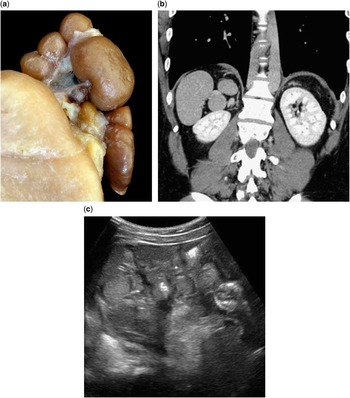
From the outset of detailed investigation of patients with complex congenital cardiac malformations, it was recognised that the arrangement of the bodily organs differed from the usual arrangement, and also from the mirror-imaged variant, usually described at that stage as “situs inversus”. Ivemark,Reference Ivemark 6 for example, in his description now recognised as groundbreaking, introduced the phrase “asplenia, a teratologic syndrome of visceral symmetry”. With even greater prescience, Putschar and Mannion stated the following: “The relationship of agenesis of the spleen to disturbed development of laterality, however, goes beyond the manifestations of obvious situs inversus. Between the normal situs, which is asymmetrical, and the situs inversus, which is the asymmetrical mirror-image of normality, a symmetrical situs sometimes exists, exhibiting symmetrical rightness or leftness on both sides”.Reference Putschar and Mannion 7 The early pioneers of paediatric cardiology, therefore, recognised fully the existence of isomerism within the thoracoabdominal organs. Thus, as the spleen is a left-sided organ, its absence is to be anticipated in the setting of right isomerism, whereas splenic tissue formed to either side of the dorsal mesogastrium (Fig 1) is the essence of left splenic isomerism.
The images show ( a ) multiple spleens as observed during autopsy, with the diagnostic feature being the presence of splenic tissue on either side of the dorsal mesogastrium. Panels ( b ) and ( c ) show how the features can be distinguished during life using either CT ( b ) or ultrasound ( c ). Note that, in the CT, the multiple spleens are right sided.

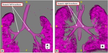
Evidence of bronchial and pulmonary isomerism, moreover, has existed for almost half a century. It was Van Mierop et al,Reference Van Mierop, Eisen and Schiebler 8 who pointed initially to the value of chest radiography in revealing the presence of bronchial isomerism, with their proposals subsequently endorsed by other investigators.Reference Landing, Lawrence, Payne and Wells 9 , Reference Partridge, Scott, Deverall and Macartney 10 Current techniques, such as CT, demonstrate the ease with which such features can now be recognised during life. The images also permit the ready distinction between right as opposed to left bronchial isomerism (Fig 2). Bodily isomerism, however, is not confined to the spleen or the lungs. Although the abdominal organs, apart from the spleen, do not show evidence of true isomerism, a short or annular pancreas is found only in the setting of left isomerism. This can lead to intestinal obstruction, or volvulus, with a short mesentery, but this finding is also frequent in the setting of right isomerism.Reference Cohen, Anderson and Cohen 11 Biliary atresia is another lesion found frequently in the setting of left isomerism, but with normal formation of the heart, again showing that isomerism is not necessarily present throughout all the systems of organs.Reference Debich, Devine and Anderson 12 Malformations of the central nervous system are also known to be present in the setting of either left or right isomerism. Although the precise role of left–right symmetry is unclear in humans, studies in mice have demonstrated that there is some degree of symmetry, particular in synaptic composition and function of neuronal networks.Reference Kawakami, Dobi, Shigemoto and Ito 13 This may also be the case in humans, although to the best of our knowledge this has yet to be studied.
The CTs show the ease with which it is now possible to distinguish left ( a ) from right ( b ) bronchial isomerism. It is also the case that, in left bronchial isomerism, the pulmonary artery supplying the lower lobes of both the lungs crosses the bronchus before its first bifurcation, making the bronchus hyparterial, whereas in right bronchial isomerism the bronchus branches on each side before it is crossed by the arteries supplying the lower lobes, making it eparterial.

These findings with regard to isomerism, nonetheless, are more than anatomic curiosities. They carry important functional implications. The cardiovascular malformations, for example, lead to obvious haemodynamic and circulatory derangements. Whether the spleen be absent or multiple, or even solitary, functional asplenia is frequent in the setting of either left or right isomerism.Reference Nagel, Williams, Stewart, Paul and Stumper 14 The malformations of the central nervous system, furthermore, can lead to developmental delay or impairment in cognitive function. The abdominal organs need to be assessed in detail, as malrotation is frequent, and the pancreas can be short. Identification of isomerism, therefore, is but the starting point of more detailed analysis, the more so because it must be recognised that there is no uniformity in the involvement of the different systems of organs.
Does isomerism exist within the heart?
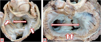
Part of the problem with the widespread recognition of isomeric features within the heart is that they are restricted to the atrial chambers. Instances of ventricular isomerism are remarkably rare. Although double aortic arch may be considered a form of arterial isomerism, such findings are also rare in the setting of so-called “heterotaxy”. It did not help, however, that when some of us first promoted the existence of cardiac isomerism,Reference Macartney, Zuberbuhler and Anderson 15 we used the term “atrial isomerism”. This prompted the suggestion that if the atrial chambers were truly isomeric then patients with left cardiac isomerism would have eight pulmonary veins!Reference Van Praagh and Van Praagh 16 This, of course, is reducing the argument to the absurd, because during development each lung forms only two pulmonary veins. Symmetry of pulmonary venous return, furthermore, is found in many patients with left isomerism. It remains the indubitable fact, nonetheless, that it is only the atrial appendages that are uniformly isomeric in the setting of so-called “heterotaxy”.Reference Uemura, Ho, Devine, Kilpatrick and Anderson 17 When assessed on the basis of the extent of the pectinate muscles relative to the atrioventricular vestibules, it is an easy matter, at least for the morphologist, to distinguish between the morphologically right and morphologically left atrial appendages (Fig 3).
The images show how, according to the extent of the pectinate muscles relative to the atrioventricular junctions, it is an easy matter for the morphologist, with the heart in his or her hands, to demonstrate the presence of isomeric right ( a ) and isomeric left ( b ) atrial appendages. Note that both hearts, as viewed from the atrial aspect, have a common atrioventricular junction (double-headed white arrow with red borders). In the heart from the patient with left isomerism ( a ), the pectinate muscles are confined within the tubular appendages, with no spillage to the dorsal parts of the atrioventricular junctions (single-headed white arrows with red borders). In the heart from the patient with right isomerism ( b ), the pectinate muscles extend to the cardiac crux on both sides (single-headed white arrows with red borders). Compare the images with those shown in Figures 4 and 5.

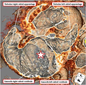
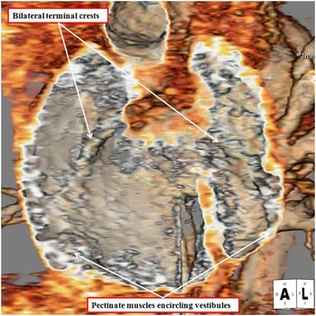
It is also the case that molecular biologists have proved that by knocking out genes responsible for producing laterality it is possible to generate animals with obvious right as opposed to left isomerism. Thus, knocking out the Pitx2 or Cited-2 genes will produce unequivocal right isomerism,Reference Bamforth, Bragança and Farthing 18 whereas knocking out the Lefty-1 gene will produce equally strong evidence of left isomerism.Reference Meno, Shimono and Saijoh 19 The question remains, however, as to whether evidence of isomeric atrial appendages can reliably be obtained during life. Recent experience with CT angiography now shows that this is achievable, and when the tests are conducted in an appropriate manner, with little more dosage of radiation required as for a standard chest radiograph (Figs 4 and 5).Reference Wolla, Hlavacek, Schoepf, Bucher and Chowdhury 20
The CT angiogram shows the view of the common atrioventricular junction of the heart from the patient with left bronchial isomerism as shown in Figure 2. Both appendages are tubular, with the pectinate muscles confined within the appendage. Compare with Figure 3a. The star shows a common atrioventricular junction.

The CT angiogram shows the view of the common atrioventricular junction of the heart from the patient with right bronchial isomerism as shown in Figure 2. Both appendages are triangular, with the pectinate muscles encircling the atrioventricular junctions on both sides. Compare also with Figure 3b.

Nevertheless, recognition of isomerism of the atrial appendages is no more than the starting point of analysis of patients known to have isomerism. Distinction between right as opposed to left isomerism points the way towards the intra-cardiac features to be sought in ongoing investigations. Thus, absence of the coronary sinus is a universal finding in patients with isomeric right atrial appendages, but not necessarily in those with absence of the spleen. This is because not all patients with isomerism of the right atrial appendages have absence of the spleen, just as not all those with isomerism of the left atrial appendages have multiple spleens.Reference Uemura, Ho, Devine and Anderson 21 Totally anomalous pulmonary connection, however, is another feature universally found in the setting of isomeric right atrial appendages, although the pulmonary veins will connect anomalously to an atrial chamber with a morphologically right appendage in about half the patients.Reference Uemura, Ho, Devine, Kilpatrick and Anderson 17 These are the only lesions uniformly present in patients with isomerism. Interruption of the inferior caval vein, with continuation through the azygos system, is much more frequent in patients with isomeric left atrial appendages. Univentricular atrioventricular connections, in contrast, usually double inlet through a common valve, are much more frequent in association with right isomerism. Pulmonary atresia or stenosis is to be anticipated with right isomerism, whereas coarctation is more frequent with left isomerism; however, these are associations at best. Bilateral caval veins are frequent in either variant, although it is self-evident that continuation through a coronary sinus is feasible only in those with isomeric left atrial appendages. Bi-ventricular and mixed atrioventricular connections, often through a common atrioventricular valve, can be found with either variant. All of this information emphasises the importance of full sequential segmental interrogation of patients known to have isomeric atrial appendages, along with assessment and description of the location of all the thoracoabdominal organs.
Conclusions
Taken overall, the evidence shows that “heterotaxy” is ill-suited as a term for the clinical entity it is currently used to describe. There is little question that the features of the syndromes thus identified are better segregated on the basis of isomerism, but recognising that not all systems show uniform arrangements. This variation is readily addressed by specific attention to each of the systems. This is more important when considering the heart, as it is only the atrial appendages, when assessed on the basis of their pectinate muscles, which are truly isomeric. Recognition of isomerism of the atrial appendages, which should now be achievable in the clinical setting, then sets the scene for full and accurate sequential segmental analysis. This, in turn, will allow for better understanding of the functional derangements that may result. Such distinction between right and left isomerism also offers a much better means of determining the genetic cues responsible for the production of so-called “heterotaxy”. Genetic modification of mice shows that it is by knocking out different cascades of genes that it is possible to produce models of rightReference Bamforth, Bragança and Farthing 18 as opposed to leftReference Meno, Shimono and Saijoh 19 isomerism. Evidence is also now increasingly emerging of the multiple and complex interactions between genes in producing congenital cardiac malformations.Reference Li, Klena and Gabriel 22 If specific genes are to be recognised as producing problems of lateralisation, it will be necessary to properly assign patients to comparable groups. This will not be achievable if all patients are grouped together as having “heterotaxy”. Chances of identifying specific genes will be greatly enhanced if the patients are segregated on the basis of left as opposed to right isomerism.