


Dental procedures and oral surgery hold numerous infectious hazards. The oral cavity is a natural habitat for a large number of microorganisms. Reference Dewhirst, Chen and Izard1 This ecological niche can be a reservoir for opportunistic and pathogenic microorganisms that can pose a risk for cross contamination and local and systemic infections. Reference Laheij, Kistler, Belibasakis, Välimaa and de Soet2 Additionally, patients undergo invasive procedures, local and systemic anesthetic injections, and irrigation with large volumes of nonsterile water. Furthermore, most dental clinics lack infection control experts, and audits on compliance with safe injection or cleaning and sterility standards are conducted infrequently. A number of studies have demonstrated low compliance with infection control practices. Reference Mutters, Hägele, Hagenfeld, Hellwig and Frank3-Reference Oosthuysen, Potgieter and Fossey5 Nonetheless, despite the numerous potential risks, few reports of outbreaks after dental procedures have been published, due perhaps in part to failure of providers and patients to draw a connection between an episode of sepsis and an antecedent procedure. Reference Radcliffe, Bixler and Moorman6,Reference Ross, Mehr and Greeley7 As a result, cross transmission in dentistry may be underreported in the literature.
Maxillary sinus augmentation (sinus floor elevation) procedures have become increasingly common before placement of dental implants in posterior maxillae that have suffered severe bone loss due to sinus pneumatization, alveolar bone atrophy, or trauma. Reference Scarano, Degidi and Iezzi8 The aim of this report is to describe the investigation of an outbreak of Enterobacter kobei bloodstream infections that developed after sinus-lifting procedures conducted in 2 private dental clinics.
Methods
Outbreak identification
In June 2018, the National Center for Infection Control (NCIC) of the Israel Ministry of Health received notification of 2 cases of E. kobei bloodstream infection that developed within hours of a sinus-lifting procedure conducted in 2 dental clinics in central Israel.
Procedure
Maxillary sinus augmentation is performed from inside the patient’s mouth, where the oral surgeon creates an incision into the gingiva. Subsequently, the surgeon cuts a “window” to the sinus, then bone-graft material is placed inside the newly created space. The graft material used can be an autograft, an allograft, or a xenograft.
Outbreak investigation
Site visits were conducted in the 2 involved dental clinics. Standard practices regarding patient care in the dental clinic were reviewed, including policies and practices of cleaning and sterilizing equipment, use of single-use medication vials, and preparation technique for local and intravenous anesthetics.
Sample collection, isolate identification and antibiotic susceptibility testing
The NCIC team collected surveillance cultures from the hands of personnel and from potential environmental sources (ie, dental water system, disinfectants, portable medication and equipment carts, environmental surfaces and intravenous medications) using sterile swabs moistened with normal saline for sampling hands and surfaces.
Isolates of E. kobei grown from blood cultures in the clinical microbiology laboratories of the treating hospitals were sent to the national reference laboratory of the NCIC for further investigation. Bioburden estimation from surveillance samples collected was determined using a filtration membrane assay in accordance with the ISO 11737 guideline. 9,10 Presence and enumeration of microorganisms in water-based solutions were determined by a microbial enumeration test in accordance with US Pharmacopeia monographs 6111 and 62, 12 without the presence of adequate neutralizer. Samples of drinking water collected from faucets were tested in accordance with the ISO 8199 guideline. 13 Drug solutions were tested using an automated microbial detection system (BACT/ALERT 3D, bioMérieux SA, Marcy l’Etoile, France) to increase sensitivity of detection for aerobic microorganisms (iFA Plus, bioMérieux SA) and anaerobic microorganisms (iFN Plus, bioMérieux SA).
Bacterial colonies were identified using matrix-assisted laser desorption/ionization time-of-flight mass spectroscopy (MALDI-TOF MS) (Bruker, Bremen, Germany). Antimicrobial susceptibility testing was performed using the VITEK 2 system (bioMérieux SA).
Molecular typing
Genotyping of E. kobei isolates was performed using repetitive element sequence-based polymerase chain reaction (REP-PCR). Prior to analysis, each isolate was streaked onto a Mueller-Hinton agar plate (Hylabs, Rehovot, Israel) and incubated aerobically at 35±2°C. REP-PCR was performed as previously described by Viau et al. Reference Viau, Kiedrowski and Kreiswirth14 Banding patterns were visualized using QIAxcel ScreenGel software (Qiagen, Hilden, Germany) combined with the QIAxcel Advanced capillary electrophoresis system (Qiagen).
The criteria used to analyze the results obtained by the REP-PCR method have been described previously. Reference Tenover, Arbeit and Goering15 The DNA fingerprint of isolates obtained by REP-PCR were compared by visual inspection of the banding patterns. Two isolates were assigned to the same REP-PCR group if their patterns differed by <3 bands, according to previously defined criteria. Reference Viau, Kiedrowski and Kreiswirth14 The study was approved by the jurisdictional institutional review board.
Results
Case descriptions
Case 1: On June 12, 2018, a 45-year-old woman with no underlying illnesses underwent maxillary sinus elevation in private dental clinic A. The procedure was performed by inserting a bone xenograft through a window opened in the upper jaw. The team present included an oral surgeon, an anesthesiologist, and a dental assistant. The procedure was conducted with both local and general anesthesia. Within 1 hour of its completion, the patient developed increasing jaw swelling. The patient was transferred to the emergency ward in a tertiary-care medical center (hospital R), where she presented with massive jaw swelling. The patient was hospitalized in a surgical ward for observation. After several hours, she developed severe hypotension and was transferred to the general intensive care unit. Blood cultures grew E. kobei. Due to unresponsive septic shock, surgical debridement was conducted on June 14, and the bone graft granules were removed. Cultures obtained from bone granules also grew E. kobei. Within 24 hours of the debridement, the patient’s condition improved, and she went on to recover and was discharged.
Case 2: On June 18, 2018, a 60-year-old woman with no underlying illnesses underwent maxillary sinus augmentation with autologous bone graft. The procedure was performed in private dental clinic B, located in a different town from clinic A. Within 24 hours of the procedure, the patient developed severe weakness. The patient presented to the emergency ward at a tertiary-care medical center (hospital S) with fever and hypotension and was admitted to the intensive care unit in septic shock. Broad-spectrum antibiotics were begun. Blood cultures grew E. kobei. The patient responded within 48 hours to systemic antibiotics, went on to recover, and was discharged.
Outbreak investigation
On June 14 and June 20, the NCIC received reports of 2 cases of sepsis developing shortly after a sinus elevation procedure in patients then hospitalized at hospitals R and S, respectively. Clinic A was temporarily closed on June 15, by order of the Ministry of Health. Site visits were conducted at clinic A and clinic B on June 18 and June 27, respectively. The healthcare providers were interviewed and surveillance cultures were obtained.
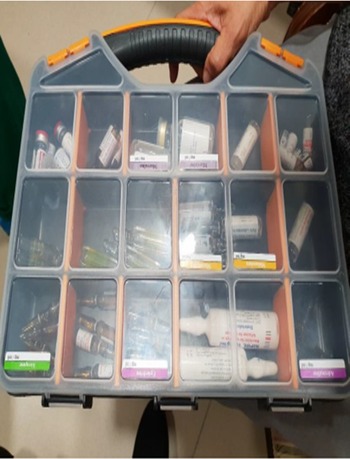
The procedures were performed in 2 dental clinics by different surgeons and dental assistants. A single anesthesiologist, who also worked at a private hospital, administered the systemic anesthesia in both cases. The intravenous medications were stored in a portable plastic cart (Fig. 1). Between June 12 and the day of the investigation interview, the anesthesiologist had treated 5 patients who underwent ambulatory surgery in 2 dental clinics, using the same cart (infection incidence, 40%). None of the 21 patients treated by the anesthesiologist in the private hospital, using the hospital’s medications, developed infection following surgery during the 2 weeks preceding the index-case infections. Patients who underwent general anesthesia in the 2 private clinics after June 1 did not report postprocedure infectious complications. In addition, a letter sent to all general hospitals nationwide describing the outbreak and soliciting information on similar cases of post-dental procedure sepsis yielded no information on additional cases.
Portable medication cart.

The anesthesiologist described and demonstrated the technique of intravenous drug preparation and administration. Hand hygiene was performed only once, at the beginning of the procedure, before donning clean gloves. Subsequently, medications needed for the procedure were selected from the cart. No multidose vials or syringes were used. Disinfection of the bottle septa before withdrawing medication was not performed, nor was disinfection of the intravenous administration set ports before use. Opened medication vials were never returned to the medication cart. Medication vials were reaccessed only for the same patient. Gloves were not changed before or after insertion of the peripheral intravenous catheter. The medication cart was transported between the private dental clinics by a private vehicle. The storage of the cart in between dental procedures was in the anesthesiologist’s office. The anesthesiologist could not recall when the cart had last been cleaned.
Surveillance data
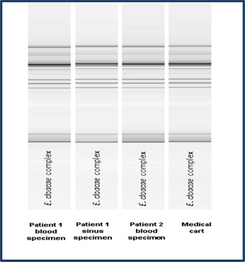
In total, 21 surveillance samples were received. Cultures from medications, fluids, and healthcare workers’ hands were culture negative or yielded nonsignificant growth (Supplementary Table 1 online). Enterobacter kobei was recovered from the interior chambers of the anesthesiologist’s medication cart. The antibiotic susceptibility profile of the isolates recovered from both patients and from the cart was identical. Resistance was detected to amoxicillin-clavulante, cefazolin, cefoxitin, and fosfomycin. The patient isolates and the environmental isolate belonged to a single clone (Fig. 2).
Repetitive sequenced-based PCR of clinical and environmental Enterobacter isolates.

Infection control measures
Following the outbreak, the anesthesiologist underwent retraining in principles of good practice in intravenous drug preparation and hand hygiene during anesthesia. He was instructed to avoid storage of medications in the portable cart and to clean the cart after each use. A meeting was held involving the appropriate bodies within the Ministry of Health to promote medication transport standards and mandatory reporting on infectious complications of outpatient procedures.
Discussion
Numerous outbreaks of bacterial and viral infections resulting from improper injection practices have been reported from various healthcare settings. Reference Dobbs, Guh and Oakes16-Reference Ross, Mehr and Carothers18 The current study reports on an outbreak following dental procedures conducted in 2 different private clinics. The common source was the medication cart used by the anesthesiologist. The investigation revealed the potential of the working environment to become contaminated and serve as a source of invasive infection. Notably, although anesthesiology infection control guidelines emphasize the importance of storage conditions and cleanliness of working surfaces, Reference Munoz-Price, Bowdle and Johnston19 no study to date has described the direct contribution of contaminated surfaces to the development of bloodstream infections associated with intravenous drug preparation.
Microbial contamination of various environmental reservoirs in dental settings is common. Reference Pasquarella, Veronesi and Napoli20 Nonetheless, serious bacterial infections derived from exogenic reservoirs in dental clinics are extremely rare. A report from Italy described an 83-year-old woman who died of Legionnaire’s disease after contracting the infection from the water lines in a dental practice. Reference Ricci, Fontana and Pinci21 A dental unit water system was also the source of a cluster of odontogenic infections among children who developed Mycobacterium abscessus infection after undergoing pulpotomy at a pediatric dentistry practice. Reference Hatzenbuehler, Tobin-D’Angelo and Drenzek22 Another study described acquisition of methicillin-resistant Staphylococcus aureus due to contaminated surfaces in a dental clinic. Reference Kurita, Kurashina and Honda23 Therefore, the occurrence of sepsis after a dental procedure should trigger immediate investigation and intervention.
Healthcare-associated infections have been traditionally described in the hospital setting. Reference Sood and Perl24 With increasing frequency of invasive procedures performed in outpatient settings, healthcare-associated infections may now occur within various ambulatory care settings as well. Reference Oyong, Coelho, Bancroft and Terashita25 Injection safety violations and equipment cleaning issues are frequent sources of these outbreaks. Identifying outbreaks in the outpatient setting can be extremely challenging. Limited patient follow-up may lead to missed detection of complications occurring after the procedure. In addition, patients developing infectious complications may be referred to different hospitals, which may delay recognition of an outbreak. Delayed recognition of an outbreak may have serious consequences for patient safety because it hampers the ability of public health officials to perform timely investigations. The Enterobacter outbreak described in this study was recognized early due to the vigilance of hospital epidemiologists affiliated with both hospitals involved. The early identification of the outbreak enabled the initiation of control measures that resulted in halting the outbreak.
Environmental surfaces have been associated with the development of nosocomial infections. Reference Suleyman, Alangaden and Bardossy26 Most reports have demonstrated the contribution of the environment in acquisition of epidemiologically important pathogens. Reference Boyce27,Reference Weber, Rutala, Miller, Huslage and Sickbert-Bennett28 Few studies to date have provided evidence of direct microbial transmission from the environment to patients undergoing general anesthesia. Reference Loftus, Koff and Burchman29-Reference Gargiulo, Sheridan and Webster31 The mechanism by which contaminated surfaces lead to bloodstream infection is unclear. A study conducted in a surgical suite demonstrated that the anesthesia work area became significantly contaminated with potential pathogens. Reference Loftus, Koff and Burchman29 Transmission likely occurred secondary to contamination of the provider’s hands during induction of anesthesia. Furthermore, bacterial contamination of the anesthesia work area was associated with contamination of peripheral intravenous stopcock sets in more than a third of the cases. Reference Loftus, Koff and Burchman29
The anesthesiologist involved in the present investigation described a number of incorrect practices during drug preparation and administration. These practices may increase the risk of contamination of parenteral medications. Reference Gargiulo, Sheridan and Webster31 Several studies have described the risk of preparing drugs directly by the provider, rather than in the pharmacy. Reference Austin, Hand and Elia32 In a recent review, the most common infection control breaches were the use of multidose vials, reuse of syringes, and poor hand hygiene during preparation and administration of medications. Reference Suvikas-Peltonen, Hakoinen, Celikkayalar, Laaksonen and Airaksinen33
During intravenous drug preparation, 2 primary factors that may lead to microbial contamination of the medications are the cleanliness of the work environment and the aseptic technique of the provider. We believe that the breaches in appropriate work habits described by the anesthesiologist and the contaminated medication cart led to bloodstream infections that developed shortly after intravenous drug administration.
World Health Organization guidelines require storage of medications in standardized and monitored conditions across all healthcare settings. 34 Yet, anesthesia conducted in private dental clinics is a unique setting. Anesthetic medications are not available in these clinics due to safety issues and a lack of appropriate storage conditions. As a result, the medications are frequently stored and transported in portable medication carts. Following the outbreak, we realized that the national guidelines should address the topic, stressing the importance of avoiding intravenous medications storage in portable carts and the need to clean and disinfect the cart after each use. There is an urgent need for broader education of anesthesiologists and practice change.
In conclusion, an outbreak of post–dental-procedure sepsis was linked to a contaminated portable medication cart, emphasizing the importance of medication storage standards and strict aseptic technique when preparing intravenous drugs used in anesthesia. Immediate reporting of sepsis following outpatient procedures enabled early identification and termination of the outbreak. Implementation of infection control measures when general anesthesia is administered in outpatient settings is critical to reducing the risk of life-threatening infectious complications.
Acknowledgments
Financial support
No financial support was provided relevant to this article.
Conflicts of interest
All authors report no conflicts of interest relevant to this article.
Supplementary material
To view supplementary material for this article, please visit https://doi.org/10.1017/ice.2021.176