INTRODUCTION
With the increasing depletion of land resources, marine resource exploitation has gradually become one of the study focuses in academia. Marine sponges have attracted the attention of scientific researchers and become an important source of bioactive metabolites due to the diversity of their symbiotic microorganisms (Kobayashi & Ishibashi, Reference Kobayashi and Ishibashi1993; Edrada et al., Reference Edrada, Heubes, Brauers, Wray, Berg, Gräfe, Wohlfarth, Mühlbacher and Schaumann2002) and the novel biochemical components in their body walls (Sakemi et al., Reference Sakemi, Ichiba, Kohmoto, Saucy and Higa1988; Keifer et al., Reference Keifer, Schwartz, Koker, Hughes, Rittschof and Rinehart1991; Fusetani et al., Reference Fusetani, Shinoda and Matsunaga1993; Müller et al., Reference Müller, Wimmer, Schatton, Böhm, Batel and Filic1999; Wellington et al., Reference Wellington, Cambie, Rutledge and Bergquist2000). Sponges are sessile organisms that capture food by filtering organic matter in the water. These organisms have a strong metabolic capacity to use various substances obtained from water, so sponges (or their symbionts) are able to biosynthesise bioactive compounds (Simpson & Garson, Reference Simpson and Garson1998; Piel et al., Reference Piel, Dequan, Gaiping, Butzke, Platzer, Fusetani and Matsunaga2004). The way they feed may be the reason that such plentiful metabolites can be found in sponge body walls. Moreover, marine sponges host abundant symbiotic microorganisms, which may also be an excellent source of active material. Isolating sponge symbiotic microorganisms and extracting microbial metabolites from them has also gradually become a research focus. Studies have shown that many substances extracted either from sponges or from their symbiotic microbes are biologically active (Krishnan et al., Reference Krishnan, Balasubramaniam, Roy, Sarma, Hairun and Sunder2014; Costantini et al., Reference Costantini, Romano, Rusolo, Capone, Guerriero, Colonna, Ianora, Ciliberto and Costantini2015). Bioactive metabolites, such as alkaloids, terpenoids and sterols, which may have significant antibacterial, antiviral or antineoplastic effects, can be extracted from sponge body walls (Carter & Rinehart Jr, Reference Carter and Rinehart1978; Bewley et al., Reference Bewley, Debitus and Faulkner1994; Reddy et al., Reference Reddy, Ravinder, Narasimhulu, Sridevi, Satyanarayana, Kondapi and Venkateswarlu2006; Lu et al., Reference Lu, Chueh, Kung, Pan, Shen and Guh2007) or from their symbiotic microorganisms (Shigemori et al., Reference Shigemori, Bae, Yazawa, Sasaki and Kobayashi1992; Zheng et al., Reference Zheng, Chen, Han, Lin and Yan2005; Anand et al., Reference Anand, Bhat, Shouche, Roy, Siddharth and Sarma2006).
Although a wide variety of active substances can be extracted from marine sponges, the total content is disappointingly low. Early studies showed that only milligrams of bioactive metabolites were extracted from hectograms or even kilograms of sponges (Perry et al., Reference Perry, Blunt, Munro and Pannell1988; West et al., Reference West, Northcote, Hood, Miller and Page2000). For further study toward the application of bioactive metabolites in the pharmaceutical industry, up to several thousand tonnes of sponge supply are required (Munro et al., Reference Munro, Blunt, Dumdei, Hickford, Lill, Shangxiao, Battershill and Duckworth1999; Duckworth & Battershill, Reference Duckworth and Battershill2003). For the present, the marine sponge resources for research mainly come from wild populations, which could greatly damage the marine ecological environment (Maldonado & Riesgo, Reference Maldonado and Riesgo2009). Furthermore, the total amount of the wild sponge population still does not meet the needs of pharmacological research, which has become an obstacle to better investigation and application of the novel active materials (Munro et al., Reference Munro, Blunt, Dumdei, Hickford, Lill, Shangxiao, Battershill and Duckworth1999; Duckworth & Battershill, Reference Duckworth and Battershill2003). Therefore, finding a reliable sponge supply is crucial to solving the sponge shortage problem. Fortunately, the problem can be solved by sponge aquaculture. Currently, the common method for sponge artificial culture is based on the explanting of wild sponge populations, which is laborious and time consuming due to the intensive work required to cut the large amounts of sponge mass and attach them to the substrates (Page et al., Reference Page, Northcote, Webb, Mackey and Handley2005; Hawes et al., Reference Hawes, Oengpepa and Center2010; Friday, Reference Friday2011; Schiefenhövel & Kunzmann, Reference Schiefenhövel and Kunzmann2012). Our unpublished observations showed that sponge explants can only survive for one generation due to the low water temperature in winter, which indicated that successive generations of explants can be very sensitive to seasonal climate changes. By contrast, sexually produced descendants may adapt better to the environment and have a larger amount of reproductive output according to the studies on wild populations (Meroz & Ilan, Reference Meroz and Ilan1995; Riesgo et al., Reference Riesgo, Maldonado and Durfort2007). It is also easier to perform the artificial culture process than to use explants. Once a proven technique for sponge culture based on sexual reproduction is established, the amount of available sponge will increase by a wide margin. Establishing this technique requires a better understanding of sponge sexual reproduction. This basic biological study is therefore particularly necessary (De Caralt et al., Reference De Caralt, Otjens, Uriz and Wijffels2007; Wahab et al., Reference Wahab, De Nys and Whalan2012).
Previous studies on sexual reproduction of demosponges have investigated many aspects, including the timing and duration of gametogenesis and embryogenesis, sexual pattern and reproductive output (Fromont & Bergquist, Reference Fromont and Bergquist1994; Riesgo et al., Reference Riesgo, Maldonado and Durfort2007; Maldonado & Riesgo, Reference Maldonado and Riesgo2009; Ereskovsky, Reference Ereskovsky and Ereskovsky2010). It can be summarized from all these studies that the reproductive strategies and their factors of influence in sponges may be very similar or different depending on species (Ettinger-Epstein et al., Reference Ettinger-Epstein, Whalan, Battershill and De Nys2007; Gerasimova & Ereskovsky, Reference Gerasimova and Ereskovsky2007; Mercurio et al., Reference Mercurio, Corriero and Gaino2007; Ereskovsky, Reference Ereskovsky and Ereskovsky2010). For example, the sexual reproduction of several species from the same genus can be similar in the sexual pattern, while being different in the duration of embryogenesis (Fromont & Bergquist, Reference Fromont and Bergquist1994). Studies on the exact genus or even species are still required.
The poecilosclerid demosponge Mycale phyllophila (Hentschel 1911) is a cosmopolitan species and can be found in shallow seas in both the northern and southern hemispheres (Hajdu, Reference Hajdu, Van Soest, Boury-Esnault, Hooper, Rützler, De Voogd, Alvarez De Glasby, Hajdu, Pisera, Manconi, Schoenberg, Janussen, Tabachnick, Klautau, Picton, Kelly, Vacelet, Dohrmann, Díaz and Cárdenas2015). It is a common species in the Fujian coastal area, where it displays good adaptation to the environmental conditions along these areas. Sponges of the Mycale genus are a source of several potential bioactive metabolites (Critcher & Pattenden, Reference Critcher and Pattenden1996; Kalinovsky et al., Reference Kalinovsky, Antonov, Afiyatullov, Dmitrenok, Evtuschenko and Stonik2002; Ortega et al., Reference Ortega, Zubía, Sánchez, Salvá and Carballo2004; Singh et al., Reference Singh, Chunxiao, Xiaoming, West, Wilmes, Chan, Hamel, Miller, Northcote and Ghosh2009; Miller et al., Reference Miller, Singh and Northcote2010), such as mycalamides, mycalazals, pelorusides and pateamines; these possess promising antiviral and antitumor activity, and can be extracted from sponges in the Mycale genus (Perry et al., Reference Perry, Blunt, Munro and Pannell1988; West et al., Reference West, Northcote, Hood, Miller and Page2000; Ortega et al., Reference Ortega, Zubía, Sánchez, Salvá and Carballo2004; Miller et al., Reference Miller, Singh and Northcote2010). Unpublished observations from our research suggested that M. phyllophila may be an excellent source of mycalazals, which are cytotoxic metabolites that have been shown to be growth inhibitors of the human prostate carcinoma cell line LNcaP (Ortega et al., Reference Ortega, Zubía, Sánchez, Salvá and Carballo2004; Carballo et al., Reference Carballo, Yañez, Zubía, Ortega and Vega2010). The sexual reproduction of some Mycale species in the Mediterranean, the Red Sea, and near Discovery Bay in Jamaica has been studied (Reiswig, Reference Reiswig1973; Meroz & Ilan, Reference Meroz and Ilan1995; Corriero et al., Reference Corriero, Liaci, Marzano and Gaino1998). However, we know little about the reproductive biology of M. phyllophila, including the reproduction pattern, the timing of the reproduction activity and the factors that affect the reproductive stages, which are all important for culturing this species. It is essential to learn more about the sexual reproduction of M. phyllophila to ensure the constant supply of sponges for further pharmaceutical research.
This study aims to provide information on the sexual reproductive cycle of the demosponge M. phyllophila through surveying the sponge population for two breeding seasons. Our investigation is the first to report on the sexual reproduction of this Mycale species in the coastal waters of China. We hope to contribute to a better understanding of sponge sexual reproduction to help solve the sponge shortage problem.
MATERIALS AND METHODS
Study site descriptions
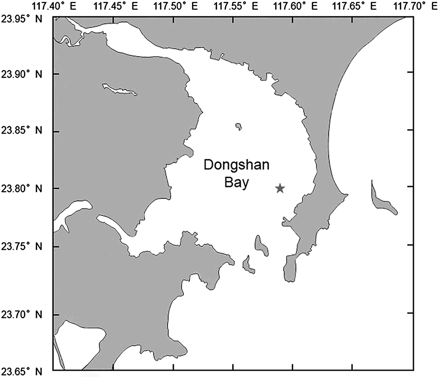
The investigated population of Mycale phyllophila was obtained from an experimental site established in Dongshan Bay (117.5928E 23.8015N, Figure 1). Dongshan Bay is the largest semi-enclosed bay in southern Fujian, located on the west bank of the Taiwan Strait and stretching inland with an irregular pear shape. Because of the particular geographic conditions, the water environment in Dongshan Bay has the features of good water quality, mild temperature variation and moderate wave disturbance (Lu et al., Reference Lu, Li, Zhang and Ma2014), which are favourable for the aquaculture of sponges. This site was chosen because of the high abundance of M. phyllophila that live on the net cages and other aquaculture facilities for the common economic species. Our experimental field consisted of several net cages used previously for abalone culture. The sponge larvae naturally attach onto plastic ropes that fishermen used to hang for abalone aquaculture. There were ropes on the net cages, which could reach depths of 1.5–3 m, and the adult sponges developed on the ropes and nets.
Study site: Dongshan Bay (Fujian, China) on the west bank of Taiwan Strait, indicated by a star.

Sample collection and preservation
To better understand the patterns of sexual reproduction in this species, specimens from 15 M. phyllophila individuals were collected at regular monthly intervals from June 2012 to January 2014. The 15 individuals included 10 tagged individuals and five random individuals. The random sampling was to help determine the mode of sexuality and avoid potential influence of repeat sampling. Water temperature was recorded at the same sites using temperature loggers (HOBO Water Temp Pro v1) at 1 h intervals. Small samples of approximately 8 cm3 in volume were cut off from each of the sponges with surgical scissors and fixed in Bouin solution immediately after collection. The specimens were kept in the Bouin fixation solution at room temperature for 24 h and were then transferred into 70% ethanol for further preservation.
Histological processing and microscopy
For light microscopy, specimens were dehydrated through a graded ethanol series and embedded in paraffin. Histological sections with a thickness of 7 µm were stained with haematoxylin-eosin and mounted on slides with resinene. An Olympus BX51 microscope connected to a digital camera and a computer was used to conduct observations. Image-Pro Plus 6.0 software was used for processing pictures and measuring sexual reproductive elements.
We estimated the density of gametes by applying the following equation (Abercrombie, Reference Abercrombie1946; Maldonado & Riesgo, Reference Maldonado and Riesgo2009),
 $$d = \displaystyle{{Ntk} \over {(D + t)}}$$
where d is the number of gametes per mm3 of tissue in reproductive sponges, N is the number of gametes counted in the 5 mm2 field of the histological sections, t is the thickness of the sections (7 µm), D is the average diameter of gametes for each month, and k is a constant (28.57) that converts the volume of observation (0.035 mm3) to 1 mm3.
RESULTS
Reproductive activities
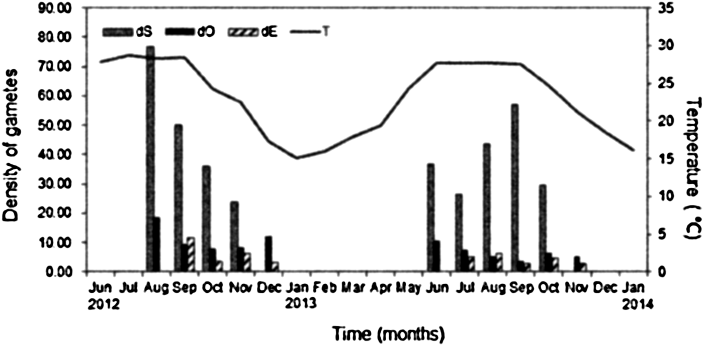
Reproductive activities peaked in the season when seawater temperature was highest (above 25°C). Oogenesis in Mycale phyllophila lasted for several months each year with observed interannual changes (Figure 2). In 2012, the first oocytes occurred in August, and oocyte production was observed for 5 months. In 2013, oocytes first appeared in the mesohyl in June, and oocyte production lasted for 6 months (Figure 2). The first observed spermatogenesis in M. phyllophila specimens also occurred in August 2012 and June 2013 and lasted for 4–5 months. Embryogenesis extended for 4 months in 2012 and for 5 months in 2013. Compared with the tagged individuals, the random samples showed the same reproduction pattern with all the sexual reproductive elements observed.
Mean density of embryos (dE), spermatic cysts (dS) and oocytes (dO) (number of gametes per mm3 of sponge tissue) and their relationship with the seawater temperature (T) during the investigated seasons.

Oogenesis
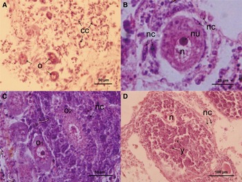
Light microscopy observations showed that 5–10 young oocytes formed a brooding chamber (Figure 3A) and that these were randomly distributed in the mesohyl. These young eggs were spherical or irregular in form and measured 30–50 µm in diameter (Figure 3A, B). A distinct nucleus with a thin cytoplasmic shell was observed inside the oocytes (Figure 3B). Archaeocytes with a nucleus and few inclusions gathered around the oocytes and acted as nurse cells. Oocytes engulfed the nurse cells, which turned into yolk bodies via phagocytosis, to grow in volume (Figure 3B). More advanced oocytes (100–150 µm) developed from early oocytes, and the number of inclusions increased gradually through the phagocytosis of nurse cells (Figure 3C). Oocytes in this stage still need to absorb nurse cells to increase their size. Oocytes reached a final mature size of 300–400 µm (Figure 3D). These late oocytes had fewer peripheral cells than younger oocytes because most of the nurse cells had been transformed into yolk over the course of oogenesis. The nucleus was pushed aside by the enlarged egg yolk, indicating that the oocytes are telolecithal eggs with an uneven distribution of yolk (concentrated at one pole) in the cytoplasm. The simultaneous appearance of oocytes in different stages within one adult sponge indicates that oogenesis is asynchronous in M. phyllophila (Figure 3C).
Oogenesis of Mycale phyllophila Hentschel, 1911: (A) primary oocytes (o) scattered in the mesohyl, note choanocyte chambers (cc) around the oocytes; (B) detail of a primary oocyte with a prominent nucleus (n) and nucleolus (nu) surrounded by nurse cells (nc), showing the process of the oocyte phagocytosing a nurse cell; (C) a more advanced oocyte (o2), and absorbed nurse cells, note a primary oocyte (o1) next to o2; (D) mature oocyte with prominent nucleus (n) surrounded by nurse cells (nc), note the yolk bodies (y) inside the mature oocyte.

Spermatogenesis
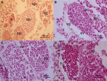
Spermatogenesis in this sponge species occurs in spermatic cysts. Asynchronous spermatic cysts are dispersed throughout the maternal mesohyl matrix (Figure 4A). Inside the spermatic cysts, there are many different cell types, demonstrating that spermatogenesis is asynchronous in M. phyllophila. Archaeocytes congregated around the spermatic cysts, which measured approximately 15 µm (Figure 4B). Enveloped by a follicular layer, clusters of spermatogonia formed spermatic cysts with a size of approximately 100 µm. Primary spermatocytes with a large nucleus came from spermatogonia that had gone through mitosis, reaching a size of approximately 10 µm. Primary spermatocytes must undergo meiosis I before becoming secondary spermatocytes, which are approximately 8 µm in size (Figure 4C). After meiosis II, secondary spermatocytes divided into smaller, 6 µm-diameter spermatids. Mature spermatozoa form from developed spermatids and are small, semi-transparent and ellipsoid with a diameter ranging from 3 to 5 µm (Figure 4D). The follicular envelope can be observed throughout the whole process of spermatogenesis, keeping its form in spermatic cysts until sperm release.
Spermatogenesis of Mycale phyllophila Hentschel, 1911: (A) different stages of spermatogenesis showing spermatogonia (sg), primary and secondary spermatocytes (sp1, sp2), and spermatozoa (s) inside the spermatic cysts; (B) a spermatic cyst containing spermatogonia (sg) and spermatids (sp) is encircled by an envelope (ev), note several archaeocytes (ac) in the mesohyl; (C) the developing spermatic cyst on the right still encloses the spermatogonia (sg), showing asynchronous development inside one spermatic cyst, note the spermatic cyst is encircled by an envelope (ev); (D) mature spermatozoa (s) on the left and spermatids (sp) on the right.

Embryogenesis and larval development
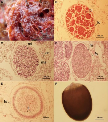
Embryos in adult sponges were small, milk-white spheres (Figure 5A). The embryos gather around the oscula for convenient larvae release. A transparent membrane can be observed surrounding each embryo. This was observed to be a layer of follicle under microscopy. The follicle acts as an egg envelope, persisting throughout embryogenesis until larvae release. As cleavage progressed, polarization and unequal cleavage resulted in blastomeres with varying sizes (Figure 5B). Micromeres were present as smaller blastomeres at the periphery; these divided quickly and differentiated into mucoid cells and cilia cells during the larvae-forming stage (Figure 5C). Macromeres in the middle of the embryo were slower to divide and became other cell types in the mesohyl. Embryogenesis was observed to be asynchronous, as embryos in different stages could be observed in the same individual. Larval morphogenesis started with the formation of the mucoid cells that came from the peripheral micromeres (Figure 5D). In more advanced larvae, cilia cells generated from mucoid cells were observed. Distinct variation was also observed inside the larvae. Diverse cell types began to appear inside the forming larvae. Cytoplasm became thinner in preparation for the development of various larval structures. Spicules were distinctly visible in the developing larvae within the maternal mesohyl (Figure 5E). Demosponge larvae are called parenchymella when they are released. At this stage, cilia were absent from both the anterior and posterior poles, and spicules were concentrated in the posterior pole (Figure 5F).
Embryogenesis and larval morphogenesis of Mycale phyllophila Hentschel, 1911: (A) embryos inside M. phyllophila adult (arrows); (B) an embryo in an early phase of development, with a few unequal blastomeres (bl) showing unequal cleavage, note the embryo was encircled by a follicle layer (fo); (C) more advanced embryo showing polarization of blastomeres, macromeres (ma) were observed mainly in the internal part of the embryo, while micromeres (mi) were observed at the periphery, note choanocyte chambers (cc) in the mesohyl; (D) a pre-larva showing mucoid cells (m) that can be differentiated from the peripheral cells of the mature embryo. Note the surrounding follicle layer (fo) and developing embryos on the left; (E) a larva which was ready to be released, displaying a complement of differentiating cells, note spicules (s) were distinctly visible; (F) a released parenchymella with spicules (arrows) concentrated in the posterior pole.

DISCUSSION
Reproductive patterns
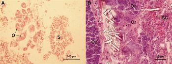
The synchronous presence of all reproductive elements and the occurrence of embryogenesis within the parent body in both the tagged and random individuals indicates that Mycale phyllophila is a hermaphroditic and viviparous species (Figure 6), as has been reported for other Mycale species (Meroz & Ilan, Reference Meroz and Ilan1995; Corriero et al., Reference Corriero, Liaci, Marzano and Gaino1998). We observed all sexually reproductive elements, but failed to capture the process of sperm release and fertilization. It is difficult to record the complete process of sexual reproduction in histological sections due to the small proportion of the sponge tissue that was sampled, especially because the reproductive elements appear randomly in the tissues. It is difficult in some species to detect any spermatogenic activity; one example is the sponge M. contarenii (Corriero et al., Reference Corriero, Liaci, Marzano and Gaino1998), which has a remarkably short period of sperm formation.
Synchronous appearance of oocytes and spermatic cysts: (A) primary oocytes (o) next to the spermatic cysts (sc); (B) primary oocytes (o1), mature oocytes (o2), and spermatic cysts (sc) appear synchronously.

Duration of the reproductive activities
The reproductive activities in M. phyllophila last for 5–6 months (Figure 2). The difference in duration from year to year may be due to interannual reproductive variability caused by environmental changes (Ettinger-Epstein et al., Reference Ettinger-Epstein, Whalan, Battershill and De Nys2007; Mercurio et al., Reference Mercurio, Corriero and Gaino2007), in that the reproductive activities of marine sponges are known to be susceptible to environmental factors, including temperature, salinity, lunar phase and nutritive resources (Elvin, Reference Elvin1976; Fell, Reference Fell1976; Fromont & Bergquist, Reference Fromont and Bergquist1994; Qu et al., Reference Qu, Zhang, Li, Yu and Jin2005).
The duration of oogenesis was 5–6 months in M. phyllophila (Figure 2), which accorded with the common time span of 5–7 months for oogenesis in demosponges (Ereskovsky, Reference Ereskovsky2000, Reference Ereskovsky and Ereskovsky2010; Whalan et al., Reference Whalan, Battershill and De Nys2007; Maldonado & Riesgo, Reference Maldonado and Riesgo2009). The long spawning period may be a reproductive strategy to ensure the continuous supply of the larvae (Meroz & Ilan, Reference Meroz and Ilan1995), which is a favourable characteristic for a potentially cultivable species.
Spermatogenesis in M. phyllophila started in the same month as oogenesis and lasted for 4–5 months (Figure 2), which is long among demosponges in which the normal duration of spermatogenesis is 1–2 months (Ayling, Reference Ayling1980; Ereskovsky, Reference Ereskovsky2000; Whalan et al., Reference Whalan, Battershill and De Nys2007; Riesgo & Maldonado, Reference Riesgo and Maldonado2008). In some other species, spermatogenic activity lasts for only several weeks. For instance, sperm formation in P. ficiformis lasts only 2.5 weeks (Maldonado & Riesgo, Reference Maldonado and Riesgo2009). Corticium candelabrum have a similar duration of 4–5 months for spermatogenesis (Riesgo & Maldonado, Reference Riesgo and Maldonado2008).
Embryogenesis in M. phyllophila started a month later than oogenesis and spermatogenesis and lasted for 4–6 months (Figure 2), which is average for demosponges (Corriero et al., Reference Corriero, Liaci, Marzano and Gaino1998; Ereskovsky, Reference Ereskovsky2000; Gerasimova & Ereskovsky, Reference Gerasimova and Ereskovsky2007). Although the temperature in December 2012 was lower than that in 2013, we were still able to detect embryos in the mesohyl. This may be because the breeding season started later in 2012 (Figure 2) due to a cold shock event in February 2012. A decrease in cold water temperature to 13°C resulted in the death of all adult M. phyllophila individuals at the study site. New M. phyllophila individuals did not reappear until June 2012, the month we started sampling. It may therefore take 1–2 months for new sponges to grow and be ready to start spawning (Simpson & Gilbert, Reference Simpson and Gilbert1974; Kaye & Reiswig, Reference Kaye and Reiswig1991; De Caralt et al., Reference De Caralt, Otjens, Uriz and Wijffels2007). Mycale phyllophila likely need a certain length of time to complete a full spawning process – a late start to reproductive activities will therefore lead to a late end.
Effect of the seawater temperature
Seawater temperature has a critical influence on the reproductive cycle of sponges. A strong relationship between sexual reproductive activities (including the onset of reproduction, the reproductive period and the reproductive output) and seawater temperature can be observed in some sponges (Mercurio et al., Reference Mercurio, Corriero and Gaino2007; Chung et al., Reference Chung, Huang, Lee and Liu2010; Ereskovsky et al., Reference Ereskovsky, Dubois, Ivanišević, Gazave, Lapebie, Tokina and Pérez2013). Mycale phyllophila is one of those sponges. The breeding season of M. phyllophila lasted from early summer to late autumn, synchronizing with the rise and fall of the water temperature. Its reproductive activity peaked in the hot season when the average seawater temperature was above 25°C (Figure 2). This indicates that, if this species is cultured in the field, it will need special care during the warm season in order to obtain greater reproductive output.
The density of spermatic cysts and oocytes reached a sudden high level in August 2012 (Figure 2). We assumed that this was a response to the cold shock event in February 2012. The sponges began to thrive following the sudden decline in population the previous spring, and more sponge recruits were needed while the weather was still suitable for reproduction to ensure the survival of the population. This is in accord with the reproductive strategies of r-strategist populations (Southwood et al., Reference Southwood, May, Hassell and Conway1974; Ereskovsky, Reference Ereskovsky2000).
Reduction in reproductive activity
The reduction in the density of reproductive elements in the M. phyllophila study population, particularly in spermatic cysts, in 2013 compared with 2012 may have been due to a decrease in fertility separate from the assumption about the efforts to thrive. The evidence for the decrease in reproductive capability in sponge individuals can be found in the study of the tropical sponge Luffariella variabilis (Ettinger-Epstein et al., Reference Ettinger-Epstein, Whalan, Battershill and De Nys2007) and the North Sea sponge Geodia cydonium (Mercurio et al., Reference Mercurio, Corriero and Gaino2007). These studies indicate that individual sponges may not be capable of continuous and perennial sexual reproduction. Furthermore, a sponge individual may fall into decline as it ages. However, embryogenesis tended to occur over an extended period in 2013 compared with 2012. A possible explanation for the reduction in overall reproduction combined with an extended reproductive period in 2013 is that the sponges tried to reduce the risk inherent to having one smaller synchronized spawning event in order to stabilize the population (Whalan et al., Reference Whalan, Battershill and De Nys2007).
ACKNOWLEDGEMENTS
The authors thank the Open and Sharing Platform of Equipment and Technology of the College of Ocean and Earth Sciences, Xiamen University, for providing certain instrument equipment and technical advice. We express our thanks to Mr Yongzhi Hong for the monthly sampling in the study site. We also want to thank the anonymous reviewers for their suggestions and constructive comments that considerably helped to improve the quality of this manuscript.
FINANCIAL SUPPORT
This research was financially supported by grants from the Scientific Research Project of Xiamen Southern Ocean Center (S.D., grant number 13GYY002NF07), (D.W., grant number 14CZP039HJ13); and the National Fund for Fostering Talents in Basic Science from National Natural Science Foundation of China (D.W., grant number J1210050).
CONFLICT OF INTERESTS
None.
ETHICAL STANDARDS
The authors assert that all procedures contributing to this work comply with the ethical standards of the relevant national and institutional guides on the care and use of laboratory animals.
ETHICS POLICY
The authors declare the work has not been previously published or submitted for publication elsewhere. All co-authors have approved the final version for publication. Employers and funding agencies have approved it for publication. All persons listed as co-authors merit authorship by virtue of their scientific contributions to the paper. Appropriate ethics conduct and approvals were adhered to where field sampling was conducted, and with regard to care of animals in the laboratory. None of the text has been copied or paraphrased from other publications, or if so is in quotation marks and the sources cited. The novelty of the paper is not exaggerated by failure to cite previous studies. The Methods and Results are a full and honest description of how the data were collected and analysed.