Introduction
Sterol 14α-demethylases (CYP51s) are the most conserved cytochrome P450 enzymes (Lepesheva and Waterman, Reference Lepesheva and Waterman2004) across phylogeny that catalyse one, essentially the same stereoselective three-step reaction of the oxidative removal of the 14α-methyl group from one or more of the five naturally occurring cyclized sterol precursors, lanosterol, 24,25-dihydrolanosterol, eburicol, obtusifoliol and C4-norlanosterol (Fig. 1). In eukaryotes, the reaction occurs in the endoplasmic reticulum and is required for biosynthesis of sterols, which serve as essential components of plasma membranes (bulky sterols, cholesterol in humans, ergosterol in fungi) and also as precursors for regulatory molecules that modulate growth, division, differentiation and development processes (sparking sterols) (Lepesheva and Waterman, Reference Lepesheva and Waterman2007; Nes, Reference Nes2011). Fast sterol production is most crucial for rapidly multiplying cells. It is also important to bear in mind that while humans can consume cholesterol from the diet, blocking of ergosterol production in unicellular human pathogens is lethal.
CYP51 reaction is an essential step upon sterol biosynthesis. The pathway involves multiple (>30) steps, beginning with the condensation of acetyl-CoA molecules that serve as initial building blocks, and proceeds to squalene, which then forms epoxide and cyclizes into the triterpene sterol skeleton (cycloartenol or lanosterol). These precursors are further modified to produce cholesterol, ergosterol, or sitosterol, which are the major membrane sterols in humans, fungi/protozoa and plants, respectively. The CYP51 reaction occurs either immediately or soon after squalene cyclization. The 14α-methyl group of the substrate (lanosterol, 24,25-dihydrolanosterol, eburicol, obtusifoliol and/or C4-norlanosterol) is converted into the alcohol, then into the aldehyde derivative and finally is removed as formic acid with the introduction of the Δ14–15 double bond into the sterol core. The CYP51 reaction includes three consecutive cytochrome P450 catalytic cycles, consuming three molecules of oxygen, six electrons and six protons. A detailed description of other reactions of the pathway can be found in Nes (Reference Nes2011). Inset: Sterol molecules are incorporated into membranes with the 3β-OH facing the water interface and the side chain extending into the hydrophobic core to interact with fatty acyl chains of phospholipids and proteins.

Although sterol biosynthesis involves multiple steps, so far only two of them have become the major targets for systemic clinical drugs. Statins (cholesterol-lowering agents), which act upstream of the pathway, at the step of mevalonate production, are presently the most frequently prescribed medications (Superko et al. Reference Superko, Momary and Li2012), while azoles, inhibitors of CYP51, serve as the most widely used antifungals (Lass-Flörl, Reference Lass-Flörl2011; Denning and Bromley, Reference Denning and Bromley2015) and are under investigation to be repurposed for treatment of human infections with protozoan parasites (Buckner and Urbina, Reference Buckner and Urbina2012).
A briefing on the history
Chronologically, the use of antifungal azoles was begun long before the mechanism of their action was elucidated. All the drugs were discovered via phenotypic screening, the first of them entering the market more than half a century ago [chlormidazole – 1954; clotrimazole − 1958; miconazole − 1971 [Janssen Pharmaceutica] (Sheehan et al. Reference Sheehan, Hitchcock and Sibley1999; Heeres et al. Reference Heeres, Meerpoel and Lewi2010)]. Initially, the azole drugs were mostly applied topically (Sawyer PR, Reference Sawyer1975a, Reference Sawyerb), with ketoconazole – 1977 [Janssen Pharmaceutica] becoming the first oral systemic antifungal (Graybill and Craven, Reference Graybill and Craven1983).
In 1978, it was reported that azoles kill fungal cells because of the damage to the fungal cell membranes that occurs ‘as a result of depletion of the major fungal sterol ergosterol’ (Van den Bossche et al. Reference Van den Bossche1978). In 1980, it was shown that ergosterol depletion ‘coincides with the increase of the content of C14-methylated sterol precursors and has to be attributed to an interference with one of the reactions involved in the removal of the 14α-methyl group of lanosterol’ (Van den Bossche et al. Reference Van den Bossche, Willemsens, Cools, Cornelissen, Lauwers and van Cutsem1980). By that time it was already observed that in artificial membranes incorporation of lanosterol instead of cholesterol substantially affects the membrane fluidity and permeability (Yeagle et al. Reference Yeagle1977).
The fact that lanosterol 14α-demethylation occurs in the microsomal fraction, requires NADPH and molecular oxygen and is inhibited by CO suggested that ‘the process must be cytochrome P450 dependent’ (Mitropoulos et al. Reference Mitropoulos, Gibbons and BE1976; Ohba et al. Reference Ohba1978). This was proven experimentally, when a specific cytochrome P450 enzyme (then named P45014DM) was purified from Saccharomyces cerevisiae (Yoshida and Aoyama, Reference Yoshida and Aoyama1984) and shown to catalyse all the three steps of the reaction: the 14α-methyl group hydroxylation, the oxidation of the 14α-methylalcohol to the 14α-methylaldehyde and then elimination of the aldehyde as formic acid with the introduction of the C14–C15 double bond into the sterol core (Aoyama et al. Reference Aoyama, Yoshida and Sato1984). Moreover, it was noticed that ‘the activity of yeast P45014DM is 100% inhibited by ketoconazole at a concentration equal to that of the enzyme’ (Yoshida and Aoyama, Reference Yoshida and Aoyama1987), a feature quite unusual for competitive inhibition.
Later, the orthologous enzyme catalysing the same reaction was purified from rat hepatic microsomes (Trzaskos et al. Reference Trzaskos, Kawata and Gaylor1986). For some time mammalian P45014DM was considered as a potential target for cholesterol-lowering drugs (Frye and Leonard, Reference Frye and Leonard1999), but strong inhibitors have never been found. Moreover, it was observed that mammalian cells are much less sensitive to azoles than fungal cells (Vanden Bossche, Reference Vanden Bossche and McGinnis1985; Vanden Bossche et al. Reference Vanden Bossche1987; Van den Bossche, Reference Van den Bossche1988).
Why the enzyme is called CYP51
Because the first CYP51 protein (Aoyama et al. Reference Aoyama, Yoshida and Sato1984) and the first CYP51 encoding gene (Kalb et al. Reference Kalb1986) were isolated from yeast, all the catalytically orthologous P450s discovered subsequently were placed into one family and named CYP51, the number that in the updated cytochrome P450 (CYP) nomenclature was initially reserved for fungal sequences (Nelson et al. Reference Nelson1993). The CYP51 family now joins members from all biological kingdoms, including >400 bacteria (actinobacteria and proteobacteria), and this enzyme is regarded as a possible evolutionarily ancestor for all other currently existing cytochromes P450 (Nelson, Reference Nelson1999; Yoshida et al. Reference Yoshida2000). Since the number of the CYP51 family members is indefinitely large and there are organisms that have more than one CYP51 gene [e.g. CYP51A and CYP51B in some fungal species (Mellado et al. Reference Mellado2001; Hargrove et al. Reference Hargrove2015)] or in Trypanosoma cruzi strains (Cherkesova et al. Reference Cherkesova2014), or multiple CYP51 genes in some plants (Lepesheva and Waterman, Reference Lepesheva and Waterman2007), attempts to add more labels to the CYP51 name appear to be unreasonable and confusing [e.g. sometimes human CYP51 is called ‘CYP51A1’, although in fact it is the B-type CYP51 (Hargrove et al. Reference Hargrove2016)].
CYP51 – targeting antifungals
CYP51 inhibitors are broadly used as fungicides in agriculture to protect crops. Amongst them are imidazoles [e.g. imazalil, since 1973 (Janssen)], pyrimidines [fenarimol, since 1971 (Eli Lilly)] and triazoles [triadimefon, since 1973 (Bayer)] (Morton and Staub, Reference Morton and Staub2008). Topical antifungal drugs that are applied in medicine and veterinary to treat localized infections of the skin, hair or nails (e.g. athlete's foot, jock itch, ringworm) are predominantly imidazoles (miconazole, clotrimazole, tioconazole, econazole, butaconazole, etc.), with terconazole (1983) and efinaconazole (2013) representing two triazole-based topical antifungals (Sheehan et al. Reference Sheehan, Hitchcock and Sibley1999; Patel and Dhillon, Reference Patel and Dhillon2013). On the contrary, the clinical antifungal azoles that are available for systemic use, except for ketoconazole, are all triazoles (Fig. 2) as it has been proposed that the triazole ring might be generally more advantageous over the imidazole ring of ketoconazole, both in terms of metabolic stability and weaker inhibition of other human cytochromes P450 (Dalvie et al. Reference Dalvie2002).
Clinical antifungal azoles used for the treatment of systemic human infections. The whole set is represented by six derivatives of two basic scaffolds, fluconazole and ketoconazole. Ketoconazole is an imidazole, the others are 1,2,4-triazoles.

Clinical azole antifungals of systemic use
Due to some advanced life-saving medical techniques and the increase in the number of immunocompromised patients (both HIV-infected and those who are on steroids, recovering after anticancer chemotherapy or undergoing solid organ and bone marrow transportation), the incidence of systemic (invasive) fungal infections in the past decades has been increasing sharply (Kauffman et al. Reference Kauffman2007; Gullo, Reference Gullo2009; De Pascale and Tumbarello, Reference De Pascale and Tumbarello2015), resulting in global mortality from fungal infections of 1.5–2 million people per year, far more than are killed by malaria or tuberculosis (Denning and Bromley, Reference Denning and Bromley2015). Yet, the number of clinical antifungals available for systemic use remains astonishingly limited. Of the three types of drugs, amphotericin B, a macrolide that removes ergosterol from the fungal membrane, is highly toxic and echinocandins that damage the fungal cell wall have a more limited spectrum of activity, both of them lacking oral formulations (Andes, Reference Andes2013; Pappas et al. Reference Pappas2016). Therefore, the six clinical azoles presented in Fig. 2 remain the most widely used antifungal medicines.
Ketoconazole (1977)
Ketoconazole (Fig. 2, MW 531, log P 3.54) was developed at Janssen Pharmaceutica as a derivative of miconazole and has served as an oral systemic drug for 40 years (Borelli et al. Reference Borelli1979; Heel et al. Reference Heel1982; Heeres et al. Reference Heeres, Meerpoel and Lewi2010). The daily dosage is 200–400 mg. Ketoconazole displays a broad spectrum of antifungal activity, good oral bioavailability, and is in general well tolerated. Moreover, over the years of use it was reported to be beneficial in anticancer chemotherapy (Trachtenberg et al. Reference Trachtenberg, Halpern and Pont1983; Trachtenberg and Pont, Reference Trachtenberg and Pont1984; Lopez-Barcons et al. Reference Lopez-Barcons2017) or therapy of organ transplant recipients receiving cyclosporine immunosuppression (Chapman et al. Reference Chapman1996; Carbajal et al. Reference Carbajal2004). The downside of ketoconazole is a short lifetime in human blood because the drug is rapidly metabolized in the liver (Dalvie et al. Reference Dalvie2002), where it interacts with many drug-metabolizing cytochromes P450, CYP3A4 in particular (Mosca et al. Reference Mosca1985; Zhang et al. Reference Zhang2002). Thus, it was reported that at a single oral dose of 200 mg maximal plasma concentration of ketoconazole (12 µ m) was reached in 1 h, yet in 12 h only 0.1 µ m (<1%) of the drug was detectable in the circulation (Huang et al. Reference Huang1986). The corresponding pharmacokinetic (PK) values reported in mice are a single oral dose of 40 mg kg−1 produced maximal plasma concentration of 25 µ m 1 h after administration, the drug being cleared from the circulation within 8 h (Borelli et al. Reference Borelli1979). Because of the danger of drug–drug interactions, serious hepatotoxicity and the risk of adrenal dysfunction ketoconazole is no longer used for long-term therapy, its systemic use being limited to the cases when patients do not respond to or do not tolerate alternative medications https://www.uptodate.com/contents/pharmacology-of-azoles.
Itraconazole (1984)
Itraconazole (Fig. 2, MW 704, log P 6.77) is the 1,2,4-triazole analogue of ketoconazole. The drug was also developed at Janssen Pharmaceutica and became the first triazole-based oral systemic antifungal. The daily dosage is 200–400 mg, duration of therapy up to 12 months. Compared with ketoconazole, itraconazole has a wider spectrum of antifungal activity, e.g. it is more efficient in the treatment of aspergillosis (Jennings and Hardin, Reference Jennings and Hardin1993; Tracy et al. Reference Tracy2016). It displays a higher lipophilicity and a lack of endocrine-related side-effects (weaker inhibition of human steroidogenic P450s) (Hardin et al. Reference Hardin1988). As a result of its lipophilicity itraconazole has a high affinity to tissues, where its concentration can be a few times higher than in plasma (Heykants et al. Reference Heykants1990; Sheehan et al. Reference Sheehan, Hitchcock and Sibley1999), including the brain, though its penetration into the cerebrospinal fluid is very limited (Felton et al. Reference Felton, Troke and Hope2014). After a single oral dose of 200 mg peak plasma levels of itraconazole in humans are reached in 3 h (~0.4 µ m), the concentration is low, but steady, the clearance time exceeding 20–30 h [0.01 µ m was reported detectable on day 3 (Hardin et al. Reference Hardin1988)]. The corresponding PK values in mice are a single oral dose of 20 mg kg−1 produced maximal plasma concentration of 3 µ m 1 h after administration and the drug was still detectable after 8 h (0.08 µ m) (Ishibashi et al. Reference Ishibashi2007). Absorption of itraconazole can be enhanced by intake of food. It has been reported that after two weeks of therapy combined with the high-fat diet, itraconazole exhibits steady-state kinetics, the average concentration in human blood reaching up to 8 µ m, although high range of variability between individual samples was observed (Denning et al. Reference Denning1989; Buchkowsky et al. Reference Buchkowsky, Partovi and Ensom2005). Similar to ketoconazole, itraconazole is extensively metabolized in the liver, predominantly by CYP3A4, and metabolites are excreted into the urine and bile (Bruggemann et al. Reference Bruggemann2009). Itraconazole is considered safer than ketoconazole but still can cause different adverse reactions including, gastrointestinal (>10% patients), cardiovascular, dermatologic, hepatic, central nervous system (1–10%) (Tucker et al. Reference Tucker1990). In general, however, the symptoms are mild and can be readily assessed by monitoring the patient clinically (Buchkowsky et al. Reference Buchkowsky, Partovi and Ensom2005).
Posaconazole (2005)
Posaconazole (Fig. 2, MW 701, log P 5.44) is the itraconazole derivative whose lipophilicity was decreased by the replacement of the two Cl atoms (2,4) in the β-phenyl ring of the molecule with two F atoms. This afforded further elongation of the distal portion of the side chain arm. Posaconazole was synthesized at Schering–Plough (currently Merck). The daily dosage is 800–1200 mg, duration of therapy can be more than 1 year (Keating, Reference Keating2005; Clark et al. Reference Clark, Grim and Lynch2015). Posaconazole is the broadest spectrum azole antifungal available, with activity against Aspergillus, Candida, Cryptococcus, zygomycota, endemic mycoses and some agents of hyalohyphomycosis (Kauffman et al. Reference Kauffman2007). Yet, it is mostly used for prophylaxis in immunosuppressed patients at high risk for invasive fungal infections and for salvage therapy in cancer patients (Clark et al. Reference Clark, Grim and Lynch2015). Kreiter et al. report that after a single oral dose of 400 mg a median posaconazole peak plasma concentration of 0.9 µ m was achieved in 10 h, and the drug was still detectable within 183 h (Krieter et al. Reference Krieter2004). Li et al. found that after a single dose of 800 mg maximal plasma level of posaconazole was reached at 6 h, varying from 1.8 µ m (high-fat meal) to 0.5 µ m (fasting) (Li et al. Reference Li2010). Because higher posaconazole concentrations are generally associated with improved treatment response (for example, patients with a mean plasma concentration of 1.25 µg mL had a 75% response rate, whereas patients with mean concentrations of 0.41–0.72 and 0.13 µg mL−1 had response rates of 53 and 24%, respectively), a plasma concentration >1.25 µg mL−1 (1.8 µ m) has been suggested as a conservative target (Walsh et al. Reference Walsh2007; Clark et al. Reference Clark, Grim and Lynch2015). The PK values reported in mice also vary. A single dose of 20 mg kg−1 produced maximal plasma concentration of 6 µ m 8 h after administration, the drug concentration at 24 h was 3 µ m (Nomeir et al. Reference Nomeir2008). A single dose of 40 mg kg−1 produced maximal plasma concentration of 6 µ m 3 h after administration, the drug concentration at 24 h decreased to 1.1 µ m (Rodriguez et al. Reference Rodriguez2009). This supports the notion that posaconazole must display saturable oral absorption (Ezzet et al. Reference Ezzet2005). Regardless of its relatively low oral bioavailability, posaconazole has broad tissue distribution (Clark et al. Reference Clark, Grim and Lynch2015), penetrates the blood–brain barrier (Calvo et al. Reference Calvo2010), and in blood cells its concentration can be >10-fold higher than in plasma (Farowski et al. Reference Farowski2010). Given the unpredictable plasma concentrations, and wide interpatient variability associated with the use of oral posaconazole, many experts recommend therapeutic drug monitoring (Clark et al. Reference Clark, Grim and Lynch2015), although now, when the intravenous formulation of posaconazole has become available (Jeong et al. Reference Jeong2016) it could be helpful in resolving the problem. Unlike ketoconazole or itraconazole, posaconazole undergoes limited hepatic metabolism (primarily via glucuronidation) and is mostly eliminated unchanged via feces (Heeres et al. Reference Heeres, Meerpoel and Lewi2010). It has been reported that posaconazole does not affect human liver P450s such as CYP1A2, 2C8/9, 2D6, 2C19 or 2E1 but still inhibits CYP3A4 (Wexler et al. Reference Wexler2004; Lass-Flörl, Reference Lass-Flörl2011). Posaconazole is generally well tolerated, and serious side-effects are rare. The most commonly reported adverse reactions are gastrointestinal distress including nausea (7–41%), vomiting (2–6%), abdominal pain (1–5%) and elevated transaminases (2%). Longer duration of treatment did not result in the incremental or new side-effects, which are generally reversible upon treatment discontinuation (Clark et al. Reference Clark, Grim and Lynch2015).
Fluconazole (1988)
Fluconazole (Fig. 2, MW 306, log P 0.99) is a water-soluble triazole derivative of miconazole, developed at Pfizer (Heeres et al. Reference Heeres, Meerpoel and Lewi2010). Both oral and intravenous formulations are available. The daily dosage varies between 150 and 800 mg (maximum 1600 mg), depending on location and severity of infection; the treatment duration may be >1 year (Debruyne, Reference Debruyne1997; Lass-Flörl, Reference Lass-Flörl2011; Pappas et al. Reference Pappas2016). Fluconazole displays excellent oral bioavailability (>90%) and achieves high levels in the cerebrospinal fluid. During the past 30 years it has become and remains the first-line agent for treatment and prophylaxis of all types of invasive candidiasis (Kullberg and Arendrup, Reference Kullberg and Arendrup2015; Pappas et al. Reference Pappas2016). Fluconazole is also used to treat blastomycosis (Brick and Agger, Reference Brick and Agger2012) and cryptococcal meningoencephalitis (Perfect et al. Reference Perfect2010). Its spectrum of antifungal activity, however, is mainly limited to yeast (Lass-Flörl, Reference Lass-Flörl2011). After a single oral dose of 150 mg the peak plasma concentration of 21 µ m was reached in 2 h; 24 h after administration it remained 8 µ m (Debruyne, Reference Debruyne1997). The PK values reported in mice are: a single oral dose of 1 mg kg−1 resulted in maximal plasma concentration of 2.3 µ m 1 h after administration (Fromtling, Reference Fromtling1988), while a 5 mg kg−1 dose led to the concentration of 23 µ m (Louie et al. Reference Louie1998). Fluconazole is a moderate inhibitor of CYP3A4, CYP2C8/9 and CYP2C19, and interacts with several drugs metabolized by these enzymes (https://www.uptodate.com/contents/pharmacology-of-azoles). The drug is minimally metabolized, about 80% being excreted unchanged in urine. Because the elimination depends on renal function, dose reduction is mandatory in renal failure (Lass-Flörl, Reference Lass-Flörl2011). Overall, fluconazole revealed a good long-term safety (Lass-Flörl, Reference Lass-Flörl2011), the side-effects are mild and might include headache (2–13%), dizziness (1%), skin rash (2%), nausea (2–7%), abdominal pain (2–6%), vomiting (2–5%), diarrhoea (2–3%) and increased liver enzymes though serious hepatotoxicity is rare. However, the long-term use of fluconazole as a clinical drug has led to the development of resistance in many fungal strains (Morschhauser, Reference Morschhauser2016).
Voriconazole (2002)
Voriconazole (Fig. 2, MW 349, log P 2.28) is a fluconazole analogue, where one of the two triazole rings is replaced with the 5-fluoropyrimidine ring. Although it took Pfizer 14 years, which involved synthesis and testing over 1200 fluconazole analogues before voriconazole was selected (Denning and Bromley, Reference Denning and Bromley2015), this modification markedly improved the spectrum of antifungal activity, so that voriconazole not only treats different forms of yeast infections but is also the agent of choice for invasive aspergillosis (Aperis and Mylonakis, Reference Aperis and Mylonakis2006; Lass-Flörl, Reference Lass-Flörl2011; Denning and Bromley, Reference Denning and Bromley2015): 52% remission, 72% survival (Heeres et al. Reference Heeres, Meerpoel and Lewi2010). Voriconazole is available in both oral and intravenous forms (Heeres et al. Reference Heeres, Meerpoel and Lewi2010). The oral dosing regimen includes a loading dose of 400 mg twice daily for 1 day, followed by 200 mg twice daily, therapy duration is often >3 months (Elewa et al. Reference Elewa2015; Pappas et al. Reference Pappas2016). The bioavailability after oral administration of voriconazole is > 90% on an empty stomach but it is negatively affected by the presence of food (Heeres et al. Reference Heeres, Meerpoel and Lewi2010), the CSF and CNS penetration is similar to fluconazole (Lass-Flörl, Reference Lass-Flörl2011). After a single oral dose of 200 mg the peak plasma concentration of 1.6 µ m was reached within 2.4 h; 24 h later it decreased to 0.3 µ m (Peng and Lien, Reference Peng and Lien2005). After multiple oral doses of 400 mg twice a day the steady-state plasma concentrations of voriconazole are reached after 5–7 days of treatment (Bruggemann et al. Reference Bruggemann2009) and are mainly within the range of 8–17 µ m, although high inter- and intrapatient variability is observed (Aperis and Mylonakis, Reference Aperis and Mylonakis2006; Elewa et al. Reference Elewa2015), suggesting that therapeutic drug monitoring may be beneficial for optimizing both efficacy and safety (Pasqualotto et al. Reference Pasqualotto2010; Elewa et al. Reference Elewa2015). The PK values in mice are a single oral dose of 40 mg kg−1 produced maximal plasma concentration of 31 µ m 1 h after administration, about 15 µ m was detectable in 12 h (Mavridou et al. Reference Mavridou2010). Voriconazole is extensively (>98%) metabolized in the liver by N-oxidation, primarily by CYP2C19 and to a lesser extent by CYP3A4 and CYP2C9, its metabolites have a little clinical effect and their excretion in urine does not depend on renal function (Aperis and Mylonakis, Reference Aperis and Mylonakis2006; Lass-Flörl, Reference Lass-Flörl2011). For voriconazole, the main safety issues are the numerous drug interactions and side-effects, which include visual disturbances (30%), liver enzyme elevation (13%) skin rush (7%) fever, nausea, vomiting, diarrhoea, headache, abdominal pain and respiratory disorders (<5%) (Heeres et al. Reference Heeres, Meerpoel and Lewi2010; Lass-Flörl, Reference Lass-Flörl2011; Elewa et al. Reference Elewa2015).
Isavuconazole (2015)
Isavuconazole (Fig. 2, MW 437, log P 5.24) is produced by Astellas as a water-soluble prodrug isavucozonium sulphate (Pettit and Carver, Reference Pettit and Carver2015; Maertens et al. Reference Maertens2016). It is the newest antifungal azole approved for clinical systemic use in the USA and Europe. The compound is a derivative of voriconazole and an isomer of ravuconazole (2,5-difluorophenyl vs 2,4-difluorophenyl ring), the drug candidate that has been in clinical trials for a long time but has not passed them yet, mostly due to the problems with its PKs. Isavuconazole has been approved as a broad-spectrum triazole for treatment of invasive aspergillosis and invasive mucormycosis. Both oral and intravenous forms are available and can be switched if necessary without losing bioavailability (Cornely, Reference Cornely2017). After a single oral dose of 200 mg the peak plasma concentration of 6 µ m is reached within 3.5 h; 24 h after administration the concentration decreases to 0.3 µ m, elimination time is extended to 76–104 h. The standard oral dosing regimen includes a loading dose of 200 mg, followed by maintenance doses of 50 or 100 mg daily so that the steady-state concentration of the drug in plasma remains 6 µ m (Pettit and Carver, Reference Pettit and Carver2015). The median length of therapy is 180 days (Wilby, Reference Wilby2017). The PK values in mice were reported for subcutaneous administration: a single dose of 13 mg kg−1 produced maximal plasma concentration of 10 µ m in 0.5 h, the concentration after 8 h was dropping to 0.5 µ m (Warn et al. Reference Warn2009). Isavuconazole appears to have fewer drug interactions than voriconazole (Pettit and Carver, Reference Pettit and Carver2015) and is predominantly metabolized in the liver by CYP3A4 and CYP3A5 followed by UGT glucuronidation, the metabolites are excreted in feces and bile (Rybak et al. Reference Rybak2015). Isavuconazole displays more predictable plasma concentrations and is believed to have improved tolerability (Wilby, Reference Wilby2017). Treatment-emergent adverse events are mild and most commonly included gastrointestinal disorders, pyrexia, hypokalaemia, headache, constipation and cough (Rybak et al. Reference Rybak2015). When compared in the same clinical trials, isavuconazole was shown to be non-inferior to voriconazole, both in terms of efficiency and safety: drug-related treatment-emergent adverse effects were reported in 42% patients receiving isavuconazole and 60% receiving voriconazole (Maertens et al. Reference Maertens2016).
To summarize, though more efficient drugs are clearly needed, so far the six clinical azoles represent the safest and most widely used systemic antifungals, the cornerstone of antifungal therapy (Wilby, Reference Wilby2017).
Repurposing of systemic antifungals for human infections with protozoa
While many protozoan pathogens [such as Plasmodium, Toxoplasma (Alveolata) or Trichomonas (Excavata)] do not synthesize endogenous sterols, scavenging cholesterol from their hosts, the parasites from the family Trypanosomatidae (order Kinetoplastida) and pathogenic free-living amoebae [e.g. Acanthamoeba (order Centramoebida)] do. These specific endogenous sterols are essential and their functions cannot be fulfilled by human cholesterol.
Trypanosomatidae parasites
In Trypanosomatidae, the sterol biosynthesis pathway resembles that in fungi (Fig. 1): squalene-2,3-epoxide is cyclized directly into lanosterol, and the major products are ergosterol and its C24-alkylated analogues. Sequencing of their genomes confirmed the presence of all the required enzymes of the pathway (El-Sayed et al. Reference El-Sayed2005). There are three major human diseases due to Trypanosomatidae: Trypanosoma cruzi causes Chagas disease (American trypanosomiasis), Trypanosoma brucei causes sleeping sickness (African trypanosomiasis) and Leishmania causes leishmaniasis. Most of the attempts to repurpose antifungal azoles were undertaken on T. cruzi and Leishmania.
Trypanosoma cruzi. T. cruzi along with its haematophagous triatomine insect vector, also known as kissing bug, were first identified as the origin of human infection by a Brazilian doctor Carlos Chagas in 1908, yet the pathogen has been infecting humans on the American continent for at least 9000 years (Aufderheide et al. Reference Aufderheide2004). Chagas disease is a major health problem that is endemic in 21 Latin American countries, with over 25 million people at risk of contracting the disease and more than 10 000 deaths per year (http://www.who.int/chagas/epidemiology). Furthermore, due to human migration and broadening of the insect vector distribution area, it is now becoming a global health issue (Bern et al. Reference Bern2011; Perez-Molina and Molina, Reference Perez-Molina and Molina2017).
Chagas disease is actually an anthropozoonosis, with >150 mammalian species forming the infection reservoir. Trypanosoma cruzi is transmitted to humans not only by kissing bugs, but also via blood transfusion, organ transplantation, contaminated food and drinks, breastfeeding and from mother to child. In mammals, T. cruzi resides both extra- and intracellularly, as non-multiplying bloodstream trypomastigotes that carry the infection throughout the body and multiplying amastigotes, respectively. Trypanosoma cruzi infects numerous organs and tissues, though damages predominantly the heart, gastrointestinal tract and nervous system. The disease occurs in two phases. The acute phase often manifests with the non-specific symptoms of general inflammation and therefore can pass undiagnosed. Years or sometimes decades later, up to 40% of patients who survive the acute phase develop irreversible cardiomyopathy, arrhythmias, megaviscera, and more rarely, polyneuropathy and stroke, and these frequently lead to death (Bern et al. Reference Bern2011; Lepesheva, Reference Lepesheva2013; Perez-Molina and Molina, Reference Perez-Molina and Molina2017).
By now it is quite clear that the term T. cruzi applies to a genetically highly variable population that represents a pool of more than 70 so-called strains, which must be in fact different species (Cherkesova et al. Reference Cherkesova2014). The strains are now often referred to as belonging to one of the six Distinct Typing Units (DTUs) called TcI–TcVI. Colombiana, TcI (Colombia), Y, TcII (Brazil), Tulahuen, TcVI (Chile) are examples of most relevant strains that cause highly virulent infections (Zingales et al. Reference Zingales2009). Depending on the strain, the disease varies in its progression, mortality rates, the severity of the acute vs chronic stages, tissue tropism, abundance of dormant forms of the parasite and susceptibility to the treatment (Filardi and Brener, Reference Filardi and Brener1987; Martinez-Diaz et al. Reference Martinez-Diaz2001), which is still limited to two drugs, benznidazole and nifurtimox. Both of them (nifurtimox in particular) are highly toxic, and have low efficacy, especially in the chronic stage (Perez-Molina and Molina, Reference Perez-Molina and Molina2017).
The first-ever report on the use of antifungal azoles for T. cruzi was on miconazole and econazole, both drugs were shown to inhibit the cellular growth of Tulahuen T. cruzi at concentrations of about 20 µ m (Docampo et al. Reference Docampo1981). In 1983, it was demonstrated that oral ketoconazole (30 mg kg−1) is active in a murine model of Chagas disease, protecting mice from death caused by the Y strain T. cruzi (McCabe et al. Reference McCabe, Araujo and Remington1983). Ketoconazole was also found efficient in preventing death in mice infection with CL, MR and Tulahuen T. cruzi (60 mg kg−1) (McCabe et al. Reference McCabe, Remington and Araujo1984). Similar to fungi, inhibition of T. cruzi multiplication by ketoconazole was accompanied by the altered composition of the sterols in the parasite cells: sharp (34-fold) accumulation of 24-methylenedihydrolanosterol (eburicol in Fig. 1) and depletion of ergosterol-like products was observed (Beach et al. Reference Beach, Goad and Holz1986). Itraconazole was found even more active, preventing death in mice infected with Y, CL and Tulahuen T. cruzi at the dosage of 15 mg kg−1 and apparently leading to the parasitological cure if mice were treated with 150 mg kg−1 for about 60 days (McCabe et al. Reference McCabe, Remington and Araujo1986). Moreover, in 2013 the outcome of 20 years of follow-up of treatment of 46 human patients with itraconazole (Apt et al. Reference Apt1998) was published, concluding that itraconazole prevented the development of ECG abnormalities and cured 33% of patients (Apt et al. Reference Apt2013). The efficacy of fluconazole, however, was found to be low (Campos et al. Reference Campos1992), and correlated well with its weak influence on the T. cruzi growth and sterol composition (Goad et al. Reference Goad1989). Treatment of mice infected with Tulahuen T. cruzi with voriconazole, 40 mg kg for 30 days, was also not particularly satisfactory, as at the end of the trial it resulted in only 75% survival rate (10% survival rate was observed at these conditions for non-treated animals), with high percentage of mice still having the parasite nests in the myocardium and skeletal muscle (Gulin et al. Reference Gulin2013).
The most promising results were produced by posaconazole. The first study was performed using Y and Bertoldo strains (Urbina et al. Reference Urbina1998) and involved both the acute (Y) and chronic (Bertoldo) murine models of Chagas. In the acute model, all the untreated animals died, while 43 doses of posaconazole (25 mg kg−1 day−1) led to a 100% parasitological cure and 100% survival. In the chronic model, the treatment with posaconazole was started 45–60 days postinfection (in the Bertoldo strain infection most of the mice survive the acute stage but then deteriorate because of the cardiac conditions) and after 43 doses (15 mg kg−1 day−1) provided 85% protection from death and 75% parasitological cure. The following study involved both immunocompetent and immunocompromised animals. The acute infection was with benznidazole-susceptible CL, partially resistant Y, and highly resistant Colombiana, SC-28 and VL-10 T. cruzi. In the model of chronic infection, the CL, Y and Colombiana strains were used. The mice were treated with posaconazole (20 mg kg−1 day−1) or benznidazole (100 mg kg−1 day−1). In both cases, posaconazole demonstrated better results than benznidazole, especially in immunocompromised hosts (Molina et al. Reference Molina2000). Posaconazole was also investigated in a murine model of Y T. cruzi infection in combination with the anti-arrhythmic drug, amiodarone and the synergetic effect (100% survival, 80% parasitological cure) was observed (Benaim et al. Reference Benaim2006). In 2010, a successful treatment of chronic Chagas in an immunosuppressed human patient has been reported (Pinazo et al. Reference Pinazo2010), and this prompted entering of posaconazole into clinical trials for chronic Chagas disease (Leslie, Reference Leslie2011). The trials named CHAGASAZOL were performed in Spain and included three groups of chronic chagasic patients that were receiving low-dose posaconazole (100 mg twice a day), high-dose posaconazole (400 mg twice a day) or benznidazole (150 mg twice a day) for 60 days (Molina et al. Reference Molina2014). The results were quite disappointing because, although none of the posaconazole patients had to stop treatment due to side-effects [vs 15% of the patients treated with benznidazole, or 32% when the dosage of 200 mg was used (Morillo et al. Reference Morillo2017)], the follow-up rtPCR test demonstrated only 10 and 20% success rates in the low- and high-dose groups, respectively. Later, the same group of authors reported that based on the fact that cure ratio was clearly related to the posaconazole dose, higher doses of the drug and longer treatment time should have produced better results (Molina et al. Reference Molina, Salvador and Sa´nchez-Montalva´2015). In our opinion, this is a very reasonable assumption, because (by analogy with fungal infections) effects of inhibitors of sterol biosynthesis are always slower and strongly depend on the drug exposure, as the endogenous sterols in the parasite cells (including its metabolically quiescent forms) must be exhausted before the drug becomes ‘cidal’ vs ‘static’. Of special interest is the observation that posaconazole exposure in humans was 5–10-fold lower than in animal models (Molina et al. Reference Molina, Salvador and Sa´nchez-Montalva´2015). A prodrug of ravuconazole (E1224, Eisai) was another azole that entered clinical trials for Chagas disease. The trials had a low success rate, and this might well be because of the issues with the compound poor PKs, the reason why ravuconazole has never passed clinical trials as an antifungal drug. The data on its isomer isavuconazole are not yet available.
In all cases, when the T. cruzi cellular sterols were analysed after the use of antifungal azoles, accumulation of eburicol (Fig. 1) prevailed, implying that this sterol is the most likely substrate of CYP51 in T. cruzi, and also that the C24 methylation (catalysed by 24-sterol methyl transferase) should be the first reaction after the squalene-2,3-epoxide cyclization. Some traces of obtusifoliol suggested that even when the 14α-methyl group is still present, partial removal of one of the two methyl groups at the C4-position can occur (see Fig. 1), e.g. (Beach et al. Reference Beach, Goad and Holz1986; Urbina et al. Reference Urbina1998).
Leishmania. More than 20 species of trypanosomatids of the genus Leishmania infect humans causing leishmaniasis, a vector-borne disease that is transmitted by sandflies. Sandflies also transmit the parasites to many mammals, such as dogs, rodents, primates, marsupials, bats, etc. (Roque and Jansen, Reference Roque and Jansen2014). The disease is spread all over the world, though is more prevalent in the countries with a warm climate. The parasite is not found only in Australia and Antarctica. Leishmaniasis is considered endemic in ~90 countries, with 0.7–1 million new cases and 20 000–30 000 deaths per year (www.who.int/mediacentre/factsheets/fs375/en/).
In the sandfly, Leishmania exists in the form of extracellular promastigote. When the infected sandfly bites humans, promastigotes that reach the puncture wound are phagocytized by macrophages and transform into the obligate intracellular amastigotes that multiply and grow, ultimately rupturing the host cell and infecting new mononuclear phagocytes, including those which circulate in the blood.
Different species of Leishmania are morphologically indistinguishable but cause different types of the disease and therefore are joined into two major groups, one causing cutaneous and mucocutaneous leishmaniasis (e.g. L. major, L. mexicana, L. braziliensis, L. panamensis) and the other causing visceral leishmaniasis also known as kala-azar or black fever (L. donovani, L. infantum, L. chagasi) (Berman, Reference Berman1997). Cutaneous leishmaniasis manifests as skin lesions on exposed areas of the body that may start as papules, nodules and end up as ulcers. The ulcers can be dry or wet, localized or disseminated (particularly in immunocompromised patients). Frequently, the lesions are self-healing, though without treatment the process may take more than a year. Mucocutaneous leishmaniasis affects the skin and mucous membranes, causing severe destruction of skin and tissues of the mouth and nasal cavity, complications can irreversibly mutilate the face. Visceral leishmaniasis is the most severe form of the disease; the parasite infects mononuclear phagocyte systems of internal organs, such as spleen, liver, bone marrow and blood. The symptoms include high fever, weight loss, enlargement of the spleen and liver, and anaemia. If left untreated, visceral leishmaniasis is fatal in over 95% of cases. Post-Kala-Azar-Dermal-Leishmaniasis (PKDL) develops in some patients alongside but more commonly after apparent cure from visceral leishmaniasis (50 and 5–10% of cases in Sudan and India, respectively) (Zijlstra et al. Reference Zijlstra2003).
The recommended treatment depends on the type and severity of the disease and includes pentavalent antimonials, pentamidine, miltefosine, amphotericin B and (for cutaneous leishmaniasis) antifungal azoles: ketoconazole, itraconazole and fluconazole (https// www.cdc.gov/parasites/leishmaniasis/health_professionals/index.html). Interestingly, the attempts to use antifungal azoles for treatment of leishmaniasis were undertaken even before the information about their effects on Leishmania sterols or animal models of infection became available. This was probably connected with the successful use of amphotericin B (since 1959), which is also the antifungal drug (since 1955) acting through the removal of ergosterol from the fungal membranes.
The first case of clinical treatment of leishmaniasis with ketoconazole was reported in 1982. Ketoconazole was administered to six patients (three with cutaneous and three with mucocutaneous) at 200 mg twice a day for 3 months. The cutaneous lesions disappeared after 2 weeks, while healing of mucocutaneous infections required 12 weeks. No relapses were seen 3 months after the treatment was completed (Urcuyo and Zaias, Reference Urcuyo and Zaias1982). Other examples of successful use of ketoconazole (Jolliffe, Reference Jolliffe1986; Saenz et al. Reference Saenz, Paz and Berman1990; Navin et al. Reference Navin1992), itraconazole (Borelli, Reference Borelli1987; Dogra and Saxena, Reference Dogra and Saxena1996; Momeni et al. Reference Momeni1996; White et al. Reference White2006), posaconazole (Paniz Mondolfi et al. Reference Paniz Mondolfi2011) and even fluconazole (Alrajhi et al. Reference Alrajhi2002; Toubiana et al. Reference Toubiana2006; Sousa et al. Reference Sousa2011; Daly et al. Reference Daly2014) for treatment of human cutaneous leishmaniasis are known. The antifungal azoles clearly accelerate the healing process and are tolerated much better than the regular medicine (Raether and Seidenath, Reference Raether and Seidenath1984). But the question, why they have low efficiency, particularly against visceral leishmaniasis, both in humans (Sundar et al. Reference Sundar, Kumar and Singh1990; Momeni et al. Reference Momeni1996) and in animal models (Al-Abdely et al. Reference Al-Abdely1999), remains unresolved.
Sterol analysis, however, indicates that although azoles inhibit Leishmania cell growth and impair sterol biosynthesis (Berman et al. Reference Berman, Holz and Beach1984), they do not stop the pathway at the step of lanosterol, eburicol or even obtusifoliol (Fig. 1). Instead, the C4,C14-methylated zymosterol (C4-norlanosterol), C14-methylated zymosterol (C4-desmethyllanosterol) and C14-methylated fecosterol (C4-desmethylobtusifoliol) accumulate (Berman et al. Reference Berman, Holz and Beach1984; Goad et al. Reference Goad, Holz and Beach1985; Beach et al. Reference Beach, Goad and Holz1988; Lepesheva et al. Reference Lepesheva2015), indicating that even though the final structure of the ergosterol nucleus cannot be formed (which must still be crucial for sparking sterol functions), the C4-desmethylated precursors are more likely to be at least partially suitable to serve as membrane components, the role which host cholesterol in Leishmania cannot play (Beach et al. Reference Beach, Goad and Holz1988). If so, much more efficient azole-based drugs or rather drug combination would be required to produce a prompt cytocidal effect in Leishmania (Lepesheva et al. Reference Lepesheva2015). Promising results of combination therapy using allopurinol and ketoconazole (Halim et al. Reference Halim1993) or amphotericin B and fluconazole (Horber et al. Reference Horber1993) (visceral leishmaniasis in human) as well as 81% reduction of the parasite burden in L. donovani-infected mice treated with combination of posaconazole and arylimidamide DB766 (Joice et al. Reference Joice2017) support this notion.
Trypanosoma brucei. Two subspecies of T. brucei, T. b. gambiense and T. b. rhodesiense, cause sleeping sickness (human African trypanosomiasis), while the subspecies T. b. brucei causes nagana in cattle but does not infect humans and therefore is often used as a model organism in animal studies. The T. brucei species are transmitted by tsetse fly bites. Wild and domestic animals can host these parasites and thus represent an important reservoir of infection. Sleeping sickness occurs in 36 sub-Saharan Africa countries. Based on the WHO report, approximately 70 million people who live/travel in the endemic area are still at different levels of risk, but, due to increased control, the number of new cases reported in 2014 dropped below 4000, and the estimated number of actual cases was only about 15 000 (http://www.who.int/trypanosomiasis_african/country/en).
In tsetse fly, T. brucei multiplies as procyclic trypomastigote in the midgut and then as epimastigote in the salivary gland, where it transforms into metacyclic trypomastigote that infects humans. In humans, it transforms into bloodstream trypomastigote, which then invades blood and body fluids, eventually crossing the blood–brain barrier. At all life stages, the parasite remains extracellular. It evades the host immune system via rapid changes of its VSG coat (Pinger et al. Reference Pinger, Chowdhury and Papavasiliou2017).
Trypanosoma b. gambiense is responsible for 97% of reported cases of sleeping sickness. It is found in western and central Africa and causes a chronic infection: the symptoms of the disease may emerge months or even years after infection when the central nervous system is affected. Trypanosoma b. rhodesiense is found in eastern and southern Africa, it causes a rapidly developing acute infection: first symptoms are observed within 1–2 weeks, and death ensues usually within months. The symptoms begin with fever, headache, muscle and joint aches, enlarged lymph nodes and then progress to sleep disturbances, progressive confusion and mental deterioration. Amongst the four drugs that are used to treat sleeping sickness, suramin and pentamidine do not cross the blood–brain barrier and so are only effective at the acute stage of infection. Eflornithine does not work against T. b. rhodesiense, and melarsoprol is extremely toxic causing death in >5% of patients (https://www.cdc.gov/parasites/sleepingsickness/treatment.html). Untreated, sleeping sickness is usually fatal.
Apparently, amphotericin B is not active against T. brucei, because this parasite can use host cholesterol as a structural component for the membranes. Based on this fact, at some point it was postulated that ‘bloodstream forms of T. brucei do not synthesize sterols de novo’ (Coppens and Courtoy, Reference Coppens and Courtoy2000). As a result, the information on the effects of antifungal azoles on T. brucei/sleeping sickness is very scarce.
Nevertheless, we found that ketoconazole inhibits the growth of both procyclic and bloodstream cells of T. brucei in a dose-dependent manner with the EC50 of about 15 µ m (Lepesheva et al. Reference Lepesheva2007). Most recently, the corresponding value of EC50 = 8.5 µ m has been reported for posaconazole (Dauchy et al. Reference Dauchy2016), and some inhibitory effect was also produced by itraconazole (Haubrich et al. Reference Haubrich2015). Moreover, using highly specific anti-T. brucei CYP51 antibodies for Western blot immunoanalysis, we confirmed that, although at a level lower than in procyclic cells, the CYP51 gene is expressed in bloodstream T. brucei (Lepesheva et al. Reference Lepesheva2010b). These findings were recently reproduced by another research team (Dauchy et al. Reference Dauchy2016), which in addition used RNAi to demonstrate the CYP51 gene essentiality in bloodstream T. brucei. Finally, a clear dose-dependent suppression of T. brucei infection in mice was observed after oral administration of an experimental T. brucei CYP51 inhibitor VNI or clotrimazole (Lepesheva et al. Reference Lepesheva2010b), and posaconazole–eflornithine combination showed substantial improvement in mice survival in the infections with different T. brucei subspecies (Dauchy et al. Reference Dauchy2016). These results imply that even if azole-based drugs alone may not be sufficient to cure sleeping sickness, their ability to cause the parasite growth retardation in vivo together with much better safety profile makes this kind of medicine quite attractive for combination therapy.
Opportunistic human parasites amongst free-living amoeba
Some details of the sterol biosynthesis pathway in the pathogenic free-living amoebas remain elusive. In 1980s, it was reported that Acanthamoeba polyphaga (Raederstorff and Rohmer, Reference Raederstorff and Rohmer1985) and two species of amoeba of the genus Naegleria (Raederstorff and Rohmer, Reference Raederstorff and Rohmer1987) synthesize sterols de novo using the pathway that is more typical for photosynthetic organisms so that squalene-2,3-epoxide is cyclized into cycloartenol, which is then converted into 24-methylenecycloartanol and then forms the CYP51 substrate obtusifoliol. Yet in 2017, another team of authors, when analysing the sterols of Acanthamoeba castellanii failed to identify cycloartenol in this organism and suggested lanosterol as a potential CYP51 substrate (Thomson et al. Reference Thomson2017). Regardless of these discrepancies [or perhaps species-related differences (Thomson et al. Reference Thomson2017)], the products of the pathway appear to be mostly ergosterol-like (see Fig. 1), similar to those in Trypanosomatidae.
Humans can be infected by free-living amoeba species from four genera. Acanthamoeba (e.g., A. polyphaga, A. keratitis, A. culbertsoni, A. hatchetti, A. castellanii) and Balamuthia (B. mandrillaris) belong to the order Centramoebida, Naegleria (N. fowleri) belongs to the order Schizopyrenida and Sappinia (S. diploidea) belongs to the order Euamoebida. These opportunistic pathogens live in water, soil and air and cause a variety of severe health complications, particularly in immunocompromised patients and contact lenses wearers. Thus, Acanthamoeba species are causative agents of blinding keratitis, sinus and lung infections, and granulomatous encephalitis. Balamuthia mandrillaris may form a skin lesion or may migrate to the brain and cause granulomatous encephalitis. Naegleria fowleri, or brain-eating amoeba, causes non-opportunistic primary amoebic meningoencephalitis, which is acute, fulminant and rapidly fatal (Trabelsi et al. Reference Trabelsi2012), and one case of encephalitis due to S. diploidea has been described (Gelman et al. Reference Gelman2001). The life-cycle of free-living amoeba has two stages: a motile active trophozoite stage and a dormant stress resistant cyst stage with minimal metabolic activity; both of them are infectious. Naegleria also has the third, flagellate stage that can exist in the cerebrospinal fluid. Because the amoebic infections are emerging diseases, they are difficult to diagnose clinically, leading to delay in treatment and resulting in a high mortality rate. The treatment usually includes combinations of different drugs, amongst them CDC lists amphotericin B, pentamidine, rifampicin, miltefosine and several antifungal azoles, both systemic (ketoconazole, itraconazole, fluconazole, voriconazole) and topical (miconazole and clotrimazole, for keratitis). Nevertheless, most cases of brain and spinal cord infection with amoeba remain fatal (https://www.cdc.gov/dpdx/freeLivingAmebic/tx.html).
The first clinical use of antifungal azoles for amoeba infection was reported in 1984 when recurrence of the Acanthamoeba keratitis in the second eye transplant was cured with systemic ketoconazole and topical miconazole (Hirst et al. Reference Hirst1984). In 1990, the combination of oral itraconazole and eye drops of miconazole was reported to cure three patients with A. keratitis (Ishibashi et al. Reference Ishibashi1990). In 1999, a lung transplant patient who had disseminated acanthamoebiasis was successfully cured with a drug combination that included oral itraconazole and ketoconazole cream (Oliva et al. Reference Oliva1999). Combination of corneal cryosurgery with oral fluconazole was found effective in the treatment of A. keratitis (Amoils and Heney, Reference Amoils and Heney1999). A patient with AIDS diagnosed with granulomatous amoebic encephalitis was treated with fluconazole and sulfadiazine, and the single lesion in the brain was surgically excised. No disease relapse was observed (Seijo Martinez et al. Reference Seijo Martinez2000). Voriconazole was first used for the treatment of disseminated Acanthamoeba infection in a lung transplant recipient, where it was administered in combination with amphotericin B (Walia et al. Reference Walia2007). Subsequently, other cases of successful treatment of amoebic keratitis (Bang et al. Reference Bang2010; Tu et al. Reference Tu, Joslin and Shoff2010; Arnalich-Montiel et al. Reference Arnalich-Montiel2012) and granulomatous amoebic encephalitis (Webster et al. Reference Webster2012) using voriconazole monotherapy or voriconazole-containing drug combinations were reported.
Most recently, direct inhibition of the A. castellanii CYP51 activity with voriconazole, itraconazole and fluconazole was studied in vitro, showing that, unlike fluconazole, voriconazole and itraconazole are highly potent inhibitors that could not be displaced by the enzyme substrate during the reaction. In cellular experiments, however, the potency of voriconazole was found more than 10-fold higher in comparison with itraconazole, which the authors suggest might be due to the poor uptake of the bulky itraconazole molecule into the cells. In this study, obtusifoliol was experimentally confirmed as the preferred amoeba CYP51 substrate (Lamb et al. Reference Lamb2015). This is in full agreement with the amoeba CYP51 sequences (see Fig. 3, the signature residue is marked with a black circle).
Amino acid sequence alignment of eukaryotic CYP51. The alignment was performed using >200 proteins. The sequences of two fungal (C. albicans and A. fumigatus), three protozoan: two Trypanosomatidae (T. cruzi and L. infantum) and amoeba (A. polyphaga), and human CYP51s are displayed as examples. The residues conserved in >99% CYP51 family members are in black, the phyla-specific residues that form the surface of the substrate-binding cavity are in grey. The residue that defines the CYP51 substrate preferences is marked with black circle (●): F – C4-monomethylated sterols, L/I – C4-dimethylated sterols). Two CYP51 family signatures are underlined, the P450 signature, involving the haem-coordinating cysteine, is marked with the dashed line.

CYP51 structure/function and inhibition
In the absence of structure/functional information on the target CYP51 enzymes, the use of phenotypic screening remained the only option, and the development of new drugs was slow and had low efficiency. With the advances in the molecular biology techniques, sequencings of the genomes of many human pathogens have been performed finally enabling inclusion of the CYP51 enzymes into the drug discovery process.
CYP51 sequences
The CYP51 enzymes from different phyla have very low amino acid sequence identity (Fig. 3). For example, the average sequence identity between trypanosomatid and fungal CYP51s is around 25%, the identity between the amoeba and trypanosomatid CYP51s is 33%, and even the identity between the Candida albicans and A spergillus fumigatus CYP51 sequences are only 46%. The average length of a eukaryotic CYP51 polypeptide chain is about 500 residues, and yet only <40 of them are completely conserved across the phyla (Lepesheva and Waterman, Reference Lepesheva and Waterman2007). Because of this low sequence identity, attempts to repurpose the antifungal azoles for treatment of protozoan infections may not be the best solution, especially since the set of systemic antifungal azoles is rather limited and the efficacy of treatment of systemic fungal infections is quite unsatisfactory (Denning and Bromley, Reference Denning and Bromley2015). New, better CYP51 inhibitors are needed.
The CYP51 genes from the species of interest can now be cloned, the proteins can be heterologously expressed in bacteria (Escherichia coli), purified and characterized. The fact that the enzyme is in its functionally active state can be verified by the CO-spectrum, because as a Cys-coordinated haemoprotein, upon the iron reduction CYP51 produce a characteristic absorbance maximum around 450 nm after binding of carbon monoxide (Omura and Sato, Reference Omura and Sato1964).
CYP51 spectral response to ligand binding
When expressed in bacteria, CYP51 enzymes display the Soret band maximum at 417 nm (Fig. 4A). This means that the purified protein is in the substrate-free form, with the haem iron in the oxidized (Fe3+) low-spin hexa-coordinated state and a water molecule playing the role of the sixth (axial) ligand, bound to the iron on the distal side of the haem plane, the side that forms the catalytic surface of the CYP51 substrate-binding cavity (Hargrove et al. Reference Hargrove2012a). When the substrate (a substrate analogue) enters the CYP51-binding cavity, it expels the water molecule from the iron coordination sphere. The iron becomes penta-coordinated high-spin, and the Soret band maximum shifts to 393 nm, producing the type 1 spectral response. When a ligand stronger than water (e.g. a heterocyclic ring nitrogen) coordinates to the CYP51 haem iron, the Soret band maximum shifts to the right, producing the type 2 spectral response, the length of the shift inversely correlating with the length of the Fe–N coordination bond (Hargrove et al. Reference Hargrove2013) (Fig. 4B, C).
CYP51 binding ligands can be identified by spectral titration. (A) Absolute absorbance spectra of water-bound (black), obtusifoliol-bound (blue, type 1 response), and azole-bound (red, type 2 response) T. brucei CYP51.The Soret band maxima are marked. Inset: the water-bound haem iron. (B, C). Type 2 response of T. cruzi CYP51 to the binding of imidazole-based VNI [PDB code 3gw9] (B) and, pyridine-based UDO [PDB code 3zg3] (C). Absolute (top) and difference (bottom) absorption spectra. The P450 concentration ~0.4 µ m, the optical path length 5 cm. Insets: the titration curves, prepared in Prism.

This feature can and has been used in optical high-throughput screening (HTS) for new CYP51-binding ligands, e.g. (Lepesheva et al. Reference Lepesheva2008; Konkle et al. Reference Konkle2009), and the apparent binding affinities of the HTS hits can be estimated by spectral titration. The results, however, have to be taken with caution because the low K ds do not necessarily mean strong enzyme inhibition (Fig. 5), and therefore tight binding ligands with the apparent K ds< 1 µ m (close to the average values calculated for the CYP51–substrate complex formation) have to be further tested as inhibitors of the CYP51 activity in the reconstituted enzyme reaction in vitro (Lepesheva et al. Reference Lepesheva2007; Lepesheva et al. Reference Lepesheva2008).
Low spectral K ds do not necessarily mean strong inhibition of the CYP51 activity. While both VNF and its α-phenyl isomer display high spectral binding affinity and comparable inhibitory effects on the initial rate of reaction (5 min), VNF is not replaced in the CYP51 active site with the substrate overtime (60 min). The reaction mixture contained 1 µ m T. cruzi CYP51, 1 µ m inhibitor and 50 µ m substrate.

CYP51 inhibition
By definition, antifungal azoles are supposed to work as competitive reversible inhibitors. Indeed, many of them (for example, fluconazole), similar to the α-phenyl isomer of VNF in Fig. 5, strongly affect the initial turnover number but are being replaced by the substrate in the CYP51 active site during the course of the reaction (Lepesheva et al. Reference Lepesheva2007). These kind of drugs can be useful (provided they have good PK properties), yet their efficacy is always going to be limited, because they cannot completely block sterol biosynthesis in a pathogen, the feature required for the ‘cidal’ vs ‘static’ inhibitory effect.
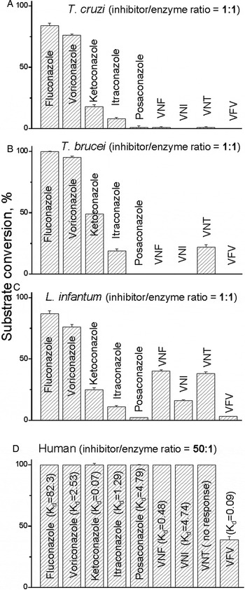
The questions why some of the compounds completely block the CYP51 activity at 1/1 molar ratio inhibitor/enzyme (Yoshida and Aoyama, Reference Yoshida and Aoyama1987; Lepesheva et al. Reference Lepesheva2007; Lepesheva et al. Reference Lepesheva2008) (Fig. 6A–C) and cannot be replaced in the enzyme-binding cavity by the substrate, thus acting as functionally irreversible inhibitors, and why this is never the case for human CYP51 (Fig. 6D) had remained enigmatic until the CYP51 crystal structures have been determined.
Inhibitory effects of systemic clinical antifungal azoles and experimental inhibitors on the activity of (A) T. cruzi, (B) T. brucei, (C) L. infantum and (D) human CYP51 orthologs; 60 min reaction. The results are presented as means ± s.e.m. In all experiments the P450 concentration was 0.5 µ m, the concentration of the sterol substrates [(A) eburicol; (B, C) obtusifoliol; (D) lanosterol] was 50 µ m. The values of the apparent spectral K ds for human CYP51 are given in μ m.

CYP51 structures and structural basis for the enzyme druggability
We have characterized and solved the structures of CYP51s from three trypanosomatid parasites [T. brucei (Lepesheva et al. Reference Lepesheva2004), T. cruzi (Lepesheva et al. Reference Lepesheva2006) and L. infantum (Hargrove et al. Reference Hargrove2011)], in the ligand-free form (Lepesheva et al. Reference Lepesheva2010b) and in complexes with different inhibitors, including clinical azoles fluconazole and posaconazole (Lepesheva et al. Reference Lepesheva2010a; Hargrove et al. Reference Hargrove2011) and experimental compounds, haem-coordinated imidazole- (Lepesheva et al. Reference Lepesheva2010a, Reference Lepeshevab; Buckner et al. Reference Buckner2012; Andriani et al. Reference Andriani2013;Friggeri et al. Reference Friggeri2014), triazole- (Lepesheva et al. Reference Lepesheva2015), pyridine- (Hargrove et al. Reference Hargrove2013) and tetrazole-based (Hoekstra et al. Reference Hoekstra2016) as well as the substrate analogue MCP (Hargrove et al. Reference Hargrove2012b), CYP51s from two fungal human pathogens [A. fumigatus (Hargrove et al. Reference Hargrove2015; Hargrove et al. Reference Hargrove2017b) and C. albicans (Hargrove et al. Reference Hargrove2017a)] and the CYP51 counterpart from human (Hargrove et al. Reference Hargrove2016). Comparative structural analysis has led us to the following conclusions.
CYP51 enzymes preserve their conserved catalytic function regardless of the extremely low amino acid sequence identity across phylogeny by maintaining strict similarity at the secondary and tertiary structural levels (Lepesheva and Waterman, Reference Lepesheva and Waterman2011). For example, nine of the invariant CYP51 family residues (shown in black in Fig. 3) are glycines or prolines that separate the major secondary structural elements, thus maintaining their length and location within the CYP51 molecule. Most of the other invariant family residues either form the haem-binding sequence (P450 signature, dashed line in Fig. 3) or are located within the substrate-binding cavity, although the surface of the cavity is mostly formed by the residues that are phyla-specific (grey in Fig. 3) (Lepesheva and Waterman, Reference Lepesheva and Waterman2011; Hargrove et al. Reference Hargrove2017a).
All the inhibitors, including the substrate analogue MCP, bind in the CYP51 active site without causing any substantial rearrangements in the protein backbone, as if ‘freezing’ the enzyme in its substrate-free conformation (Fig. 7). The inhibitors simply occupy the available space and acquire the shape of the CYP51 substrate-binding cavity. Because large-scale conformational rearrangements in the protein moiety are very likely to be required upon the CYP51 catalysis (binding of the physiological substrate and/or formation of the specific complex with the electron donor protein cytochrome P450 reductase), it appears that the stronger this ‘freezing’ effect is, the stronger the inhibitory potency must be. In addition to the formation of the Fe–N coordination bond (which affects the iron redox potential), this freezing effect is achieved through multiple interactions between the inhibitor molecule and the protein residues. Some of these interactions have a greater impact than others.
Binding of inhibitors does not cause any large-scale rearrangements in the backbone of the CYP51 molecule. (A, B) Superimposed protozoan CYP51 structures, (A) protein chains of ligand-free T. brucei (black) and inhibitor-bound T. brucei, T. cruzi and L. infantum CYP51. The substrate entry is circled. (B) Inhibitors bound in the protozoan CYP51 active site. (C) Inhibitors bound to the fungal CYP51 active site. The haem is depicted in grey. (D) Formulas of the inhibitors (posaconazole, voriconazole and fluconazole are shown in Fig. 2). The colour code of each crystal structure corresponds to the colour of the inhibitor name (PDB ID) beneath the formulas. The correspondent PDB codes of the crystal structures are shown in brackets, the codes of fungal structures are italicized.

Hydrogen bonds
H-bonds are at least one order of magnitude stronger than Van der Waals contacts. For example, we found that the carboxamide fragment of an experimental inhibitors VNI (Lepesheva et al. Reference Lepesheva2010b) and its derivative VFV (Lepesheva et al. Reference Lepesheva2015) in the protozoan CYP51 structures forms two H-bonds that connect two functionally essential regions of the CYP51 molecule, the B′ and I helices (Fig. 8A). On the other hand, VNI that acts as a highly potent functionally irreversible inhibitor of protozoan CYP51s is a rather moderate reversible inhibitor of fungal enzymes (Hargrove et al. Reference Hargrove2012a). Accordingly, in the structure of A. fumigatus CYP51 VNI is oriented differently (see Fig. 7C) and does not form any H-bonds with the protein (Hargrove et al. Reference Hargrove2015). Another example, the tetrazole-based investigational antifungal drug candidates from Viamet (VT1 and VT2 in Fig. 7D) form the H-bond with the fungal-specific histidine (His374 in A. fumigatus and His377 in C. albicans CYP51, respectively) (Hargrove et al. Reference Hargrove2017a, Reference Hargroveb). Both of these compounds act as functionally irreversible inhibitors of the A. fumigatus and C. albicans enzymes (Hargrove et al. Reference Hargrove2017a, Reference Hargroveb). Besides, they have also been found effective in vivo against isolates of Candida glabrata and Candida krusei, which were clinically resistant to other antifungal treatments (Schell et al. Reference Schell2017). Comparison between fluconazole and voriconazole as inhibitors of A. fumigatus CYP51 is probably even more impressive. The two molecules differ only in the composition of a single ring, a smaller triazole ring in fluconazole (weak reversible inhibitor) and a larger 5-fluoropyrimidine ring in voriconazole (see Fig. 2). The pyrimidine ring of voriconazole forms the H-bond with A. fumigatus CYP51 Y122, and voriconazole, regardless of its small size, is one of the most potent inhibitors of A. fumigatus (Hargrove et al. Reference Hargrove2015).
VFV bound in the CYP51 active site. The 2F o–F c electron density map around the haem and the inhibitor is contoured at 2.0 σ and shown as a grey mesh. The carbon atoms of VFV are green, the carbon atoms of 22 CYP51 residues that form Van der Waals contacts with the inhibitor are light grey, and the carbon atoms of the haem are dark grey. The atoms of oxygen, nitrogen, and sulphur are read, blue, and yellow, respectively. The haem iron is presented as an orange sphere. The active site-defining secondary structural elements (semitransparent cartoon) are labelled for clarity. The H-bonds between the enzyme and inhibitor are displayed as red dashes.

Surface binding subsite around the substrate entrance
Posaconazole represents an example of the compound that acts as a functionally irreversible inhibitor for all tested CYP51s from human pathogens. In the CYP51 structures, its long arm is exposed above the protein surface (see Fig. 9), forming multiple interactions around the substrate entry (Lepesheva et al. Reference Lepesheva2010a; Hargrove et al. Reference Hargrove2017a). It appears that these interactions prevent opening of the substrate access channel. A similar effect was displayed by a potent experimental inhibitor LFD (shown in Fig. 7D), although the compound was only tested as an inhibitor of and co-crystallized with T. cruzi CYP51 (Friggeri et al. Reference Friggeri2014).
Surface representation of T. cruzi CYP51 bound to posaconazole. The long arm of the inhibitor protrudes above the entrance into the substrate access channel.

The C/I-helices area
The helices C and I (they contact the biphenyl arm of VFV in Fig. 8) form the deepest portion of the CYP51 substrate-binding cavity, which is unique for CYP51s and is not present in other P450s. This area supports the aliphatic arm of the sterol molecule (Hargrove et al. Reference Hargrove2012b), and the compounds that bind here (VNF and NEE being two examples, seen in Fig. 7B), regardless of their small size, still cannot be replaced by the substrate in the CYP51 reaction (Lepesheva et al. Reference Lepesheva2010a; Andriani et al. Reference Andriani2013). Both of these compounds were found to produce a strong anti-parasitic effect in T. cruzi cells, although unfortunately, NEE was not active in mouse models of Chagas disease and VNF has never been tested in vivo. Deeper insights into this phenomenon might help select small but potent CYP51 inhibitors with e.g. the enhanced ability to penetrate the blood–brain barrier.
CYP51 structure-based drug design
The fact that no conformational changes in the backbone of the CYP51 substrate-binding cavity are involved in inhibitor binding makes this enzyme an attractive target for structure-based drug design. Indeed, our first attempts on the VNI scaffold modification (VNT and VFV in Fig. 7) strongly support this notion as all three compounds superimpose perfectly in the protozoan CYP51 active site (Lepesheva et al. Reference Lepesheva2015).
The reasons for the modifications were as follows. Although we found that VNI is a highly promising molecule (it cured, with 100% efficiency and 100% survival, both the acute and chronic Chagas disease in mice infected with Tulahuen T. cruzi (Villalta et al. Reference Villalta2013)), the compound was less potent as inhibitor of CYP51 from Leishmania infantum (Fig. 6C) and CYP51A from the Y strain T. cruzi (Cherkesova et al. Reference Cherkesova2014).
VNT was made to test the general belief that replacement of the metabolically more vulnerable imidazole ring with the triazole ring may have a positive effect on the compound PKs. VFV was made with the goal to broaden the antiprotozoan spectrum of activity by filling the deepest (CYP51-specific) portion of the substrate-binding cavity with the additional aromatic ring. Both derivatives retained their potency to inhibit the activity of Tulahuen T. cruzi CYP51 in vitro (Fig. 6A) and were more active than posaconazole in cellular experiments, killing Tulahuen T. cruzi amastigotes within cardiomyocytes with the EC50 values within 1 nm (Lepesheva et al. Reference Lepesheva2015). In the same experiments, posaconazole displayed the EC50 of 5 nm (Villalta et al. Reference Villalta2013). Furthermore, both compounds showed the weaker inhibitory effect on CYP3A4 [the IC50 405, 2900 and 3600 nm for VNI, VNT and VFV, respectively; the corresponding values for ketoconazole and posaconazole are 8 and 120 nm (Hargrove et al. Reference Hargrove2012a)]. The lifetime of VNT in mice plasma was indeed longer; however, its concentration did not exceed 5 µ m, thus being about 8-fold lower than the maximal plasma concentration of VNI (Fig. 10). Because of this and also since VNT was too selective for Tulahuen T. cruzi CYP51 (Fig. 6B, C), it was not tested in vivo. VFV, on the other hand, met our expectations. It cured Tulahuen T. cruzi infection in mice and was found more potent than VNI in the mouse model of visceral leishmaniasis: 89% vs 60% reduction of the parasite burden (Lepesheva et al. Reference Lepesheva2015). It also showed better results in curing the Y strain T. cruzi infection in mice (Guedes-da-Silva et al. Reference Guedes-da-Silva2017). Moreover, after a single oral dose of 25 mg kg−1, VFV displayed a longer clearance time in mouse plasma (Fig. 10A) and revealed higher than VNI affinities to tissues (Fig. 10B). Non-toxic, non-mutagenic, VNI and VFV are promising new drug candidates for entering clinical trials.
Pharmacokinetics of VNI and derivatives. (A) Plasma concentration curves after a single oral dose of 25 mg kg−1. (B) VNI and VFV tissue distribution 4 h after administration (single dose) and 16 h after administration (two doses).

Human CYP51 is naturally resistant to inhibition
There is still a general concern that inhibitors of pathogenic CYP51s might actually affect the human counterpart. This is, however, a controversial issue, because inhibitors of human CYP51 could potentially serve as cholesterol-lowering agents. Nevertheless, all the attempts to use human CYP51 as a drug target have failed, and none of the known antifungal azoles or experimental inhibitors of CYP51 from human pathogens inhibit the activity of human CYP51 (Fig. 6D) (Hargrove et al. Reference Hargrove2016). We believe that this natural resistance of human CYP51 to inhibition results from the higher flexibility of the substrate-binging cavity. Unlike the microbial CYP51 structures, human CYP51 displays the loop-like (low-energy) area in the middle portion of the I-helix (the core helix in the P450 structural fold). The structure suggests that this loop-like area is formed because of the long sequence of polar residues (-HTSSTTS-, CYP51 signature 2 area in Fig. 3), which is found in all animal CYP51 sequences but is interrupted by at least one or two hydrophobic residues in the CYP51 enzymes from other phyla. In the human structure, the OH groups of these polar residues disrupt the normal main chain α-helical H-bonding (Hargrove et al. Reference Hargrove2016). The notion of the higher flexibility of the human CYP51 active site is supported by structural dynamic simulation experiments (Yu et al. Reference Yu2016), together suggesting that all animal CYP51 enzymes must be naturally resistant to inhibition.
Concluding remarks and future perspectives
The fact that CYP51 had served blindly but quite successfully as a drug target for more than half a century addresses the question of the enzyme druggability. However, better and more efficient drugs are badly needed. Current progress in understanding the CYP51 structure/function has made it possible to include the target enzyme into the drug discovery process, thus making it quite straightforward. Optical HTS, as well as a virtual screening of large libraries of drug-like molecules, can be used to identify novel inhibitory scaffolds. The tight-binding ligands can be selected by spectral titration, and those of them that act as functionally irreversible inhibitors in the reconstituted CYP51 reaction should proceed to cellular experiments and/or animal models. The issues of toxicity, bioavailability and PKs can be resolved rationally by minor, structure-guided optimization of the molecules. It is our strong belief that a larger set of more efficient, pathogen-oriented CYP51 inhibitors will help to reduce the mortality rates of fungemias, cure Chagas disease and amoebiasis, and must be effective in combination therapy for visceral leishmaniasis and sleeping sickness. Team efforts from academia and pharmaceutical companies are highly advisable to accelerate the process.
Financial support
Financial support was provided by the National Institutes of Health Grant R01 GM067871 (to G.I.L.).
Conflict of interest
None.
Ethical standards
Not applicable.