Introduction
Laryngotracheal stenosis can be clinically devastating. Rehabilitation of patients to a state of normal respiratory and phonatory function is a formidable challenge. The fact that there are currently numerous reconstruction procedures indicates that, at present, there is no standard treatment method. Kimura et al. Reference Kimura, Mukohara, Tsugawa, Matsumoto, Sugimura and Murata1 described a technique of tracheal stenosis treatment that involved splitting the anterior wall of the trachea and inserting a costal cartilage graft. The complications of this technique included anastomotic breakdown, restenosis, graft resorption, chondritis, pneumothorax and granulation tissue formation.Reference Kimura, Mukohara, Tsugawa, Matsumoto, Sugimura and Murata1, Reference Biller, Lawson and Weisberg2 The following grafts have also been used: the hyoid–sternohyoid myo-osseus flap,Reference Ward, Canalis and Fee3 tracheal cartilage autograft,Reference Backer, Mavroudis, Dunham and Lauren4 composite nasal septal graftReference Toohill5 and auricular cartilage graft.Reference Heatley, Randell and Garner6 The variety of grafts developed for laryngotracheal lumen augmentation suggests that, at present, no ideal graft has been found.
Researchers have developed a variety of tracheal prostheses made of synthetic biomaterials such as steel wire, glass, Lucite® and gortex. Alloplastic materials have always had limited success, due to frequent dislodgement, infection and rejection.Reference Agora, Spector and Hinchliffe7
The ideal grafting material for tracheal reconstruction should: (Reference Kimura, Mukohara, Tsugawa, Matsumoto, Sugimura and Murata1) provide adequate structural support to maintain airway patency; (Reference Biller, Lawson and Weisberg2) integrate into the recipient tissue to avoid the need for removal; (Reference Ward, Canalis and Fee3) produce minimal inflammatory response to avoid scar formation and stenosis; (Reference Backer, Mavroudis, Dunham and Lauren4) rapidly mucosalise to allow for improved mucociliary transport; and (Reference Toohill5) be readily available.Reference Gubbels, Richardson, Trune and Bascom8
Titanium is a hypoallergenic material which has been used extensively in orthopaedic and maxillofacial surgery, with excellent long-term results.Reference Eck, Bridwell, Ungacta, Lapp, Lenke and Riew9, Reference Kuttenberger and Hardt10 Titanium mesh has been used for sternal reconstruction following median sternotomy, giving effective stabilisation in this mobile area.Reference Ioannis, Dilip and Unnikrishnan11 In the airway, titanium has been previously used for internal fixation of laryngeal injuriesReference Verschueren, Bell, Bagheri, Dierks and Potter12, Reference Mello, Francisco and Ricardo13 and in medialisation thyroplasty.Reference Hans14 Titanium mixed with nickel to form an alloy (Nitinol) has been used in the formation of self-expandable tracheal stents. These stents have been successfully used for the management of benign and malignant tracheal stenosis.Reference Vinograd, Klin, Brosh, Weinberg, Flomenblit and Nevo15, Reference Yauagihara, Mizuro, Wada and Hitomi16
Our aim was to assess the use of titanium mesh, instead of traditional homografts, in single-stage reconstruction of the anterior laryngotracheal wall in cases of benign laryngotracheal stenosis.
Patients and methods
The study group comprised 12 tracheostomised patients suffering from chronic benign laryngotracheal stenosis, who presented to the department of otolaryngology and head and neck surgery at the Alexandria Medical School, Egypt.
Pre-operative evaluation
All patients were subjected to pre-operative clinical and radiological evaluation by computed tomography scanning. Endoscopy of the larynx and trachea under general anaesthesia was performed in order to assess the exact site, length and diameter of the stenosed segment.
Patients with glottic or supraglottic involvement were excluded from the study.
Operative procedure
Reconstruction was performed under general anaesthesia, with endotracheal intubation through the tracheostomy orifice. A transverse anterior neck incision that included the tracheostomy orifice was utilised. The upper skin flap was elevated in a subplatysmal plane to the level of the thyrohyoid membrane.
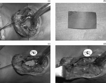
The strap muscles were exposed and carefully separated in the midline, exposing the anterior laryngotracheal wall. The isthmus of the thyroid gland was divided (if this had not been done in the previous tracheostomy), followed by splitting of the cricoid cartilage and the upper tracheal rings (Figure 1a). If indicated, the split was extended superiorly to involve the lower half of the thyroid cartilage just below the anterior commissure, or inferiorly to communicate with the tracheostomy orifice. Any scar tissue was removed.
Operative procedure. (a) Splitting the cricoid cartilage (C) and upper tracheal rings (T). (b) The titanium mesh before reshaping. (c) The titanium mesh after reshaping, sutured to the anterior laryngotracheal wall with Silastic stent (*) stenting the lumen and fixed to the skin by running sutures. (d) Strap muscles (S) sutured in the midline, covering the outer surface of the mesh.

A Silastic stent was then inserted to expand the lumen. The stent was fixed in place by non-absorbable sutures exiting from the skin on both sides of the neck. The upper end of the stent lay below the level of the vocal folds and the lower end lay just above the tracheostomy orifice.
The split ends of the trachea were fixed in its expanded position by a 0.4 mm titanium mesh (Mondeal Medical Systems, Tuttlingen, Germany). The mesh was cut and fashioned at the time of the surgery according to the length and diameter of the newly formed airway. The mesh was sutured to the laryngotracheal wall by Vicryl sutures (Figure 1b and 1c). The strap muscles were used to cover the titanium mesh (Figure 1d). The inner aspect of the mesh was left bare for re-epithelisation from the surrounding airway epithelium.
A Penrose drain was inserted and the skin was closed in two layers leaving the old tracheostomy orifice. A tracheostomy tube of appropriate size was inserted at the end of the procedure.
Post-operative care
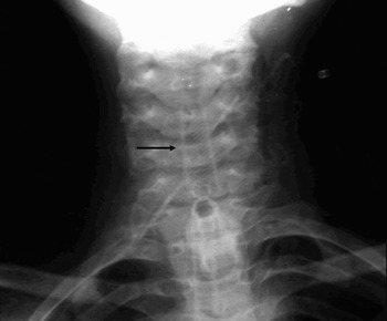
All patients received parental antibiotics, oral anti-inflammatory agents and corticosteroids for seven days. A plain neck X-ray was performed to assess the position of the titanium mesh (Figure 2).
Post-operative plain X-ray showing the titanium mesh (arrow) in place.

The patients were readmitted six weeks later and the Silastic stent removed endoscopically under general anaesthesia. The lumen was inspected and any granulation tissue was removed. One cubic centimetre of mitomycin-C (0.5 mg/ml) was applied topically for five minutes. A trial of decannulation was commenced gradually by inserting a smaller tracheostomy tube for two days, followed by complete decannulation. Patients were followed up regularly, every two weeks for two months and then every month. Any signs or symptoms of airway distress were reported. At the end of the follow-up period, the patient's respiratory reserve was classified subjectively as good (i.e. normal daily activity without dyspnoea), fair (i.e. dyspnoea on normal daily activity) or guarded (i.e. dyspnoea at rest).
Results
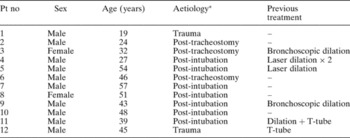
Twelve patients with benign chronic laryngotracheal stenosis were included in this study. There were 10 men (83.33 per cent) and two women (16.67 per cent), aged 19–54 years (±11.78). The aetiology of stenosis was post-intubation in 58.3 per cent (7/12) of cases (Table I). Six patients (50 per cent) had a previous failed endoscopic treatment (Table I). The stenosis was in the cervical trachea in seven cases (58.3 per cent), laryngotracheal in four cases (33.3 per cent) and subglottic in one case (8.4 per cent) (Table II).
Characteristics of study cases

* Of laryngotracheal stenosis. Pt no = patient number
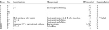
Summary of operative outcomes

LT = laryngotracheal; T = tracheal; SG = subglottic; GT = granulation tissue; FU = follow up period; LFU = lost to follow up; Y = yes; N = no
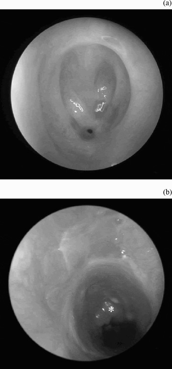
The operative procedure was successfully completed in all patients, with no intra-operative or immediate post-operative complications reported. The average operating time was one hour. The titanium mesh was well tolerated by all patients. On removal of the Silastic stent after six weeks, good neo-epithelisation was found on the inner mesh surface in the 10 patients in whom decannulation was possible. Some non-significant granulation tissue was present, mainly at the lower end of the stent, just above the tracheostomy orifice (Figure 3).
Laryngotracheoscopic views. (a) Pre-operative, showing scar tissue nearly obliterating the upper tracheal lumen; (b) post-operative, showing satisfactory lumen with apparently healthy neo-epithelisation on the inner surface of the titanium mesh (a small granuloma (*) is seen at the site of the old tracheostomy).

Decannulation was impossible in two cases. In one patient, this was due to excessive granulation tissue, with collapse of the suprastomal part of the trachea. In the second patient, the mesh prolapsed into the lumen, leading to airway obstruction; the mesh was removed endoscopically and a Montgomery T-tube was inserted.
On follow up, four patients showed mild to moderate respiratory distress. Endoscopy revealed varying degrees of granulation tissue formation. Endoscopic debulking and mitomycin-C application was performed, with immediate improvement of respiration (Table II).
The follow-up period ranged from three to 36 months (Table II). At the end of the follow-up period, 10 patients were decannulated (83.3 per cent). Respiratory reserve was good in seven patients and fair in three patients. Two patients (16.7 per cent) were unable to be decannulated.
Discussion
Laryngotracheoplasty is now standard care for symptomatic laryngotracheal stenosis. The reasons for unsuccessful decannulation after a laryngotracheoplasty may be multifactorial, depending on the techniques and grafts used. Excessive granulation tissue may develop, requiring further procedures. Cartilaginous grafts may become infected, be resorbed or collapse into the tracheal lumen. Bulky regional skin–muscle flaps may dehisce under tension or collapse into the tracheal lumen. Medial migration of the split ends of the anterior cartilaginous tracheal rings ensues, with subsequent restenosis. Donor site morbidity may compound these problems.Reference Kimura, Mukohara, Tsugawa, Matsumoto, Sugimura and Murata1–Reference Ward, Canalis and Fee3
The search for novel methods of treatment for tracheal stenosis has led many groups to investigate the feasibility of using synthetic material to reconstruct airway defects.
Over a three-year period, we performed laryngotracheoplasty using titanium mesh in 12 patients suffering from chronic laryngotracheal stenosis. The mesh was easily designed and sutured to the split laryngotracheal ends. It was covered by strap muscle in order to support the outer surface and to provide a scaffold for new epithelisation. After six weeks, the mesh was found to be incorporated into the recipient tissue, with neo-epithelisation on the inner surface of the mesh, in 10 out of 12 patients, in whom decannulation was possible.
Casiano et al. Reference Casiano, Patete and Lindquist17 used pored titanium plates with a strap muscle flap for reconstruction of the anterior laryngotracheal wall in nine patients suffering from laryngotracheal stenosis. Seven (78 per cent) patients were able to be decannulated, six needed no further intervention and one acquired granulation tissue formation requiring further endoscopic debulking. An advantage of titanium mesh is that it causes only a mild inflammatory reaction due to its hypoallergenic nature. Also, the presence of pores allows for mucociliary clearance and for regrowth of new epithelium through these pores, allowing the mesh to become easily incorporated into the recipient tissue.
Schultz et al. Reference Schultz, Vautier, Chluba, Marcellin and Debry18 histologically examined the cervical trachea of 11 mice, 15–41 days after titanium tracheal prosthesis implantation. They found a mild inflammatory reaction, with fibroblast colonisation of the titanium pores and a cylindrical ciliary epithelial layer developed on the endoluminal side of the prosthesis. According to these authors, these findings supply surgical and histological validation for the usefulness of a porous titanium tracheal prosthesis in the management of tracheal stenosis.
We routinely used Silastic intraluminal stenting for six weeks. This helped support the reconstructed area; also, removal of the stent after six weeks gave the chance to examine the lumen and to clean any secretions or granulation tissue. ZalzalReference Zalzal19 stated that stenting is required following laryngotracheal reconstruction in order to: (Reference Kimura, Mukohara, Tsugawa, Matsumoto, Sugimura and Murata1) counteract scar contracture; (Reference Biller, Lawson and Weisberg2) provide a scaffold to allow epithelium to cover the lumen of the airway; and (Reference Ward, Canalis and Fee3) hold the reconstructed area in place and prevent mechanical disruption caused by movement of the laryngotracheal complex during breathing and swallowing.
The main complication encountered in our series was granulation tissue formation, which occurred in five patients. In one patient, this was excessive, preventing decannulation. In the other four patients, granulation tissue recurred after decannulation. This was managed by endoscopic debulking, corticosteroids and local application of mitomycin-C. Topical application of mitomycin-C has been noted to reduce the recurrence of granulation tissue after laryngotracheoplasty and stent removal.Reference Ward and April20 Granulation tissue formation has been reported following use of such different grafting materials as costal cartilage,Reference Cotton and O'Conner21 hyoid boneReference Freeland22 and thyroid cartilage.Reference Thawely and Ogura23
In one of our patients (8.3 per cent), the titanium mesh prolapsed into the tracheal lumen, leading to airway obstruction immediately after removal of the stent. The mesh was easily removed endoscopically and a Montgomery T-tube was inserted.
Decannulation was possible in 10 out of our 12 patients (83.3 per cent) but failed in two patients, one of whom subsequently had a T-tube placed. The decannulated patients' respiratory reserve was good in seven cases and fair in three cases. Different graft materials have varying rates of successful decannulation. For costal cartilage grafts, the incidence of successful decannulation has varied in different series, ranging from 66.5 to 90 per cent.Reference Cotton and O'Conner21, Reference Cotton, Richardson and Seid24 For muscle flaps, different studies have found successful decannulation rates ranging from 86 to 93 per cent.Reference Eliachar25, Reference Eliachar26
• The management of laryngotracheal stenosis represents a serious surgical challenge
• This paper describes the use of titanium mesh instead of traditional homografts in reconstruction of the anterior laryngotracheal wall in 12 tracheostomised patients with benign chronic laryngotracheal stenosis
• Endoscopically, good epithelisation was found on the inner surface of the mesh in 10 cases and decannulation was possible
• Titanium mesh was found to be a good alternative for augmentation of the anterior laryngotracheal wall, offering rigid support and a low incidence of complications
In our opinion, titanium mesh fulfills the criteria for ideal grafting material.Reference Gubbels, Richardson, Trune and Bascom8 It provides adequate structural support in order to maintain airway patency. Because of its micropores, it does not impede mucociliary clearance, and it rapidly integrates into the recipient tissue so does not need to be removed. As titanium is hypoallergenic, it produces minimal inflammatory response, thus avoiding scar formation and stenosis. Finally, it is readily available. In addition, the use of titanium mesh prevents donor site morbidity, which can complicate the use of homografts.
Conclusion
Titanium mesh was found to be a good alternative for augmentation of the anterior laryngotracheal wall. It offered rigid support, with the advantages of less inflammatory reaction, no impedance to mucociliary clearance, rapid integration with recipient tissues and no donor site morbidity. The incidence of complications was less than that for other traditional homografts. If needed, endoscopic removal could be easily performed.