INTRODUCTION
Chagas disease is a neglected tropical disease caused by an infection with the protozoan Trypanosoma cruzi that affects ∼13 million people in the American continent (World Health Organization, 2005). Currently, the geographical distribution of this infectious disease is not limited to the Americas, as cases of infection have been reported in Europe, Australia and Japan, which has been attributed to organ transplants and immigration from Central and South America (Schmunis, Reference Schmunis2007; Develoux et al. Reference Develoux, Lescure, Jaureguiberry, Jeannel, Elghouzzi, Gay, Paris, Le Loup, Danis and Pialoux2010). In Latin America, T. cruzi is mainly transmitted to humans by the feces of triatomine bugs (family Reduviidae, subfamily Triatominae) during blood feeding, but the occurrence of acute Chagas disease outbreaks has also been associated with infection by oral routes (Coura, Reference Coura2006). Thus, determining the biological aspects of the interaction of T. cruzi with vertebrate and invertebrate hosts may provide potential new targets for vector control intervention and Chagas disease therapy.
T. cruzi has a complex life cycle that involves the interactions of distinct evolutive forms with both the vector digestive tract and mammalian cells. Therefore, the parasite exploits strategies to survive within the host and sustain the infection (Tyler and Engman, Reference Tyler and Engman2001). One essential event required for the bona fide parasite-host interface is the recognition of molecules between parasite and host cells. Lectins and carbohydrates have been reported to mediate receptor-ligand binding during vector- and mammalian cell-T. cruzi interplay (Alves et al. Reference Alves, Albuquerque-Cunha, Mello, Garcia, Nogueira, Bourguingnon, de Souza, Azambuja and Gonzalez2007; Villalta et al. Reference Villalta, Scharfstein, Ashton, Tyler, Guan, Mukherjee, Lima, Alvarez, Weiss, Huang, Machado and Tanowitz2009). Retention of epimastigotes in the triatomine midgut has been associated with the recognition of high-mannose glycans by carbohydrate-binding proteins of the parasite (Bonay and Fresno, Reference Bonay and Fresno1995; Bonay et al. Reference Bonay, Molina and Fresno2001). Glycoinositolphospholipids at the surface of epimastigotes are also involved in adhesion to the insect epithelial cells (Nogueira et al. Reference Nogueira, Gonzalez, Gomes, de Souza, Garcia, Azambuja, Nohara, Almeida and Zingales2007). In fact, it has been demonstrated that the successful attachment of T. cruzi to the perimicrovillar membrane (PMM) is crucial for the establishment of Reduviidae insect infection (Gonzalez et al. Reference Gonzalez, Nogueira, Feder, de Souza, Feder, Nogueira and Gonzalez1998, Reference Gonzalez, Hamedi, Albuquerque-Cunha, Nogueira, De Souza, Ratcliffe, Azambuja, Garcia and Mello2006; Alves et al. Reference Alves, Albuquerque-Cunha, Mello, Garcia, Nogueira, Bourguingnon, de Souza, Azambuja and Gonzalez2007). This initial step seems to coordinate biological events, such as epimastigote growth and differentiation into trypomastigotes and, therefore, is a focus of intervention strategies. The characterization of the PMM has revealed several potential molecules, mainly carbohydrates and carbohydrate-binding molecules that may be implicated in the mechanism of interaction with T. cruzi (Albuquerque-Cunha et al. Reference Albuquerque-Cunha, Gonzalez, Garcia, Mello, Azambuja, Almeida, de Souza and Nogueira2009). Glycosaminoglycans (GAGs), such as heparan sulfate (HS) and chondroitin sulfate (CS), have also been detected in different organs of Rhodnius prolixus, including the intestinal tract (Costa-Filho et al. Reference Costa-Filho, Souza, Martins, dos Santos, Silva, Comaru, Moreira, Atella, Allodi, Nasciutti, Masuda and Silva2004; Souza et al. Reference Souza, Sarquis, Gomes, Moreira, Lima and Silva2004).
In fact, sulfated GAGs have emerged as key molecules involved in the attachment and invasion processes of several microorganisms (Wadström and Ljungh, Reference Wadström and Ljungh1999; Sava et al. Reference Sava, Zhang, Toma, Theilacker, Li, Baumert, Holst, Linhardt and Huebner2009; Boyle et al. Reference Boyle, Richards, Gilson, Chai and Beeson2010). GAGs are linear polysaccharides composed of disaccharide repeats that consist of uronic acid (glucuronic acid and iduronic acid) or galactose and an amino sugar (N-acetylglucosamine and N-acetylgalactosamine) (Dreyfuss et al. Reference Dreyfuss, Regatieri, Jarrouge, Cavalheiro, Sampaio and Nader2009; Ly et al. Reference Ly, Laremore and Linhardt2010). The ubiquitous distribution of GAGs at the surface of mammalian and insect cells (Souza et al. Reference Souza, Sarquis, Gomes, Moreira, Lima and Silva2004; Dreyfuss et al. Reference Dreyfuss, Regatieri, Jarrouge, Cavalheiro, Sampaio and Nader2009) has indicated their putative role in T. cruzi interaction. We have previously demonstrated that trypomastigotes and amastigotes possess HBPs that mediate parasite attachment and invasion in mammalian cells (Calvet et al. Reference Calvet, Toma, De Souza, Meirelles and Pereira2003; Oliveira-Jr et al. Reference Oliveira, Alves, Calvet, Toma, Bouças, Nader, Côrtes, Krieger, Meirelles Mde and Pereira2008; Bambino-Medeiros et al. Reference Bambino-Medeiros, Oliveira, Calvet, Vicente, Toma, Krieger, Meirelles and Pereira2011). Therefore, we postulated that sulfated GAGs at the surface of midgut epithelial cells may also play a role in the adhesion of epimastigotes to the triatomine midgut.
In this study, we attempted to isolate and characterize the heparin-binding proteins (HBPs) of the epimastigote forms of T. cruzi. We confirmed the specificity and stability of protein binding between HBPs and GAGs and determined their potential biological function in the parasite-vector interaction.
MATERIALS AND METHODS
Glycosaminoglycans
Heparin (Hep), from bovine lung, was purchased from INORP Laboratories (Buenos Aires, Argentina). Heparan sulfate (HS), from bovine pancreas, was a kind gift from Dr P. Bianchini (Opocrin Research Laboratories, Modena, Italy). Chondroitin 4-sulfate (C4S), from whale cartilage, was obtained from Seikagaku Kogyo Co. (Tokyo, Japan). All biotinylated GAGs (Hep, HS, C4S) were prepared as previously described (Bouças et al. Reference Bouças, Trindade, Tersariol, Dietrich and Nader2008).
Rhodnius prolixus
Triatomines were reared and maintained in the laboratory as previously described (Garcia et al. Reference Garcia, Azambuja, Contreras and Morel1984). Fifth-instar larvae, which were starved for 30 days after the last ecdysis, were allowed to feed on citrated human blood through a membrane feeder. Insects were dissected 10 days after feeding, and the interaction of parasites and midgut epithelial cells was performed as previously described (Alves et al. Reference Alves, Albuquerque-Cunha, Mello, Garcia, Nogueira, Bourguingnon, de Souza, Azambuja and Gonzalez2007).
Parasites
An epimastigote form of T. cruzi, clone Dm28c, was grown at 28°C in LIT medium (Camargo et al. Reference Camargo1964) containing 10% FBS. For the biochemical assays and insect-parasite interaction studies, the parasites were cultivated at a density of 104 parasites/ml and harvested during the exponential growth phase.
Metabolic radio-isotope labelling of the parasite proteins
Epimastigotes (2×10⁷ cells/ml) were starved (1 h) of methionine and incubated (1 h, 37°C) with 100 mCi/ml of [35S]methionine. The metabolic labelling was stopped by the addition of 1% FBS. After washing in PBS, the proteins of labelled parasites were obtained using Triton X-114 (TX-114) (Bordier, Reference Bordier1981).
Protein extraction
Parasites (1011) were washed with PBS, and detergent-soluble proteins were obtained by TX-114 phase separation. After parasite extraction, soluble proteins were obtained after condensation at 37°C followed by centrifugation (12 000 g, 15 min, 25°C). The hydrophobic phase was subjected to affinity chromatography.
Affinity chromatography
Hydrophobic proteins were applied to a heparin affinity column as previously described (Oliveira-Jr et al. Reference Oliveira, Alves, Calvet, Toma, Bouças, Nader, Côrtes, Krieger, Meirelles Mde and Pereira2008). After washing, the retained proteins were eluted with a stepwise application of increasing NaCl concentrations (0·25 M, 0·5 M, 1·0 M, 2·0 M and 3·0 M) in the equilibrium buffer. The proteins were dialysed and concentrated using a Centriprep YM-10 (Millipore, MA, USA). Finally, the eluted fractions were stored at −20°C until further analysis.
SDS-PAGE and Western Blot
The total extracted protein (40 μg) and proteins eluted from the heparin column (20 μg) from epimastigotes were separated by SDS-PAGE (12%). After electrophoresis, the gels were stained with silver nitrate and Coomassie blue. SDS-6H (Sigma-Aldrich Chemical Co., St Louis, MO, USA) was used as a molecular mass marker.
For Western blot, the separated proteins were transferred to a nitrocellulose membrane and blocked (16 h, 4°C) with PBS containing 5% skim milk and 0·5% Tween-20. The membranes were incubated with 2 μg/ml of biotinylated GAGs (heparin, HS or C4S) for 1 h at 25°C and with streptavidin-conjugated horseradish peroxidase (1:100) for 1 h at 25°C. The complex was visualized using an ECL-kit (Santa Cruz Biotechnology, CA, USA).
Rhodnius prolixus-epimastigote interaction
The in vitro interaction of insect midguts and epimastigotes was performed as described previously (Alves et al. Reference Alves, Albuquerque-Cunha, Mello, Garcia, Nogueira, Bourguingnon, de Souza, Azambuja and Gonzalez2007). The dissected insect midguts were mixed with 106 epimastigotes in 200 μl of PBS. The involvement of HBPs and sulfated GAGs in the insect-parasite interaction was analysed by incubating the insect midguts with HBPs isolated from epimastigotes or parasites treated with 1·0–0·01 μg/ml (1 h, 4 °C) heparin, HS or CS prior to interaction. The midguts were spread onto glass slides and the number of attached parasites was randomly quantified by light microscopy. Ten insect midguts were used for each experimental group.
Surface plasmon resonance (SPR) assay
The SPR assays were performed on a carboxyl sensor chip surface (COOH) coated with immobilized neutravidin (Biocap; Nomadics, USA) that had been previously activated with biotinylated heparin (1 μg/ml). The HBPs were assessed in whole epimastigotes (106 cells) that had previously been washed in PBS. The assays were performed at 25°C in 100 μl of running buffer (PBS) and at a flow rate of 10 μl/min. The binding specificity was assessed by incubation (1 h, 4°C) of epimastigotes with 0·1–0·001 μg/ml sulfated GAGs (heparin, HS or CS). After the parasites were fixed in 4% paraformaldehyde, the binding assays were monitored in real time by a sensorgram and recorded as resonance units (RU). In these cases, the dissociation RU values represent the average of response between 633 and 925 seconds in all assays. A negative control was performed by flowing different concentrations of bovine serum albumin (BSA) directly across the heparin-coated sensor chip. Constants for association (Ka) and dissociation (Kd) were measured. These experiments were conducted in an optical biosensor apparatus, SensiQ Pioneer, and the data were analysed using the Qdat software (Nomadics, USA).
Scanning electron microscopy
After interaction of R. prolixus with untreated and GAG-treated epimastigotes, the insect midgut was fixed (1 h, 4°C) with 2·5% glutaraldehyde in 0·1 M sodium cacodylate buffer, pH 7·2, followed by post-fixation with 1% osmium tetroxide in a similar buffer. The samples were dehydrated in a graded series of acetone, critical-point dried, coated with gold in a sputter coater and analysed with a JEOL scanning microscope (JSM6390LV).
Statistical analysis
The results were analysed for significance using ANOVA and Tukey's test according to the StatsDirect statistical software, version 2.2.7 for Windows 98 (Armitage et al. Reference Armitage, Berry, Matthews and Armitage2002). Differences between treated and control insects were considered not statistically significant when P>0·05. The probability levels are specified in the text.
RESULTS
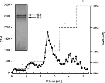
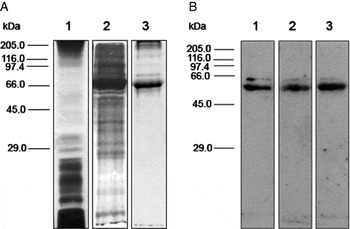
The [35S]methionine metabolic labelling assay combined with Triton X-114 phase separation and heparin-affinity chromatography was performed to evaluate the expression of heparin-binding proteins in the epimastigote forms of T. cruzi. This strategy yielded an enriched fraction of hydrophobic proteins that bound to heparin, which were eluted from an affinity column with a stepwise NaCl concentration gradient. Using this procedure, the peak of protein elution was obtained with 1·0 M NaCl, where 2 protein bands with molecular masses of 65·8 kDa and 59 kDa were mainly identified by SDS-PAGE (Fig. 1). Proteins with similar molecular masses were observed when unlabelled hydrophobic proteins were subjected to the same purification steps used for the labelled proteins. These unlabelled proteins were analysed by Western blot using biotinylated GAGs and are shown in Fig. 2. Our results demonstrated that both the 65·8 kDa and 59 kDa proteins are recognized by GAGs that consist of N-acetylglucosamine and uronic acid (heparin and HS) as well as N-acetylgalactosamine and glucuronic acid (C4S) (Fig. 2). Although both proteins interacted with the biotinylated GAGs, the 59 kDa protein seemed to be preferentially recognized by all GAGs evaluated (Fig. 2).
Affinity chromatography of heparin-binding proteins (HBPs) from epimastigotes labelled with [35S]methionine. Whole hydrophobic proteins were obtained by the TX-114 method. Proteins were eluted using different concentrations of NaCl – 0·25 M (1), 0·5 M (2), 1·0 M (3), 2·0 M (4) and 3·0 M (5), showing a peak of elution at 1·0 M. The data are presented as counts per minute (cpm). Analysis by SDS-PAGE revealed 2 bands with molecular masses of 65·8 and 59 kDa (insert).

Electrophoresis analysis of Trypanosoma cruzi epimastigote proteins. (A) Total hydrophobic proteins (1) or HBPs (2 and 3) were separated in each lane in the SDS-PAGE and revealed by silver (1 and 2) or Coomassie blue (3) staining. (B) HBPs were also separated by SDS-PAGE, transferred to a nitrocellulose membrane and revealed after incubation with biotinylated heparin (1), heparan sulfate (2) and chondroitin sulfate (3) by chemiluminescence. Molecular mass standard proteins (kDa) are indicated on the left.

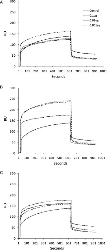
In addition, SPR assays were designed to directly evaluate the HBPs at the surface of epimastigotes and to characterize the receptor-ligand interaction. The presence of HBPs at the parasite surface was confirmed by the injection of the parasites onto the sensor chip surface, previously recovered with biotinylated heparin, and the sensorgram displayed dissociation values of 61·62±2·87 RU (Fig. 3). These disassociation values were approximately 7·3-fold higher than the values obtained after injection of heparin (dissociation 8·4±1·7 RU), indicating parasite attachment to the immobilized heparin on the sensor chip surface.
Sensorgrams showing the presence of HBPs on the surface of Trypanosoma cruzi epimastigotes by surface plasmon resonance. SPR assays were performed with 106 epimastigotes in a final volume of 100 μl and a flow rate of 10 μl/s. Biocap sensor chips were covered with biotinylated heparin, and epimastigotes were passed over its surface. The inhibition assays were performed following incubation of the epimastigotes with glycosaminoglycans (GAGs), i.e., heparin (A), heparan sulfate (B) and chondroitin sulfate (C). The parasites were assayed without pre-incubation with GAGs or after a pre-incubation with 0·1 μg/ml, 0·01 μg/ml or 0·001 μg/ml GAGs. The interaction assays were performed in PBS. The resonance signals were analysed after subtraction of a reference line using the Qdat software. The assays were performed in triplicate, and the mean value for resonance units was obtained based on the results of 3 independent assays.

The specificity of epimastigote binding to heparin was confirmed by GAG competition assays, where the parasite was previously incubated with increasing concentrations of heparin, HS and CS. Pre-incubation of epimastigotes with these sulfated GAGs led to an inhibition of parasite binding to immobilized heparin, which was measured by the association and dissociation RU values (Fig. 3). The inhibition of parasite binding was assessed by low dissociation RU values as compared to the control assay. The significant binding of RU was 37·6±9·49 (P⩽0·00002) for heparin, 37·14±8·07 (P⩽ 0·00009) for HS and 48·3±3·14 (P⩽0·0013) for CS, achieving a binding inhibition of 39%, 40% and 22%, respectively. Additionally, control assays using 3 different concentrations (0·1, 0·01 and 0·001 μg/ml) of BSA were processed in parallel in the same SPR assays. The assessed dissociation (2·0±0·3, 1·2±0·0 and 0·4±0·3 RU, respectively) values were consistent with the lack of relevant binding between BSA and heparin, showing the specificity of the binding assays performed with epimastigotes.
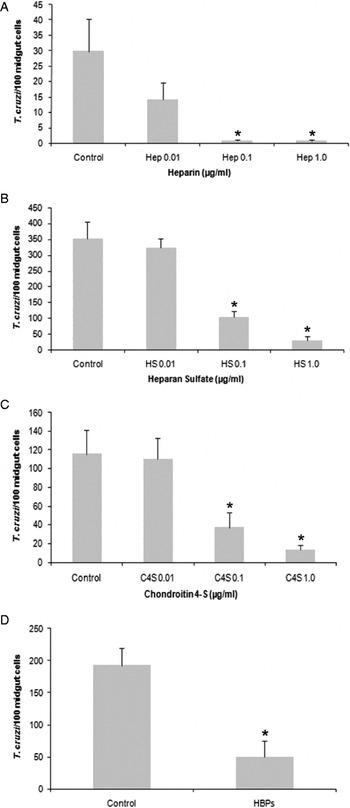
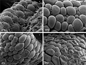
Because GAGs, specifically HS and CS, have been identified in different tissues of Triatoma brasiliensis and R. prolixus (Souza et al. Reference Souza, Sarquis, Gomes, Moreira, Lima and Silva2004), including the intestinal tract where epimastigotes attach, multiply and produce free, infective, metacyclic forms, we evaluated the role of HBPs in the epimastigote-vector interaction. Insects were dissected 10 days after feeding, and the luminal face of the midgut was exposed to facilitate the in vitro interaction of epithelial midgut cells with epimastigotes. Pre-incubation of the epithelial cells with an enriched fraction of HBPs, obtained by affinity chromatography, yielded a 3·8-fold inhibition in the adhesion of epimastigotes to posterior midgut epithelial cells. Specifically, the adhesion rate of 192·5±27·4 epimastigotes/100 midgut epithelial cells in the controls (untreated parasites) was reduced to 50·3±24·4 in pre-treated epithelial cells (Fig. 4). In preparations obtained from the controls and epimastigotes pretreated with 0·01 μg/ml of heparin, HS or C4S, similar rates of parasite adhesion per 100 midgut cells were observed (P>0·05). In contrast, attachment of only 102·08±18·08 (P<0·0001) and 29·8±12·02 (P<0·0001) parasites per 100 midgut epithelial cells were recorded when the flagellates were incubated with either 0·1 or 1·0 μg/ml HS, respectively, while the control group exhibited an adherence of 351·14±53·34 parasites under the same conditions. Similar results were observed after parasite incubation with 0·1 μg/ml (37·3±15·57) or 1·0 μg/ml (13·3±5·43) C4S compared to controls (115·5± 27·71). Similarly, parasite incubation with either 0·1 or 1·0 μg/ml heparin strongly reduced the attachment of T. cruzi to only 0·84±0·29 and 0·67±0·46 epimastigotes per 100 midgut cells (P< 0·001), respectively (Fig. 4). This inhibition was clearly visualized by scanning electron microscopy (SEM). As expected, SEM micrographs revealed a large number of microvilli on the surface of the midgut epithelial cells. After the interaction between parasite and midgut cells in vitro, epimastigotes were mostly attached by the flagellum to the epithelium of the intestinal tract, though an association with the parasite body was also observed. Large numbers of epimastigotes were found to be associated with midgut epithelial cells in the control. In contrast, few epimastigotes were found to be adhered to the epithelial cells after pre-incubation of the parasites with GAGs (Fig. 5).
Role of epimastigote HBPs and sulfated GAGs on the in vitro attachment of Trypanosoma cruzi to the posterior midgut epithelium of Rhodnius prolixus. Epimastigotes were previously incubated with different concentrations of GAGs (0·01, 0·1 and 1·0 μg/ml) in LIT medium containing 2·0×107 parasites/ml, washed and added to the interaction medium. Alternatively, posterior midguts were incubated with isolated HBPs from 1011 epimastigotes prior to interaction with T. cruzi. Adhered epimastigotes were counted per 100 epithelial cells in 10 different fields of each midgut preparation using a Zeiss microscope with a reticulated ocular. Each group shows the mean±s.e. of parasites attached in 10 midguts. Non-treated (Control), heparin (Hep; A), heparan sulfate (HS; B), chondroitin 4-sulfate (C-4S; C) and HBPs (D). Non-treated (control) experiments were performed without GAGs or HBPs in the same pre-incubation conditions. (*) P⩽0·05.

Scanning electron microscopy of the epimastigote-Rhodnius prolixus interaction. Competition assays were performed to evaluate the participation of sulfated GAGs in the parasite-host cell recognition. (A) Midgut epithelial cells were incubated with epimastigotes (control). Pre-treatment of epimastigotes with 1 μg/ml heparin (B), heparan sulfate (C) or chondroitin sulfate (D) led to an inhibition of parasite attachment to the midgut epithelial cells. Scale bar=10 μm.

DISCUSSION
Arthropods are considered to be important vectors in the transmission of human infectious diseases (Romi, Reference Romi2010; Williams et al. Reference Williams, Bader, Kearney, Ritchie and Russel2010). The transmission of infection by the parasite is mediated through the control and manipulation of biological and molecular events in the vector and mammalian hosts (Lefèvre and Thomas, Reference Lefèvre and Thomas2008; Matthews, Reference Matthews2011). Therefore, a large number of investigations have focused on understanding the biology of host-parasite interactions. In this context, studies analysing the relationship between T. cruzi and its vector, triatomines (Hemíptera: Reduviidae: Triatominae), have been emphasized in recent years (Zimmermann et al. Reference Zimmermann, Folly, Gomes, Alviano, Alviano, Silva-Filho, Atella and Lopes2010; Garcia et al. Reference Garcia, Castro, Figueiredo and Azambuja2010; Ennes-Vidal et al. Reference Ennes-Vidal, Menna-Barreto, Santos, Branquinha and d'Avila-Levy2011). An important stage in the developmental cycle of T. cruzi within the invertebrate host is the interaction between the surface of the parasite and molecules that are present in the intestinal tract of triatomines. In this article, we report the presence of HBPs in epimastigotes and their important role in the recognition and adherence of the parasite to the epithelium of the intestinal tract of R. prolixus.
Structures that bind to host cell GAGs have been identified in many pathogens (Herrera et al. Reference Herrera, Ming, Ortega-Barria and Pereira1994; Scagliarini et al. Reference Scagliarini, Gallina, Dal Pozzo, Battilani, Ciulli and Prosperi2004; Azevedo-Pereira et al. Reference Azevedo-Pereira, Pereira, Oliveira-Junior, Brazil, Côrtes, Madeira, Santos, Toma and Alves2007; Linder et al. Reference Linder, Soehnlein and Akesson2010). In T. cruzi, HBPs have been reported to promote attachment to and invasion of mammalian host cells (Ortega-Barria and Pereira, Reference Ortega-Barria and Pereira1991; Herrera et al. Reference Herrera, Ming, Ortega-Barria and Pereira1994; Calvet et al. Reference Calvet, Toma, De Souza, Meirelles and Pereira2003). We have previously demonstrated that trypomastigotes and amastigotes present HBPs that modulate parasite adhesion and invasion in cardiomyocytes (Oliveira-Jr et al. Reference Oliveira, Alves, Calvet, Toma, Bouças, Nader, Côrtes, Krieger, Meirelles Mde and Pereira2008; Bambino-Medeiros et al. Reference Bambino-Medeiros, Oliveira, Calvet, Vicente, Toma, Krieger, Meirelles and Pereira2011). However, these proteins had not been detected in epimastigotes, parasites found in the invertebrate host, and their involvement in the parasite-vector interaction had not been evaluated thus far. Our data show that epimastigotes also possess proteins with heparin-binding properties. Heparin-affinity chromatography revealed 2 major hydrophobic proteins capable of recognizing not only heparin but also HS and CS. Interestingly, HBPs have been previously identified in trypomastigotes and amastigotes (Oliveira-Jr et al. Reference Oliveira, Alves, Calvet, Toma, Bouças, Nader, Côrtes, Krieger, Meirelles Mde and Pereira2008), suggesting that these proteins are constitutively produced throughout the T. cruzi life cycle.
Thus, considering that HBPs have a key role in adhesion and invasion of mammalian cells, we evaluated their participation in the attachment of epimastigotes to midgut epithelial cells of R. prolixus, as GAGs, such as HS and CS, have been identified in the intestinal tract of this vector (Costa-Filho et al. Reference Costa-Filho, Souza, Martins, dos Santos, Silva, Comaru, Moreira, Atella, Allodi, Nasciutti, Masuda and Silva2004; Souza et al. Reference Souza, Sarquis, Gomes, Moreira, Lima and Silva2004). Inhibition of epimastigote adhesion was achieved after pre-incubation of the midgut cells with HBPs isolated from epimastigotes. In addition, the inhibition of parasite adhesion after pre-treatment of epimastigotes with GAGs corroborates the competence of ligand-receptor binding. SEM showed that GAG treatment abrogates parasite adhesion, as a substantial reduction in the levels of epimastigote adhesion was observed after GAG pre-incubation.
The most striking find was the participation of CS in the adherence of epimastigotes to midgut epithelial cells. The involvement of CS in host-parasite recognition seems to be specific for epimastigotes because this GAG does not interfere with the adhesion and invasion of T. cruzi in mammalian cells (Calvet et al. Reference Calvet, Toma, De Souza, Meirelles and Pereira2003; Bambino-Medeiros et al. Reference Bambino-Medeiros, Oliveira, Calvet, Vicente, Toma, Krieger, Meirelles and Pereira2011). Therefore, the recognition of multiple GAGs by HBPs, evidenced by Western blot assay, may improve the success of the infection in the invertebrate host by promoting a tight association between the parasite and vector midgut cells. The involvement of GAGs in the modulation of cell adhesion has also been reported for the Plasmodium sp. life cycle. Highly sulfated HSPGs mediate the invasion of Plasmodium sporozoites in hepatocytes through the major surface proteins CSP and TRAP (Pradel et al. Reference Pradel, Garapaty and Frevert2002; Coppi et al. Reference Coppi, Tewari, Bishop, Bennett, Lawrence, Esko, Billker and Sinnis2007). In addition, the participation of chondroitin sulfate A (CSA) has been reported in the adhesion of Plasmodium sp.-infected erythrocytes to the placenta, which causes severe pregnancy-associated malaria (Nunes and Scherf, Reference Nunes and Scherf2007). In the Anopheles mosquitoes, both HS and CS have been characterized in the salivary gland (Sinnis et al. Reference Sinnis, Coppi, Toida, Kinoshita-Toyoda, Xie, Kemp and Linhardt2007). Interestingly, it has been demonstrated that ookinetes recognize the chondroitin sulfate proteoglycans at the apical midgut microvilli during epithelial cell invasion (Dinglasan et al. Reference Dinglasan, Alaganan, Ghosh, Saito, van Kuppevelt and Jacobs-Lorena2007), suggesting that distinct proteoglycans may have different binding affinities.
Besides demonstrating the role of HBPs in R. prolixus-epimastigote recognition, we also examined the stability of the receptor-ligand interaction by biosensor analysis. The SPR data effectively showed the binding of epimastigotes to immobilized heparin on the Biocap chip, indicating the surface localization of proteins with the ability to bind heparin. In addition, the reduction in the association/dissociation values in the GAG competition assay reinforced the conclusion that the HBPs recognize GAGs, which is in good agreement with the SEM data. Furthermore, the maintenance of the dissociation rates above the sensorgram baseline is evidence of the stable interaction between epimastigote proteins and heparin. Although whole-cell biosensor analysis has already been applied to evaluate receptor-ligand interactions (Fang et al. Reference Fang, Ferrie, Fontaine, Mauro and Balakrishnan2006; Oli et al. Reference Oli, McArthur and Brady2006), this is the first time that this technology has been proposed to detect specific proteins on the surface of epimastigotes and correlate the physiological and stable interaction that occurs at the interface of epimastigotes to the midgut cells of the insect.
Biosensing surface procedures have been widely used to elucidate the adhesion and invasion phenomena in parasite-host relationships, especially concerning parasite proteins that recognize and bind to GAGs. Direct interaction between the measles virus and heparin was assessed by the biosensor method to prove that GAG binding to the haemagglutinin protein of this virus prevents the infection of SLAM-negative cells lines (Terao-Muto et al. Reference Terao-Muto, Yoneda, Seki, Watanabe, Tsukiyama-Kohara, Fujita and Kai2008). In addition, this technology was useful for the direct measurement of the interactions between GAGs and the Plasmodium circumsporozoite protein, which supported the hypothesis of the mechanism of invasion in liver cells. Heparin bound most strongly among the GAGs tested, and heparin decasaccharide is the smallest circumsporozoite protein-binding sequence (Rathore et al. Reference Rathore, McCutchan, Garboczi, Toida, Hernáiz, LeBrun, Lang and Linhardt2001).
In conclusion, our data demonstrate the presence of HBPs (59 kDa and 65·8 kDa) on the surface of epimastigotes that have the ability to recognize GAGs, such as heparin, HS and C4S. We also provide evidence of a stable receptor-ligand interaction and highlight the role of HBPs in targeting epimastigotes to the midgut of R. prolixus. Additional studies are needed to elucidate the nature of the GAG binding sites involved in the epimastigote-vector interaction.
ACKNOWLEDGMENTS
The authors thank Alanderson Nogueira, Caroline Amaral Pereira and Renata Soares Dias de Souza for technical support and the Platform of Electron Microscopy of the Institute Oswaldo Cruz for the use of its facilities.
FINANCIAL SUPPORT
This work was supported by grants from Fundação Oswaldo Cruz (FIOCRUZ), Conselho Nacional de Desenvolvimento Científico e Tecnológico (M.C.S.P., Grant number 477292/2010-0; C.R.A, Grant number 509737/2010-2), Programa Estratégico de Apoio à Pesquisa em Saúde (M.C.S.P., Grant number 403504/2008-2; C.R.A, Grant number 300731/2010-8), Fundação de Amparo à Pesquisa do Estado do Rio de Janeiro – FAPERJ (C.R.A, Grant numbers E-26/103.060/2008 and E-26/110.257/2010), Coordenação de Aperfeiçoamento de Pessoal de Nível Superior – CAPES (C.R.A, Grant EDITAL – 11/2009) and Fundação de Amparo à Pesquisa do Estado de São Paulo (L.T., Grant number 07/59801-1).