INS LEGACY OF ALCOHOLISM RESEARCH
This review is based on the symposium presentation delivered at the 2017 annual North American meeting of the International Neuropsychological Society (INS) in celebration of the 50th anniversary since its institution in 1967. In the spirit of the meeting’s theme, “Binding the Past and Present,” the lecture and this review recognized the past by focusing on early, rigorous neuropsychological studies of alcoholism and their influence on research currently conducted using imaging methods enabling hypothesis testing of brain substrates of observed functional deficits. Of particular note is the legacy in alcoholism studies of the men honored by two of the five named INS awards and past INS presidents: Nelson Butters and Laird Cermak. An overarching principle taken from their early work is the recognition of alcohol dependence as a medical disorder marked by physiological, functional, and structural brain insult, some of which is at least partly reversible with sobriety. Research themes taken from the past are Butters’ quest for brain structure–function relations and Cermak’s focus on memory processes and memory disorders.
Accordingly, one focus of this review is on studies of short-term, or working, memory as they apply to alcoholism with expansion into current concepts of meta-memory; additional functions will be addressed in pursuit of historical hypotheses regarding broad characterization of alcoholism-related functional impairment and in recognition of current studies of function. The structure of the review follows the talk’s three learning objectives: to recognize that alcoholism disrupts wide-reaching yet selective brain structures and functions; to appreciate that functional brain changes are a form of neuroadaptation; and to learn that sustained sobriety can result in at least partial reversal of brain structural and functional insult associated with excessive, chronic drinking.
DEFINE ALCOHOL USE DISORDER AND RECOGNIZE THAT ALCOHOL DEPENDENCE DISRUPTS SELECTIVE BRAIN STRUCTURES AND FUNCTIONS
What Is Alcoholism and How Common Is It?
Over the years, problem drinking has been called alcoholism, alcohol abuse, alcohol dependence, and now its formal diagnosis defined with DSM-5 criteria (American Psychiatric Association, 2013) is alcohol use disorder (AUD). Its current conceptualization is as a spectrum disorder, which is a problem pattern of alcohol use leading to clinically significant impairment or distress, manifested by 2 or more of 11 symptoms in a 12-month period. Several symptoms are particularly relevant to neuropsychological considerations because they are also symptoms of frontal lobe impairment: (1) had times when drinking more or longer than intended; (2) wanted a drink so badly to not be able to think of anything else; (3) continued use despite social or interpersonal problems; (4) continued use despite physiological or psychological problems.
There are many diseases and conditions that produce frontal lobe symptoms, including an extensive list of more than a dozen conditions provided in a neurology Web site (http://emedicine.medscape.com/article/1135866-differential). Curiously, alcoholism was excluded despite its higher prevalence than most other entries in the differential diagnoses—indication of how often AUD is overlooked in clinical settings outside of addiction treatment facilities. Indeed, alcoholism remains a major health problem in the world. In the United States, estimates from nearly a generation ago (Arciniegas & Beresford, Reference Arciniegas and Beresford2001) indicated that alcoholism occurs in 8–12% of the adult population. Individuals with alcohol-related problems account for 14–20% of admissions to private hospitals, 30–35% of those entering teaching hospitals, and upward of 50% of those treated in Veterans Affairs (VA) medical centers; approximately one-third to half of treatment-seeking alcoholics have detectable cognitive or motor impairments. Estimates from 2015 note that over the previous month, nearly 27% of people in the United States reported binge drinking incidents and 7% reported heavy alcohol consumption; 6.2% had AUD; yet, only 8.3% of adults needing treatment received treatment at a specialized facility (https://www.niaaa.nih.gov/alcohol-health/overview-alcohol-consumption/alcohol-facts-and-statistics).
Why Do Some People Drink Alcohol to Excess?
A fundamental question about alcoholism is why do some people consume alcohol to excess even though it interferes knowingly with their well-being? Alcohol changes the brain. Acute alcohol intoxication reversibly affects brain function, whereas chronic alcohol abuse affects the brain in enduring ways. These effects themselves may contribute to the loss of control. Initially, alcohol is the cause producing the effect of brain structural and functional compromise, and that brain change can itself become the cause to seek alcohol. In this way, alcoholism can become a self-perpetuating disorder, resulting from neuroadaption (cf., Koob & Le Moal, Reference Koob and Le Moal2006).
In addition to alcohol itself as an agent to promote addiction, some individuals carry a genetic liability for addiction, possibly selective to alcoholism. As noted by Vanyukov and Tarter (Vanyukov & Tarter, Reference Vanyukov and Tarter2000), Falconer (Reference Falconer1965) introduced the concept of liability in the context of human genetics, indicating a likelihood rather than a determined fate. A discussion of the genetics and family history of alcoholism is beyond the scope of this review; rather, scholarly discourses are presented elsewhere (e.g., Bierut et al., Reference Bierut, Dinwiddie, Begleiter, Crowe, Hesselbrock, Nurnberger and Reich1998; Edenberg & Foroud, Reference Edenberg and Foroud2014; Schuckit, Reference Schuckit1985b; Vanyukov & Tarter, Reference Vanyukov and Tarter2000).
Interrogation of Neural Systems with Neuropsychological Testing and Brain Imaging
To identify brain structural and functional deficits in alcoholism and brain–behavior relations, we have taken two approaches. Some studies start with finding brain structural or functional abnormalities using quantitative MR imaging modalities, and then ask what are the functional ramifications of observed regional brain abnormalities. Other studies start with neuropsychological deficits and seek underlying brain abnormalities as underlying mechanisms.
Profile of Functional Sparing and Impairment
To examine neuropsychological functions, we set out to establish the pattern of sparing and impairment in recently detoxified alcoholics using tests known from the lesion literature to have established specificity to detect impairment in selective functions and the potential to localize brain structures underlying impairment. Use of a large battery assessing multiple functions enabled assessment of the diffuse impairment hypothesis of alcoholism characterizing alcoholism-related deficits as non-specific (e.g., Parsons & Leber, Reference Parsons and Leber1981). Test scores were expressed as age-corrected Z-scores based on performance by sex-matched controls, where low scores are in the direction of impairment. Use of both verbal and nonverbal tests of executive skills, working memory, declarative memory, and unimanual motor ability enabled assessment of functions of each cerebral hemisphere and a test of the right hemisphere hypothesis of alcoholism, which posited that the right hemisphere and its nonverbal functions are more vulnerable than left hemisphere and its verbal functions to the untoward effects of alcoholism (e.g.,Ellis & Oscar-Berman, Reference Ellis and Oscar-Berman1989; Oscar-Berman, Reference Oscar-Berman2000; Parsons & Nixon, Reference Parsons and Nixon1998).
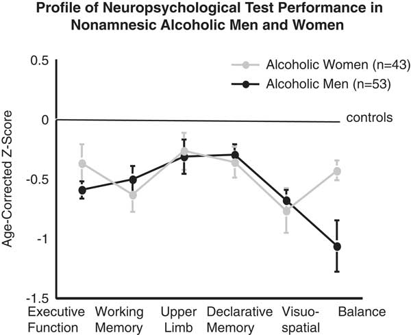
The resulting performance profile was similar to that identified by Victor, Adams, and Collins (Victor, Adams, & Collins, Reference Victor, Adams and Collins1989) using the Wechsler-Bellevue Intelligence Scale in their series of patients with alcoholic Korsakoff’s syndrome (KS), who exhibited enduring deficits in working memory, visuospatial ability, and psychomotor speed despite evidence for some recovery in world knowledge, semantic categorical comprehension, and assembly of familiar objects. Influenced by the work of Parson, Glenn, and Nixon in women (Fabian & Parsons, Reference Fabian and Parsons1983; Glenn & Parsons, Reference Glenn and Parsons1992; Nixon, Tivis, Ceballos, Varner, & Rohrbaugh, Reference Nixon, Tivis, Ceballos, Varner and Rohrbaugh2002; Nixon, Tivis, & Parsons, Reference Nixon, Tivis and Parsons1995; Parsons & Nixon, Reference Parsons and Nixon1998), we also sought a profile of impairment in alcoholic women and found that apart from static balance, alcoholic men and women showed similar deficit patterns (Sullivan, Fama, Rosenbloom, & Pfefferbaum, Reference Sullivan, Fama, Rosenbloom and Pfefferbaum2002) (Figure 1).
Profile of neuropsychological test performance in nonamnesic alcoholic men and women. Scores are expressed as age-corrected Z-scores based on sex norms and show a similar profile of impairment for men (black) and women (gray) with the exception of balance. From Sullivan et al. (Reference Sullivan, Rosenbloom, Lim and Pfefferbaum2000, Reference Sullivan, Fama, Rosenbloom and Pfefferbaum2002).

Furthermore, neither the diffuse impairment hypothesis nor the right hemisphere hypothesis of alcoholism-related impairment was supported, conclusions reached decades ago by Tarter (Tarter, Reference Tarter1975) and supported by the observation of similarly impaired verbal and nonverbal skills in two samples of alcoholics (Tivis, Beatty, Nixon, & Parsons, Reference Tivis, Beatty, Nixon and Parsons1995). Nonetheless, nonverbal tests may be more sensitive to enduring deficits (Brandt, Butters, Ryan, & Bayog, Reference Brandt, Butters, Ryan and Bayog1983; Fein, Bachman, Fisher, & Davenport, Reference Fein, Bachman, Fisher and Davenport1990) (for review, Oscar-Berman et al., Reference Oscar-Berman, Valmas, Sawyer, Ruiz, Luhar and Gravitz2014), possibly because they are often more difficult and use less familiar materials than verbal tests. These profiles indicated also where to look in the brain for underlying mechanisms of impairment.
Frontocerebellar Circuitry Hypothesis of Alcoholism
The salient deficit in static balance indicated in the neuropsychological performance profile of nonamnesic alcoholic men began the search for neural substrates of ataxia, initially tested with the modified Fregly-Graybiel Walk-a-Line Test (Fregly, Graybiel, & Smith, Reference Fregly, Graybiel and Smith1972; Sullivan, Rosenbloom, & Pfefferbaum, Reference Sullivan, Rosenbloom and Pfefferbaum2000). Known neuropathological substrates of ataxia led the search to the cerebellar vermis and ultimately evolved into our frontocerebellar circuitry hypothesis of alcoholism (Sullivan, Reference Sullivan2003; Sullivan & Pfefferbaum, Reference Sullivan and Pfefferbaum2005; Zahr, Pfefferbaum, & Sullivan, Reference Zahr, Pfefferbaum and Sullivan2017). Untoward effects of alcoholism on this far-reaching neural circuitry have foundation in speculations of early studies conducted in AUD uncomplicated by concomitant conditions and AUD complicated by dietary deficiencies or organ compromise (Tarter, Reference Tarter1975) and a later positron emission tomography study showing frontocerebellar glucose metabolism deficits in alcoholics with ataxia (Gilman et al., Reference Gilman, Adams, Koeppe, Berent, Kluin, Modell and Brunberg1990).
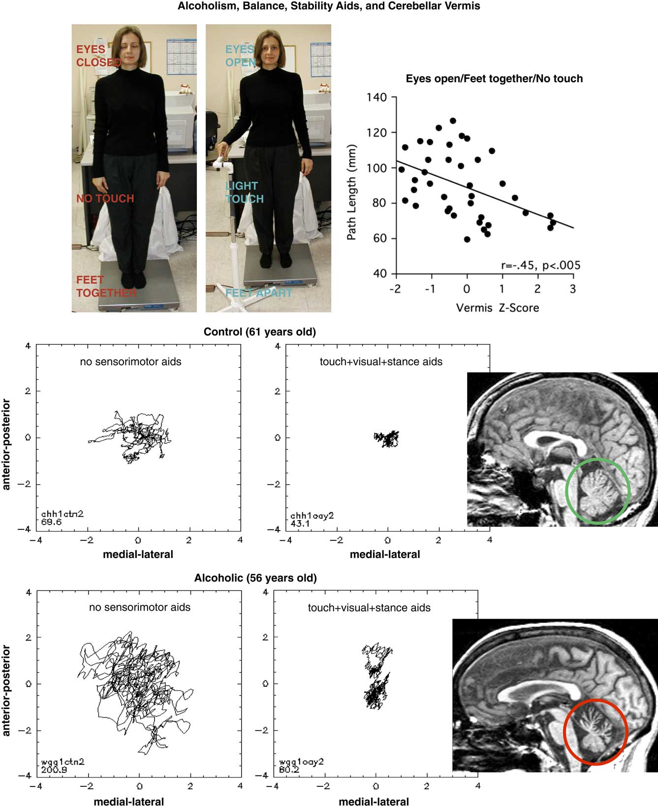
Pursuit of a refined characterization of balance instability used a balance platform, which yields a stabilogram and quantifies the kinetics of the sway path that result when trying to stand still under different sensory and motor conditions, that is, with or without sensory or stance aids—eyes closed, no touch, and feet together (Figure 2). With sensorimotor aids, the sway paths of control men and women were significantly shorter than those of alcoholic men and women, despite sobriety. With sensory and stance aids, however, even the sway of the alcoholics of both sexes was quelled (Sullivan, Rose, & Pfefferbaum, Reference Sullivan, Rose and Pfefferbaum2006a, Reference Sullivan, Rose and Pfefferbaum2006b). Thus, the force plate approach for detecting stance instability was sensitive enough for its detection in alcoholic women whose impairment was relatively elusive to the roadside-like ataxia testing.
Alcoholism, balance, stability aids, and cerebellar vermis. Top left. Woman on the balance platform. Bottom left. Example of balance platform stabilograms for control (top) and alcoholic (bottom) with and without cues. Note the sway path of “wobble” of the alcoholic as he tries to stand still. Bottom right. Structural MRI of the cerebellum (green circle) from a control (top) and alcoholic (bottom). Note the markedly smaller tissue of the cerebellar vermis of the alcoholic. Top right. Relation of vermis size and sway path length. Modified from Sullivan and Pfefferbaum (Human imaging studies of brain circuitry disrupted by alcoholism. In Noronha A, Cui C, Harris RA, Crabbe JC (Eds.). Neurobiology of Alcohol Dependence. Oxford: Elsevier, pp. 131–151, Chapter 8, 2014; Figure 8.1 on page 134); original data in Sullivan et al. (Cerebral Cortex 2006).

The next step was to seek a relation between sway path length and volume of the cerebellar vermis as a potential brain substrate of the instability in quiet standing. Correlations indicated that the smaller the anterior superior vermis, the longer the sway path. Furthermore, alcoholics who maintained sobriety longer had shorter sway paths. These patterns occurred in non-KS, recovering alcoholic men (Sullivan et al., Reference Sullivan, Rose and Pfefferbaum2006) and women (Sullivan, Rose, & Pfefferbaum, Reference Sullivan, Rose and Pfefferbaum2010a, Reference Sullivan, Rose and Pfefferbaum2010b).
Cross-sectional study cannot address whether static postural instability, or any other functional impairment, in recovering alcoholics was present before the onset of AUD, was exacerbated by AUD, or occurred because of AUD. With respect to balance specifically, early studies of nonalcoholic, adolescent sons of alcoholic fathers found them to have excess upper body sway, possibly related to hyperactivity (Hegedus, Tarter, Hill, Jacob, & Winsten, Reference Hegedus, Tarter, Hill, Jacob and Winsten1984). Paradoxically, young adult men with familial risk for alcoholism exhibited less body sway under controlled conditions of acute alcohol ingestion than did matched men who did not carry the familial liability (Schuckit, Reference Schuckit1985a). Both studies considered static ataxia as a potential biomarker or genetic trait of a predisposition for alcoholism.
Graded Brain Volume Deficits in Limbic Circuitry of Alcoholics
The deficit profile identifying compromised component processes of memory indicated where to look for neural substrates of compromise and led to Papez circuit and the limbic system. In addition, it was essential to define an appropriate diagnostic comparison to gauge the extent of the memory impairment in “uncomplicated” alcoholism. The comparison group comprised alcoholics complicated by KS, which is marked by dense anterograde amnesia, the result of Wernicke’s encephalopathy (WE) and thiamine deficiency. This diagnostic comparison is in the research traditions of Boston (e.g., Cermak, Butters, & Goodglass, Reference Cermak, Butters and Goodglass1971), Pittsburgh (Tarter, Reference Tarter1975), and Oklahoma (Parsons & Nixon, Reference Parsons and Nixon1993), where performance by uncomplicated alcoholics could be compared with non-alcoholic controls, who determined healthy norms, whereas alcoholics with KS determined amnesic performance. At the same time, the uncomplicated alcoholics served as a diagnostic comparison to the KS. Working within the context of classical neuropathological studies of alcoholics with KS, which is marked by lesions in the mammillary bodies and thalamus, we used in vivo MRI to ask what patterns of volume deficits define these two groups of alcoholics. These comparisons were the basis for testing whether the two alcoholic groups differed in deficit pattern or, alternatively, in deficit severity.
Accordingly, MRI data were acquired in groups of healthy controls, uncomplicated alcoholics, and alcoholics with KS, who we assumed from neuropathological studies (e.g., Victor et al., Reference Victor, Adams and Collins1989) would have regional limbic system volume deficits underlying their global amnesia. It was not clear what to expect in the uncomplicated alcoholics. First, the mammillary bodies showed marked bilateral volume deficits in KS, and KS with dementia had even smaller volumes. Uncomplicated, nonamnesic alcoholics also had bilateral mammillary body volume deficits but to a milder degree, and this occurred in the absence of an amnesic syndrome (Sullivan et al., Reference Sullivan, Lane, Deshmukh, Rosenbloom, Desmond, Lim and Pfefferbaum1999). Similarly, bilateral hippocampus (Sullivan, Marsh, Mathalon, Lim, & Pfefferbaum, Reference Sullivan, Marsh, Mathalon, Lim and Pfefferbaum1995), thalamus, pons (Sullivan & Pfefferbaum, Reference Sullivan and Pfefferbaum2009), cerebellar hemispheres, and vermis (Sullivan, Deshmukh, Desmond, Lim, & Pfefferbaum, Reference Sullivan, Deshmukh, Desmond, Lim and Pfefferbaum2000)—substrates of gait, balance, working memory, and components of executive functioning—each showed graded volume deficits from uncomplicated alcoholism to alcoholism complicated by KS relative to healthy controls (Sullivan & Pfefferbaum, Reference Sullivan and Pfefferbaum2009) (also see Blansjaar, Vielvoye, van Dijk, & Rijnders, Reference Blansjaar, Vielvoye, van Dijk and Rijnders1992). That all regional volume deficits observed were bilateral provided no evidence for the right hemisphere hypothesis of alcoholism.
Because the hippocampal finding in the KS patients was so controversial (cf., Squire, Reference Squire1982; Squire, Amaral, & Press, Reference Squire, Amaral and Press1990), we challenged it by measuring the extent of their volume deficit against Alzheimer’s disease (AD) patients, with known pathology of medial and lateral temporal cortex. Although the volume deficits of the AD group were greater than those of the KS in the temporal cortex, the alcoholic KS had hippocampal volume deficits equivalent to those of the AD group, thus providing convergent validity to our original findings (Sullivan & Marsh, Reference Sullivan and Marsh2003).
Graded Neuropsychological Deficits in Alcoholics
These graded effects led to the next pursuit: Do uncomplicated alcoholics carry an occult history of WE, the precursor of KS (cf., Blansjaar et al., Reference Blansjaar, Vielvoye, van Dijk and Rijnders1992; Tarter & Ryan, Reference Tarter and Ryan1983)? Are traces of WE signs also related to neuropsychological performance in alcoholics without KS, given that WE is marked by lesions of periventricular nuclei, mammillary bodies, and thalamus, resulting in the cardinal triad of ophthalmoplegia, ataxia, and confusional state and often harolding the profound anterograde memory impairment of KS (Victor et al., Reference Victor, Adams and Collins1989). Indeed, Wernicke-Korsakoff’s syndrome (WKS) is definitively known to be the result of thiamine depletion or severe deficiency, which was once a common concomitant of alcoholism (Harper, Reference Harper1979; Harper et al., Reference Harper, Sheedy, Lara, Garrick, Hilton and Raisanen1998).
The question of a nutritional mechanism of memory impairment in alcoholics has a history in the classical book, Alcoholic Korsakoff’s Syndrome: An Information-Processing Approach (Butters & Cermak, Reference Butters and Cermak1980), where Butters and Cermak speculated that “Because most chronic alcoholics may eat enough to maintain a sufficient level of thiamine, the short-term verbal memory deficits associated with diencephalic-limbic [dorsomedial nucleus of thalamus and/or mammillary bodies] lesions may be apparent in only a small percentage of chronic alcoholics” (page 158).
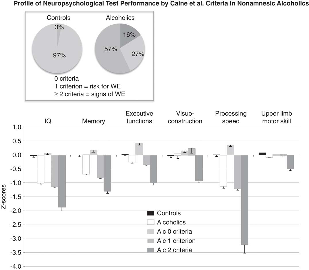
Studies of the pathologist Clive Harper reported that the incidence of neuropathological signs of WE observed postmortem was far higher in so-called uncomplicated alcoholics than recognized in vivo (Harper, Reference Harper1979). Pursuing these observations, Caine and colleagues devised 4 criteria for chart review of their postmortem cases: evidence for dietary deficiency, oculomotor abnormalities, cerebellar dysfunction, and altered mental state. Cases having two of four of these signs correlated with presence of neuropathological signs of WE (Caine, Halliday, Kril, & Harper, Reference Caine, Halliday, Kril and Harper1997). A decade later, Pitel et al. (Reference Pitel, Zahr, Jackson, Sassoon, Rosenbloom, Pfefferbaum and Sullivan2011) operationalized the Caine et al. criteria for in vivo use in alcoholics and controls; historical interview enumerated missed meals and quantitative examination tested for nystagmus, ataxia, and cognitive compromise determined with the Mattis Dementia Rating Scale (Mattis, Reference Mattis1988). Although no alcoholic presented with any of the three classical WE signs of ophthalmoplegia, ataxia, or delirium, a large proportion of the alcoholic group met one or two criteria, based on Caine et al. (Reference Caine, Halliday, Kril and Harper1997) and operationalized by Pitel et al. (Reference Pitel, Zahr, Jackson, Sassoon, Rosenbloom, Pfefferbaum and Sullivan2011). In fact, more than half the group met one criterion; 16% met two criteria; and only approximately one quarter of the group were free of these signs (Figure 3).
These alcoholics and controls were also administered a comprehensive neuropsychological test battery. The pattern of functional impairment across the entire group of alcoholics without consideration of Caine criteria indicated mild to moderate deficits in three of the six functional domains examined. Dividing the alcoholics by Caine criteria, however, presented a different picture, where alcoholics meeting no criteria performed at normal levels in all domains, those meeting one criterion showed modest selective compromise, and those meeting two criteria were the most widely affected, being significantly impaired on all 6 domains (Pitel et al., Reference Pitel, Zahr, Jackson, Sassoon, Rosenbloom, Pfefferbaum and Sullivan2011) (Figure 3).
Profile of neuropsychological test performance in nonamnesic alcoholics in relation to Caine et al. criteria. Scores are expressed as age-corrected Z-scores based on laboratory norms (black bars near 0). White with gray outline bars=Z-scores of alcoholics without regard for Caine et al. criteria; light gray bars=alcoholics meeting 0 Caine criteria; medium gray bars=alcoholics meeting 1 Caine criterion; dark gray bars=alcoholics meeting 2 Caine criteria. From Pitel et al. (Reference Pitel, Zahr, Jackson, Sassoon, Rosenbloom, Pfefferbaum and Sullivan2011).

This finding was recently extended with an enlarged group of uncomplicated alcoholics (Fama et al., Reference Fama, Hardcastle, Sassoon, Zahr, Pfefferbaum and Sullivan2017). Blood samples for thiamine levels obtained in a subset of the initially studied alcoholics revealed that higher thiamine levels were predictive of better memory performance, and that this relation was selective to memory and not to other performance domains. Thus, occult WE may be a factor underlying alcohol-related neuropsychological deficits (cf., Bowden, Reference Bowden1990), thereby supporting the earlier speculations of Butters and Cermak (Reference Butters and Cermak1980), Tarter (Tarter, Reference Tarter1975), and later by Oscar-Berman (Oscar-Berman, Reference Oscar-Berman2000).
In summary, rather than concluding that the effect of AUD on brain structure is diffuse, these studies support disruption of frontocerebellar and frontolimbic circuitry with wide-reaching networks. Complementing neurocircuitry disruption are constellations of selective functional impairments that include executive functions, attentional processes, response inhibition, episodic memory, prosody, visuospatial abilities, decision-making, and postural stability, yet leaving language, procedural learning, and upper motor skills largely intact.
APPRECIATE THAT ALCOHOLISM-RELATED FUNCTIONAL CHANGES ARE A FORM OF NEUROADAPTATION THAT MAY UNDERLIE DYSFUNCTION, MAKING ALCOHOLISM A SELF-PERPETUATING DISORDER
In keeping with the focus on memory, examples of neuroadaptation and alcohol-related change will be considered through functional neuroimaging studies of working memory. Information stored through working memory processes is held only temporarily for ultimate consolidation into a more permanent form or for manipulation to transform information from one concept to another, and is a mnemonic process commonly impaired in chronic alcoholism (Stavro, Pelletier, & Potvin, Reference Stavro, Pelletier and Potvin2013). Functional imaging methods can readily be used to interrogate working memory to reveal neural substrates of change, to determine whether alcoholics recruit the same networks as controls to perform a task at control levels, and to examine whether alcoholics have intrinsic functional connectivity similar to controls.
Functional MRI: BOLD Using Task-Activated Paradigms
The most commonly used noninvasive, functional imaging method, functional MRI (fMRI), is based on a hemodynamic response, the blood oxygen-level dependent response (BOLD), that is typically calculated in task-activated paradigms as a difference in responses between experimental and control conditions (e.g., Glover, Reference Glover2011). In a seminal task-activated fMRI study, alcoholics and controls performed a 2-back, spatial working memory task while being scanned. Subjects attempted to remember the location of 0s from trial to trial and to press a key every time a 0 appeared in the same location two trials back. For the comparison task, subjects pressed the response key every time a zero appeared in the center location; therefore, no memory was required. The alcoholics were practiced to perform as well as the controls, which is essential to allow interpretation of group differences in activation patterns should they emerge. After all, if the groups performed differently on the target task, it would be no surprise to see differences in activation patterns.
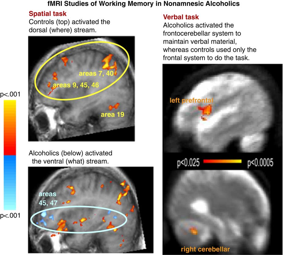
In this study (Pfefferbaum et al., Reference Pfefferbaum, Desmond, Galloway, Menon, Glover and Sullivan2001), while performing the spatial working memory task, controls activated the dorsal stream traditionally used in spatial processing tasks, whereas alcoholics activated the ventral stream used in object identification tasks (cf., Ungerleider & Mishkin, Reference Ungerleider and Mishkin1982). In a verbal working memory task, participants were given either six letters or one letter to remember over a 5-s interval and saw a probe letter, to which they responded with a button box key yes if it matched or no if it did not. To perform this simple task, alcoholics activated frontal and cerebellar regions, a task accomplished by controls with only the frontal system (Desmond et al., Reference Desmond, Chen, De Rosa, Pryor, Pfefferbaum and Sullivan2003) (Figure 4).
Results from two task-activated fMRI studies. Top left: In a spatial working memory task, controls (top) activated cortical regions of the dorsal stream, typically invoked for spatial processing (where). Bottom left: Alcoholics activated the ventral stream, typically invoked for object naming (what); from Pfefferbaum et al. (Reference Pfefferbaum, Desmond, Galloway, Menon, Glover and Sullivan2001). Right: Alcoholics activated the frontocerebellar system to maintain verbal material in working memory, whereas controls used only the frontal system to do the task. From Desmond et al. (Reference Desmond, Chen, De Rosa, Pryor, Pfefferbaum and Sullivan2003).

These were the first of several such studies showing a common theme in alcoholism (e.g., De Rosa & Sullivan, Reference De Rosa and Sullivan2003; Tapert et al., Reference Tapert, Brown, Kindermann, Cheung, Frank and Brown2001), summarized by Oscar-Berman and Marinkovic (Reference Oscar-Berman and Marinkovic2007): that the positive outcome of shift and expansion in functional anatomy can be normal performance but may occur at the price of usurping reserve that reduces processing capacity for conducting multiple tasks simultaneously and efficiently.
fMRI: BOLD Using a Resting-State Paradigm to Identify Intrinsic Functional Networks
These task-activated results led to functional connectivity analysis. Typical task-activated fMRI experiments ask, “What regions respond while engaged in a task?” and use brain activation to identify areas of functional specialization. To understand functional integration, we ask, “What regions respond together?” establishing brain connectivity by identifying brain regions with highly correlated, that is, synchronous, BOLD activity compared with regions that do not show this correlated activity.
Task-free fMRI when done in the so-called “resting state” has revealed intrinsic functional networks, the most prominent being the Default Mode Network (DMN), characterized by a pattern of activation synchrony involving the precuneus or posterior cingulum, parietal cortex, and medial prefrontal cortex (Raichle et al., Reference Raichle, MacLeod, Snyder, Powers, Gusnard and Shulman2001; Raichle & Snyder, Reference Raichle and Snyder2007). At rest, alcoholics showed the same pattern of activation synchrony as controls but had less robust synchrony between most nodes of the DMN than controls (Chanraud, Pitel, Pfefferbaum, & Sullivan, Reference Chanraud, Pitel, Pfefferbaum and Sullivan2011). One exception was for the middle frontal cortex, which had higher activation in the alcoholics than controls.
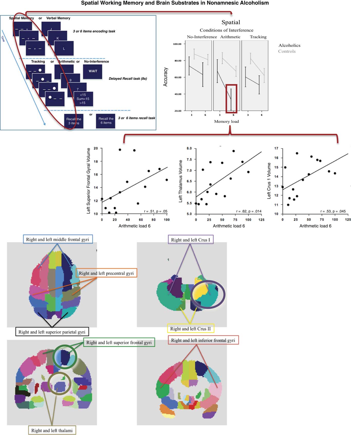
This finding led to testing whether robustness of resting-state connectivity could predict subsequent performance on a Brown-Peterson distractor task (Brown, Reference Brown1958; Cermak & Ryback, Reference Cermak and Ryback1976; Peterson & Peterson, Reference Peterson and Peterson1959) for spatial working memory with an established frontocerebellar structural substrate (Chanraud, Pitel, & Sullivan, Reference Chanraud, Pitel and Sullivan2010) (Figure 5). Using the middle frontal cortex as the seed region, that is, the point with which BOLD synchrony is tested with every other voxel in the brain, significant functional connectivity emerged with the cerebellum. Critically, higher correlation of this resting functional connectivity between the middle frontal and Crus II of the cerebellum predicted subsequent performance in the alcoholics but not controls (Chanraud et al., Reference Chanraud, Pitel, Pfefferbaum and Sullivan2011). This pattern of group differences in the context of functional connectivity differences between frontal and cerebellar targets fit criteria deeming the activation pattern as consistent with compensation (Chanraud, Pitel, Muller-Oehring, Pfefferbaum, & Sullivan, Reference Chanraud, Pitel, Muller-Oehring, Pfefferbaum and Sullivan2013).
Spatial working memory and brain substrates in nonamnesic alcoholism. Upper left: Brown-Peterson short-term (working) memory paradigm using three or six spatial or verbal items for recall and a spatial or verbal distractor task or rest conditions. Upper right: Accuracy scores showing poorer performance by the alcoholic than control group, with the greatest performance difference with the largest memory load (six items) with arithmetic interference. Middle: Correlations between better performance on the six-item spatial task with arithmetic distraction condition with larger volumes of the left superior frontal gyrus, thalamus, and Crus I in the alcoholics. Bottom: Atlas locations of the targets for correlations. From Chanraud et al. (Reference Chanraud, Pitel, Rohlfing, Pfefferbaum and Sullivan2010).

Arterial Spin Labeling as a Direct Measure of Cerebral Perfusion
Another form of noninvasive functional imaging is MR arterial spin labeling (ASL), which measures cerebral perfusion, or cerebral blood flow (CBF), itself rather than the BOLD signal measured with fMRI. Pulsed continuous ASL (PCASL) is a three-dimensional acquisition protocol (Alsop, Makovetskaya, Kumar, Selim, & Schlaug, Reference Alsop, Makovetskaya, Kumar, Selim and Schlaug2005; Detre & Alsop, Reference Detre and Alsop1999), which is reliable (Pfefferbaum et al., Reference Pfefferbaum, Chanraud, Pitel, Shankaranarayanan, Alsop, Rohlfing and Sullivan2010) and sensitive to change with task demands (Pfefferbaum et al., Reference Pfefferbaum, Chanraud, Pitel, Muller-Oehring, Shankaranarayanan, Alsop and Sullivan2011). Perfusion imaging can determine whether disease-related differences identified with fMRI are caused by impaired microvascular perfusion. To the extent that resting-state paradigms are used for identifying intrinsic functional networks whereas task-activated paradigms are well-suited for identifying nodes and networks, we used PCASL to test whether perfusion imaging could detect and replicate fMRI-identified activation patterns. Accordingly, subjects performed the spatial working memory distractor paradigm to test recall of six spatial locations following a retention interval filled with arithmetic problems (Chanraud, Pitel, & Sullivan, Reference Chanraud, Pitel and Sullivan2010); in other conditions, subjects “rested.”
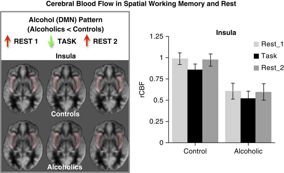
Like healthy controls, alcoholics showed the DMN pattern in selective regions (medial frontal, temporal, and cingulate cortices, posterior precuneus, and hippocampal/amygdalar complex) with high perfusion in the rest condition, low perfusion in the task condition, and a return to high perfusion in the second rest condition. By contrast, other regions (superior and inferior cerebellum, middle precuneus, parietal, occipital, and calcarine cortices), known to be associated with working memory (Chanraud et al., Reference Chanraud, Pitel, Rohlfing, Pfefferbaum and Sullivan2010; Desmond, Gabrieli, Wagner, Ginier, & Glover, Reference Desmond, Gabrieli, Wagner, Ginier and Glover1997; Tapert et al., Reference Tapert, Brown, Kindermann, Cheung, Frank and Brown2001), showed a task-activated CBF pattern of low-high-low with rest-task-rest conditions. Although both groups showed the DMN pattern in the insula, the level of perfusion was impaired in the alcoholics (Figure 6); yet, in alcoholics, greater task accuracy correlated with higher insular perfusion at rest (Sullivan et al., Reference Sullivan, Muller-Oehring, Pitel, Chanraud, Shankaranarayanan, Alsop and Pfefferbaum2013).
CBF and the insula. Left: Axial images with the insula outlined in red showing perfusion differences related to rest and task conditions. Right: Quantification of the insular CBF indicate a default-mode pattern of insular activation for both groups although deficient in the alcoholic group. From Sullivan and Pfefferbaum (Human imaging studies of brain circuitry disrupted by alcoholism. In Noronha A, Cui C, Harris RA, Crabbe JC (Eds.). Neurobiology of Alcohol Dependence. Oxford: Elsevier, pp. 131–151, Chapter 8, 2014; Figure 8.6 on page 143); original data in Sullivan et al. (Reference Sullivan, Muller-Oehring, Pitel, Chanraud, Shankaranarayanan, Alsop and Pfefferbaum2013).

We then used the insula, which is considered the hub of the salience network (Greicius, Krasnow, Reiss, & Menon, Reference Greicius, Krasnow, Reiss and Menon2003; Seeley et al., Reference Seeley, Menon, Schatzberg, Keller, Glover, Kenna and Greicius2007; Sridharan, Levitin, & Menon, Reference Sridharan, Levitin and Menon2008), as a seed for CBF-synchrony analysis. This analysis revealed functional connectivity between the insula and nodes of the salience network (anterior cingulate, medial prefrontal, temporal, and parietal cortices) in the controls but not alcoholics. Functions of the insula may have particular relevance to alcoholism given its role in impulse control, self-regulation, error monitoring, and reward processing. As a principal node of the salience network, the insula enables switching between intrinsic and task networks, notably, the executive control network (Menon & Uddin, Reference Menon and Uddin2010). This set of results led to the speculation that low insular CBF impairs connectivity among functional salience network nodes and reduces ability to switch from interoceptive desires, such as cravings (impulsive system), to cognitive control over these desires (executive system). In this way, the insula might be considered an “addiction site.” Later in this review, we will see that the insula plays a role in a critical yet under-studied component of memory function in alcoholics.
Evidence that alcoholism is a self-perpetuating disorder has foundation in observations of AUD-related modification of neurocircuitry affecting, for example, the salience network in its switching capacity to over-ride compulsive urges. Other forms of neuradaptation include shifts and expansions of functional brain regions to accommodate or compensate for AUD-injured sites.
LEARN THAT SUSTAINED SOBRIETY CAN RESULT IN IMPROVEMENT IN BRAIN STRUCTURE AND FUNCTION, INDICATIVE OF DAMAGE REVERSAL OR COMPENSATORY MECHANISMS THAT CAN BE IDENTIFIED WITH FORMAL NEUROPSYCHOLOGICAL TESTING AND LONGITUDINAL, QUANTITATIVE, STRUCTURAL, AND FUNCTIONAL BRAIN IMAGING
So far, only deficits related to excessive, chronic alcohol consumption have been considered. But, alcoholism follows a dynamic course, with the possibility of brain structural and functional recovery with sustained sobriety, which brings this review to its last section, regarding the potential for recovery of brain structure and function.
Recovery of Brain Macrostructure
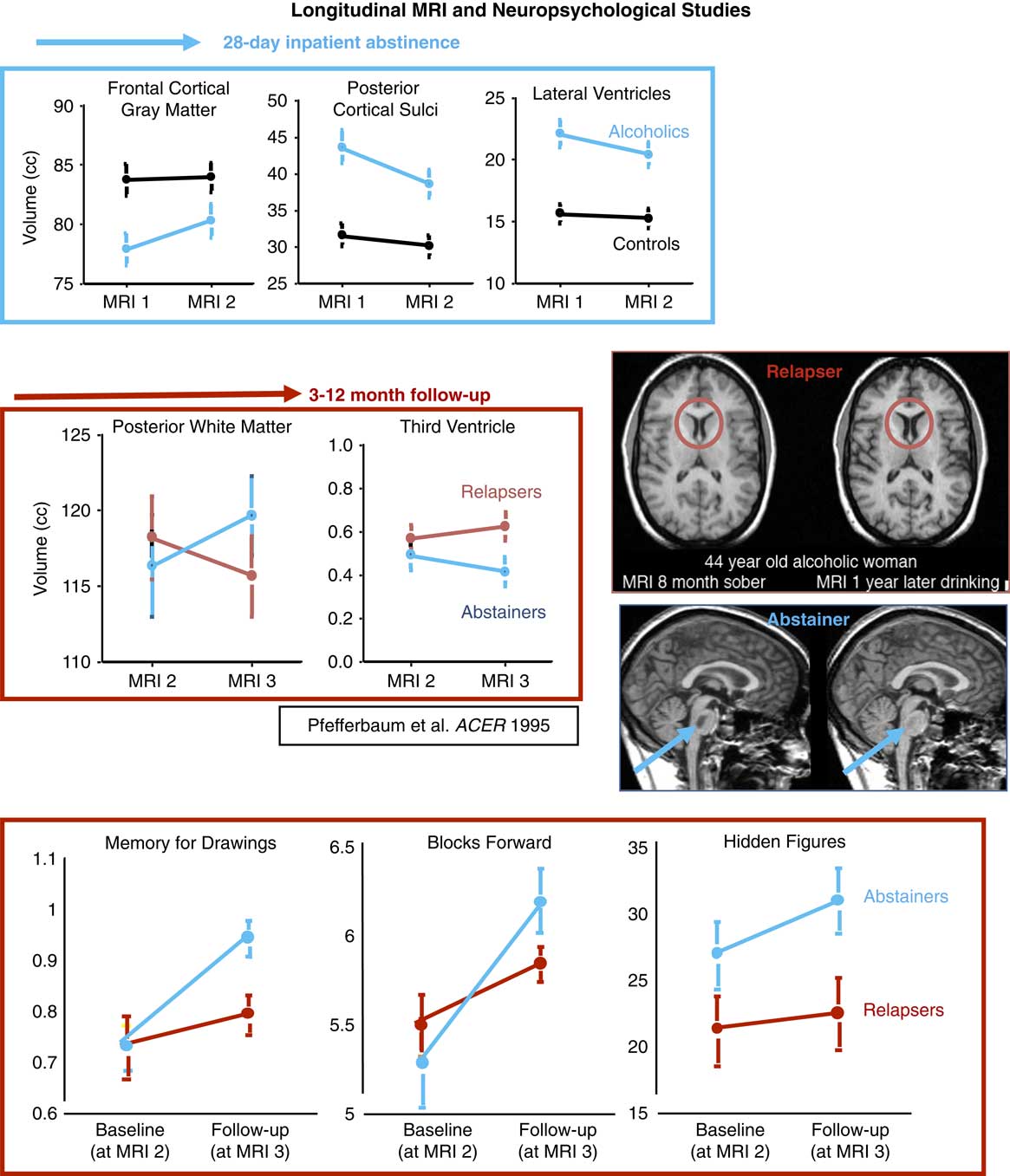
Remarkably, ventricular expansion, a macrostructural signature of prolonged, chronic, dependent-level alcohol consumption, is reversible, at least in part, with sustained abstinence. This reversal was first detected in vivo with computed tomography (Carlen, Wilkinson, Wortzman, & Holgate, Reference Carlen, Wilkinson, Wortzman and Holgate1984; Carlen, Wortzman, Holgate, Wilkinson, & Rankin, Reference Carlen, Wortzman, Holgate, Wilkinson and Rankin1978). Serial MRIs of individual cases of recovery reveal brain structural improvement (Rosenbloom & Pfefferbaum, Reference Rosenbloom and Pfefferbaum2008), visible in the lateral ventricles (Figure 7). A striking example of recovery involved a 41-year-old alcoholic woman, scanned after 2 months of sobriety and then again 1 year later. In addition to ventricular shrinkage, she exhibited resolution of radiological evidence for central pontine myelinolysis, which if left untreated, can be fatal (Lampl & Yazdi, Reference Lampl and Yazdi2002; Sullivan & Pfefferbaum, Reference Sullivan and Pfefferbaum2001).
Longitudinal MRI and neuropsychological studies of recovery and relapse. Top blue box: MRI volume data of alcoholics approximately 1-week post-detoxification and then again after 28 days of enforced abstinence in an inpatient VA treatment facility. The alcoholics showed volume gains with abstinence despite no volume change in the controls. Middle red box: Alcoholics who remained abstinent 3 to 12 months after discharge showed further improvement in brain volume changes, whereas relapsers showed worsening; from Pfefferbaum et al. (Reference Pfefferbaum, Sullivan, Mathalon, Shear, Rosenbloom and Lim1995). Bottom red box: Neuropsychological testing conducted at the 3- to 12-month follow-up revealed improved performance in both groups, suggesting practice effects, which were greater in the abstainers than relapsers; from Sullivan et al. (Reference Sullivan, Rosenbloom, Lim and Pfefferbaum2000).

In addition to single case studies, an ever-increasing number of longitudinal MRI studies on groups of alcoholics report significant changes in regional brain volumes (e.g., Cardenas, Studholme, Gazdzinski, Durazzo, & Meyerhoff, Reference Cardenas, Studholme, Gazdzinski, Durazzo and Meyerhoff2007; Rohlfing, Sullivan, & Pfefferbaum, Reference Rohlfing, Sullivan and Pfefferbaum2006; Shear, Jernigan, & Butters, Reference Shear, Jernigan and Butters1994; Wang et al., Reference Wang, Demirakca, van Eijk, Frischknecht, Ruf, Ucar and Ende2016; Yeh, Gazdzinski, Durazzo, Sjostrand, & Meyerhoff, Reference Yeh, Gazdzinski, Durazzo, Sjostrand and Meyerhoff2007). An early study imaged alcoholics approximately a week after detoxification and again approximately 3 weeks later, at the end of a 28-day VA inpatient treatment regime with enforced abstinence. Controls, scanned over the same interval, showed no change in regional brain volumes, whereas the abstaining alcoholics showed improvement, enlargement of frontal cortical volume and shrinkage of cortical sulci and lateral ventricles (Figure 7).
Follow-up of this cohort after discharge revealed that approximately half relapsed and half maintained abstinence. The abstainers showed further improvement, whereas relapsers showed further shrinkage of white matter and expansion of the third ventricle (Pfefferbaum et al., Reference Pfefferbaum, Sullivan, Mathalon, Shear, Rosenbloom and Lim1995). Further follow-up indicated that the more relapsers had drunk over the 5-year interval, the greater their volume loss of cortical gray matter (Pfefferbaum, Sullivan, Rosenbloom, Mathalon, & Lim, Reference Pfefferbaum, Sullivan, Rosenbloom, Mathalon and Lim1998) (Figure 7). These results have now been replicated with higher-resolution imaging of regional brain macrostructure (e.g., Segobin et al., Reference Segobin, Chetelat, Le Berre, Lannuzel, Boudehent, Vabret and Pitel2014) and imaging protocols focused on white matter microstructure (e.g., Pfefferbaum et al., Reference Pfefferbaum, Rosenbloom, Chu, Sassoon, Rohlfing, Pohl and Sullivan2014).
Recovery of Component Processes of Memory Functions with Alcohol Abstinence
For decades, systematic studies of short-term, or working, memory have revealed significant improvement with sustained sobriety in uncomplicated alcoholics but not in KS alcoholics (Cermak & Ryback, Reference Cermak and Ryback1976). Our early study of abstinent, non-KS alcoholics showed greater improvement in nonverbal immediate and short-term memory than did relapsers (Sullivan, Rosenbloom, Lim, & Pfefferbaum, Reference Sullivan, Rosenbloom, Lim and Pfefferbaum2000). Improvements in visuospatial ability also noted in the abstainers could have contributed to spatial memory improvements (Figure 7). Critically, improvements in memory correlated with shrinkage of the third ventricle volume, suggesting a neural substrate for recovery (Rosenbloom, Pfefferbaum, & Sullivan, Reference Rosenbloom, Pfefferbaum and Sullivan2004; Sullivan, Rosenbloom, Lim, et al., Reference Sullivan, Rosenbloom, Lim and Pfefferbaum2000).
These findings echo both earlier and more recent controlled studies of memory recovery with short-term or sustained sobriety showing a range of recovery, from little to extensive, after short-term (e.g., 1 week) to long-term (e.g., 1 year or more) sustained abstinence from alcohol (e.g., Bartels et al., Reference Bartels, Kunert, Stawicki, Kroner-Herwig, Ehrenreich and Krampe2007; Bates, Voelbel, Buckman, Labouvie, & Barry, Reference Bates, Voelbel, Buckman, Labouvie and Barry2005; Bell, Vissicchio, & Weinstein, Reference Bell, Vissicchio and Weinstein2016; Butters & Cermak, Reference Butters and Cermak1980; Fabian & Parsons, Reference Fabian and Parsons1983; Fein, Torres, Price, & Di Sclafani, Reference Fein, Torres, Price and Di Sclafani2006; Glenn, Parsons, & Sinha, Reference Glenn, Parsons and Sinha1994; Leber, Jenkins, & Parsons, Reference Leber, Jenkins and Parsons1981; Mann, Gunther, Stetter, & Ackermann, Reference Mann, Gunther, Stetter and Ackermann1999; Pitel et al., Reference Pitel, Rivier, Beaunieux, Vabret, Desgranges and Eustache2009; Rosenbloom et al., Reference Rosenbloom, Pfefferbaum and Sullivan2004; Rourke & Grant, Reference Rourke and Grant1999; Yohman, Parsons, & Leber, Reference Yohman, Parsons and Leber1985) (for reviews, see Oscar-Berman et al., Reference Oscar-Berman, Valmas, Sawyer, Ruiz, Luhar and Gravitz2014; Pitel, Eustache, & Beaunieux, Reference Pitel, Eustache and Beaunieux2014). Remaining to be established are parameters of recovery, including length of sobriety, age at alcoholism initiation, age at testing, style of drinking, concomitant nutritional deficiencies and non-alcohol drug use, sex, physiological, ethnic, and genetic characteristics of alcoholics (Stavro et al., Reference Stavro, Pelletier and Potvin2013).
Meaning of Neuroadaptation to the Recovering Alcoholic
How does the alcoholic perceive the fact that brain structure and function change for the worse with chronic drinking and that they change again with sobriety? How aware is the alcoholic of his or her impairment? There can be significant mismatches between the subjective view of one’s performance and the objective measurement of actual performance. This phenomenon can be operationalized using a metamemory paradigm, which may inform us about the extent to which alcoholics are aware of their deficits.
Work of a predecessor of INS provides insight regarding functional recovery after head injury. Kurt Goldstein, a German neurologist and psychiatrist who fled Germany in the 1930s, went to NY and then Boston, where he studied and treated veterans who had sustained head injuries in The Great War (Goldstein, Reference Goldstein1942). His work on visual agnosia together with his initial interest in philosophy likely set the stage for his interest in brain–mind relations. A primary principle proffered was that brain injury affects the organism as a whole, rendering the individual forever different from the pre-injury state (Goldstein, Reference Goldstein1995). Thus, recovery can never be complete.
This concept is consistent with that of Oscar-Berman and Marinkovich (Oscar-Berman & Marinkovic, Reference Oscar-Berman and Marinkovic2007) in noting that achieving normal performance in one function can occur at the expense of other functions (cf., Sullivan & Tapert, Reference Sullivan and Tapert2013). This concept cast a long shadow on cognitive rehabilitation efforts and has been eschewed by those who believe that recovery means a return to a premorbid state. Goldstein posited that, as for compensation, there is no such thing. Rather, a head injured patient might undergo restitution and recuperation, achieving “new normal” where health “is restored if a relation between preserved and disturbed performances is reached (page 331)” (Goldstein, Reference Goldstein1995). But such adaptation could result in a brain-injured patient’s adapting to the deficit state and rejecting change. This subjective bias would need to be overcome to embrace change through resolving the mismatch between the subjective and objective conditions.
The Goldstein idea is much like that of the First Principle of Neuropsychological Rehabilitation put forth by Prigatano (Reference Prigatano1999), stating that, “The clinician must begin with patient’s subjective or phenomenological experience to reduce their frustration and confusion in order to engage them in the rehabilitation” (page 3). This conception is even consistent with Step 1 of Alcoholics Anonymous (Wilson, 1939, Reference Wilson2014), which states, “We admit we were powerless over alcohol.” What we might conclude today, however, is to admit requires accurate assessment of the problem, and to fail at this can be mistaken as denial rather than as a cognitive impairment itself.
Feeling-of-Knowing: A Test of Metamemory
Relevant to addressing these queries is metamemory, which is an umbrella mnemonic function that refers to personal knowledge about one’s own ability to remember or recall information (Flavell, Reference Flavell1971). Metamemory draws on cognitive processes to monitor and control memory and can be examined by testing prospectively predictions about how well one might perform a memory test and by testing retrospectively one’s confidence of actual recall or recognition performance (Le Berre et al., Reference Le Berre, Pinon, Vabret, Pitel, Allain, Eustache and Beaunieux2010, Reference Le Berre, Muller-Oehring, Kwon, Serventi, Pfefferbaum and Sullivan2016). Assessment of both the prospective and retrospective memory components is achieved with the feeling-of-knowing (FOK) paradigm (Hart, Reference Hart1965). In short, subjects learn unrelated word pairs, then after a delay judge whether they think they would be able to recall the second word of the pair later on (prospective memory), and finally do a four-choice recognition task and make retrospective confidence judgments (RCJ) about their actual performance on the recognition task (for review of the method, Le Berre & Sullivan, Reference Le Berre and Sullivan2016).
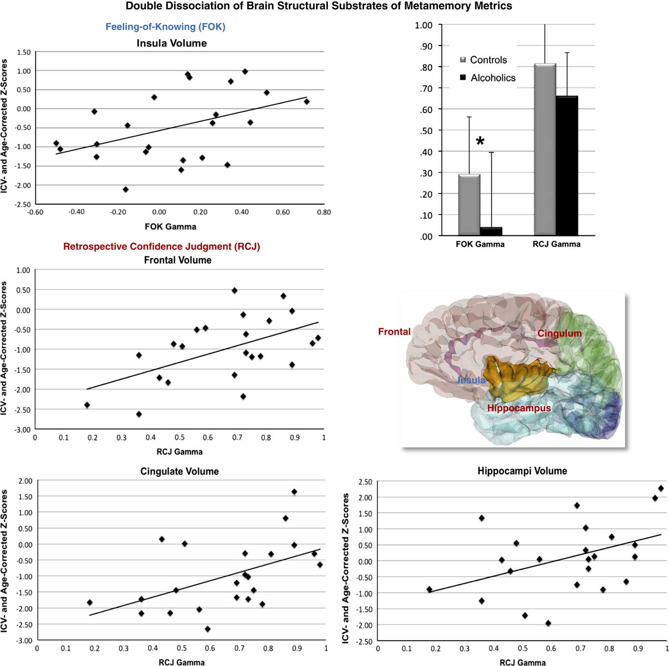
In calculating the agreement between predictions of future recognition performance and actual recognition performance, the alcoholics overestimated their recognition ability despite being accurate in their estimation of how they performed (Le Berre et al., Reference Le Berre, Muller-Oehring, Kwon, Serventi, Pfefferbaum and Sullivan2016, Reference Le Berre, Pinon, Vabret, Pitel, Allain, Eustache and Beaunieux2010). Structural MRI with parcellation to identify brain substrates of FOK and RCJ performance in the alcoholics revealed a double dissociation, where FOK accuracy was selectively related to insular volume but not to frontolimbic volumes, whereas retrospective confidence accuracy was selectively related to frontolimbic structural volumes but not to insular volumes (Le Berre et al., Reference Le Berre, Muller-Oehring, Kwon, Serventi, Pfefferbaum and Sullivan2016) (Figure 8). Task-activated fMRI revealed that in the metamemory vs. control task contrast greater activation occurred in the right superior frontal cortex in the alcoholics than controls but that better episodic memory performance and greater FOK accuracy correlated with lower activation in the right superior frontal cortex, a node of the default mode network, in the controls but not alcoholics. Within the alcoholics, greater FOK accuracy correlated with greater activation in the right insula, a correlation not observed in the controls (Le Berre et al., Reference Le Berre, Müller-Oehring, Schulte, Serventi, Pfefferbaum and Sullivan2017).
Double dissociation of brain structural substrates of metamemory metrics. Better scores on the FOK test correlated selectively with larger insular volumes, whereas better scores on the RCJ test correlated selectively with larger frontal, cingulate, and hippocampal volumes in the alcoholics. From Le Berre et al. (Reference Le Berre, Muller-Oehring, Kwon, Serventi, Pfefferbaum and Sullivan2016).

Anosognosia or Denial?
In summary, internationally based evidence (Le Berre et al., Reference Le Berre, Muller-Oehring, Kwon, Serventi, Pfefferbaum and Sullivan2016, Reference Le Berre, Pinon, Vabret, Pitel, Allain, Eustache and Beaunieux2010) was forthcoming for a functional dissociation and mismatch between subjective metamemory processes, with impaired prospective memory showing overestimation of potential mnemonic abilities, and spared retrospective assessment, indicating awareness of actual abilities. Furthermore, evidence for neural substrates of the metamemory impairment from structural MRI data revealed a double dissociation, where poorer prospective memory correlated selectively with smaller insular cortical volumes, whereas better retrospective memory correlated selectively with larger frontolimbic structures. Extending support for a role of the insula in metamemory were fMRI data, which revealed that better performance related to greater insular activation in alcoholics.
Thus, faulty FOK performance by alcoholics is marked by an over-estimation of prospective memory abilities presenting a mismatch between subjective experience and objective abilities, possibly related to the alterations in brain systems invoked to do the best one can. This pattern is consistent with a mild anosognosia (for discussion and review, see Le Berre & Sullivan, Reference Le Berre and Sullivan2016), which initially described the inability of a stroke patient to recognize the loss of limb function, with the subjective experience that the limb remains attached (Babinski, Reference Babinski1914). Agnosognosia has also been characterized by the phrase, “You don’t know that you don’t know.” Taken together, these results provide initial support for neuropsychological and neural mechanisms involving insular cortical structure and function of unawareness, perhaps misinterpreted as the psychological defense mechanism of denial rather than as an actual functional impairment, possibly anosognosia, in addiction (cf., Sullivan, Reference Sullivan2012).
It is now objectively established with controlled, longitudinal study that brain structure and cognitive and motor functioning can improve with sustained sobriety. Remaining unknown, however, are the mechanisms and means of neural recovery and whether recovery can ever be complete. The latter requires knowledge about pre-AUD neurostructural and neurobehavioral conditions, which can be determined only with prospective study.
CONCLUSION
Brain structural and functional changes occur in alcoholism. Our INS forefathers’ rigorous experimental work and keen observations initiated and enriched our understanding of alcoholism with its sometimes elusive, constellation of impairments. That legacy work has enjoyed verification with replication and extension through neuroimaging technologies unavailable in those early days, and now enabling legacy hypotheses to be tested in vivo. Among the myriad brain structure–function hypotheses now testable concern identification of and distinction between mechanisms of compensatory vs. recovery of functions with sustained sobriety and retraining efforts.
ACKNOWLEDGMENT
This review is based on a plenary talk presented at the 45th annual meeting of the International Neuropsychological Society in celebration of the 50th anniversary of the establishment of the society. The research from our laboratory presented in this review was accomplished with the invaluable collaboration with my longstanding colleagues, Adolf Pfefferbaum, MD, who put the “neuro” into neuropsychology, and Rosemary Fama, PhD, my partner in the quest for dissociable components of functions and processes. Support for this work is from the National Institute on Alcohol Abuse and Alcoholism (AA010723, AA017168, AA017923, AA021697-NCANDA) and the Moldow Women’s Hope and Healing Fund. Conflicts of interest: The author has no conflicts of interest with respect to the contents of this manuscript. Ethical considerations: The studies described using human data were conducted with permission from Internal Review Boards and the written permission of patients or their conservators and in compliance with the principles outlined in the Helsinki Declaration.