Introduction
Avoiding errors in planning and patient set-up is crucial to the accurate delivery of radiotherapy treatment.Reference Malicki1, Reference van der Merwe, Van Dyk and Healy2 A planning computed tomography (CT) of patients in treatment position is the first clinical and technical step before radiation therapy. Both the clinicians and technicians are involved in the choice of the best immobilisation and positioning system. In addition, to better visualise internal organs physicians can use contrast medium or devices, sometimes using invasive procedures.
On this occasion, patients experience, for the first time, the position they are required to adopt for treatment. It is important to consider the patient’s muscular tension, due to anxiety or discomfort, among the potential sources of systematic set-up errors in the simulation phase.Reference Lewis, Merckaert and Liénard3
In their paper, Budach et al.Reference Budach, Bölke and Fietkau4 analyse the time needed for specific radiotherapy procedures. They found the mean time required for planning CT (with contrast medium or not) is 22 minutes. The simulation phase requires 23 more minutes.
In our practice, we perform the planning CT acquisition and virtual simulation in 45 minutes. During this time, the patient becomes familiar to the position and the immobilisation required for treatment. At this stage, the personnel always inform the patient about the importance of relaxing the musculature. Nevertheless, we report here two cases in which the muscular contraction caused systematic set-up errors, requiring us to perform a second CT simulation for these two patients.
In both cases, we asked them to consciously relax the musculature that was contracted during the first CT. With this instruction, the patients assumed a reproducible position.
Case study 1
Patient #1 is a 58-year-old man with a hypopharyngeal mass of the posterior wall of the pharynx, stage IVA (TNM 8th classification).
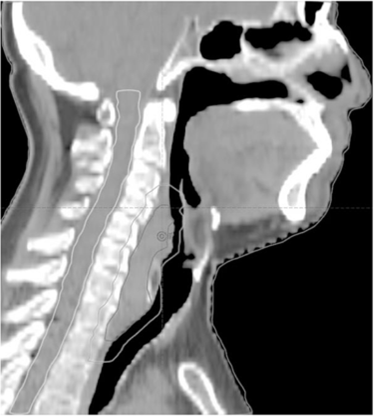
A diagnostic magnetic resonance reported a possible infiltration of the prevertebral fascia. We used a personalised head-shoulder mask for immobilisation and asked the patient not to swallow during the CT acquisition as well as during treatment. We performed two CT scans: one for the identification of isocentre and the other with radiopaque markers on the isocentre’s projection that we use for radiotherapy-planning (see Figure 1).
Patient’s #1 planning CT, sagittal view.

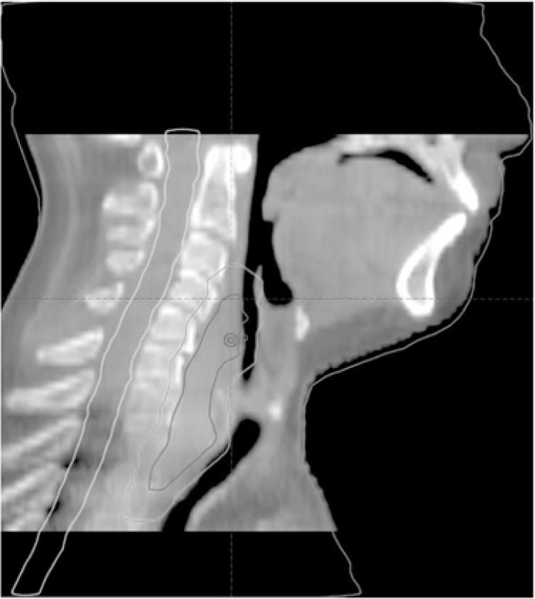
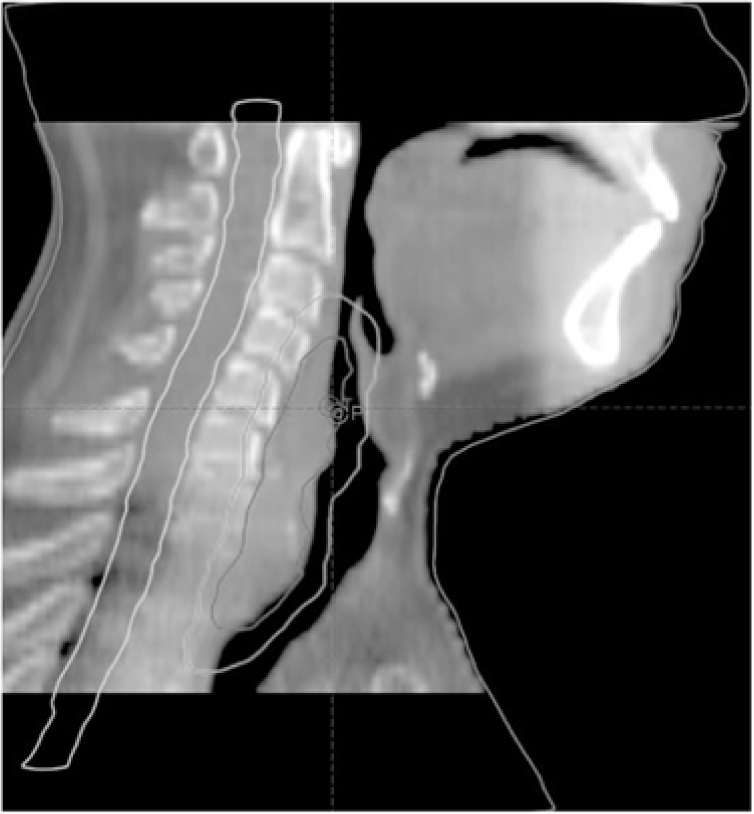
On the first cone beam CT (CBCT) acquired before the first treatment, the bony references were correctly overlapping those of the reference CT, while the gross tumour volume (GTV) was displaced 5 mm caudally. The planning target volume hardly included the displaced GTV (see Figure 2). After three CBCT on consecutive days showing similar results, we performed a second treatment plan using the same mask and the previously identified isocentre. CT scans revealed that notwithstanding the probable fixation of the tumour mass to the prevertebral fascia, a displacement of the larynx in craniocaudal direction had also involved the GTV. In the planning CT, we observed that the tongue was in contact with the palate, and probably exerting a pressure caused the elevation of the larynx (see Figure 1). In the second CT, we asked the patient to relax the larynx and tongue in a natural rest position. The resulting CT images were perfectly matching the anatomy detected with the three CBCT. In conclusion, this error was entirely due to the forced contraction of the tongue over the palate, causing the elevation of the larynx and the tumour displacement from the position it had in a neutral position. We re-planned on the new CT scan, and the control CBCT showed agreement on bony markers and soft tissues (see Figure 3).
Patient’s #1 CBCT with tumour displacement after bone alignment.

Patient’s #1 CBCT after re-planning (alignment on bone references and the corresponding soft tissue inclusion in planned volumes).

Case study 2
Patient #2 is a 68-year-old man with a macroscopic recurrence of prostatic adenocarcinoma anterior to the rectal wall.
Before the CT scan, the patient was asked to perform a rectal enema and to maintain the bladder half-filled. We opacified the urethra introducing contrast medium with a urinary catheter and inserted a small rectal tube into the first five centimetres of the anus to better identify these structures. The legs were positioned using a leg and foot holder immobilisation device.
In all the CBCT of the first 5 days of treatment, we observed a systematic rotational error of 3 degrees. Analysing the images, the right gluteus muscle appeared thicker than the contralateral. We repositioned the patient on the CT scan placing only a small rectal tube. We asked the patient to relax the musculature of the legs and the gluteus consciously.
We used the left and central tattoos as references of the isocentre and needed to identify a new point as isocentre’s projection on the right side because the gluteus was, this time, relaxed. After re-planning on the new CT, the position of the patient, using the new tattoo, was reproducible at all the following set-up controls.
Conclusions
We believe that the first skill required for the personnel involved in the simulation phase is to communicate with the patient making them aware of the importance of the degree of relaxation and intervening to facilitate its achievement. Another skill is the ability to detect muscular contraction or asymmetry on the scan used to identify the isocentre. It takes time and attention reviewing the images of the first CT acquisition before dealing with the identification of the isocentre. In the case of a prompt acknowledgement of the error, we would have avoided the need for a second CT of the patients and an additional workload of the physician and the physicist.
Author ORCIDs
A. Merlotti 0000-0002-6412-6660
Acknowledgements
None.
Conflicts of interest
None.
Author’s contribution
AM and AR have participated in the article preparation. All authors have approved the final article.