Implications
This research identifies nutritional-independent mechanisms that may affect the growth of diseased or stressed nursery pigs. These findings are important because they further our understanding of how poor swine health impacts productivity and they also reaffirm the importance of maintaining a healthy herd. The results from this research may lead to the development of useful biomarkers for assessing herd health or the selection of pigs with desirable immunological traits.
Introduction
IGF-1 is an important anabolic hormone that mediates growth and development in animals. Increases in serum IGF-1 are associated with enhanced growth performance and better carcass traits in livestock species (Clutter et al., Reference Clutter, Spicer, Woltmann, Grimes, Hammond and Buchanan1995; Owens et al., Reference Owens, Gatford, Walton, Morley and Campbell1999; Li et al., Reference Li, Wu, Ren, Zhao and Liu2013). Circulating IGF-1 is primarily produced by hepatocytes and secreted into the bloodstream where it travels to brain, muscle, bone and cartilage to stimulate cellular proliferation and differentiation (Dupont and Holzenberger, Reference Dupont and Holzenberger2003). The most abundant IGF-binding protein (IGFBP) in the blood is IGFBP-3 that sequesters IGF-1 to prolong its bioavailability and stability during transportation throughout the bloodstream (Mohan and Baylink, Reference Mohan and Baylink2002). Hepatic expression of IGF-1 is predominantly regulated by growth hormone (GH) that binds to growth hormone receptors (GHR) on hepatic tissue and activates a cascade of reactions leading to increased IGF-1 expression (Bichell et al., Reference Bichell, Kikuchi and Rotwein1992).
Previous research has indicated that serum concentrations of IGF-1 are associated with certain infectious diseases and health ailments in animals. In pigs, it has been observed that reduced concentrations of serum IGF-1 are associated with infection of Mycoplasma hyopneumoniae, porcine reproductive and respiratory syndrome virus (PRRSV; Roberts and Almond, Reference Roberts and Almond2003) and Salmonella typhimurium (Jenkins et al., Reference Jenkins, Turner, Dritz, Durham and Minton2004). However, the nutritional-independent mechanisms responsible for these changes are not well understood.
Recent findings indicate there is a complex interaction between the IGF system and the cytokine immune response (Thissen and Verniers, Reference Thissen and Verniers1997; Savage, Reference Savage2013; Suh et al., Reference Suh, Zhao, Derico, Choi and Lee2013). Previous studies have demonstrated that production of inflammatory cytokines and acute-phase proteins (APPs) are inversely correlated with IGF-1, IGFBP-3 and GHR measurements in various tissues (Thissen and Verniers, Reference Thissen and Verniers1997; Kim et al., Reference Kim, Lee, Lee and Lee2012; Suh et al., Reference Suh, Zhao, Derico, Choi and Lee2013). Further research has demonstrated that tumour necrosis factor-α (TNF-α) and interleukin (IL)-1β can inhibit in vitro expression of IGF-1 and GHR in hepatocytes, even in the presence of GH (Wolf et al., Reference Wolf, Böhm, Brand and Kreymann1996; Thissen and Verniers, Reference Thissen and Verniers1997). These cytokines and APPs may play a critical role in regulating the IGF system in diseased pigs, independent of nutritional parameters (Roberts and Almond, Reference Roberts and Almond2003). It is predicted that elevated levels of inflammatory cytokines and APPs, as a result of disease or stress, may compromise piglet development owing to unfavourable interaction with the IGF system. However, there is still very limited knowledge of the interaction between cytokines, APPs and the IGF system in pigs with clinical and subclinical diseases. The objective of this study is to determine whether the gene expression of growth factors (IGF-1, IGFBP-3 and GHR) in the liver is associated with the gene expression of immunomodulators (C-reactive protein (CRP), serum amyloid A (SAA), haptoglobin (Hp), interferon (IFN)-α, IFN-γ, TNF-α, IL-1β, IL-6, IL-10 and IL-18) in the liver as well as with the presence of Salmonella spp., Lawsonia intracellularis, Brachyspira spp., enterotoxigenic Escherichia coli (ETEC), methicillin-resistant Staphylococcus aureus (MRSA), swine influenza virus (SIV) and PRRSV in nursery pigs from commercial farms.
Material and methods
Experimental design
The treatment of all research animals was in accordance with the Ontario Animals for Research Act and the use of animals in this study was approved by the Animal Care Committee at the University of Guelph. The study recruited 168 pigs from 8 commercial swine farms in southern Ontario. Seven sows were randomly enrolled at each farm and three pigs of small, medium and large sizes were selected from each sow. Each pig was assigned an identification tag and followed from weaning to 5 weeks post-weaning. All male piglets recruited in this study had been castrated. Producers were administered a survey to collect information about the enrolled sows (farrowing date, parity, litter size, numbers of live born, stillborn), farm management (pig flow, number of gilts, sows, nursery pigs and grower-finishers, and type of farrowing room floor), health status (recent diseases, mortality in different stage of production), type of feed, vaccination and in-feed drug use. Individual pig weights were measured at weaning and at 5 weeks post-weaning, and blood and faeces were collected at weaning, 2 weeks post-weaning and 5 weeks post-weaning. The average daily gain (ADG) was determined as the difference between weights (weaning and 5 weeks post-weaning), divided by the days between measurements.
The pigs were transported to the postmortem facilities at the University of Guelph where they were euthanized at 5 weeks post-weaning. All pigs were transported during the morning in early autumn for an approximate duration of 20–60 min. Samples from the liver, intestinal lymph nodes, ileum and spleen were placed immediately into RNA later (Ambion, Grand Island, NY, USA) and allowed to sit at 4°C for 24 h before being stored at −80°C. Separate portions of the ileum and colon were also collected to test for the presence of L. intracellularis and Brachyspira spp., respectively. In addition, nasal swabs were collected before euthanasia to be tested for SIV and MRSA.
Detection of viral and bacterial agents
For the isolation of SIV, the nasal swab samples were filtered with 0.45 μm pore size filter (Millipore, Billerica, MA, USA) and 100 μl of the swab preparation was inoculated onto two wells of a tissue culture of Madin-Darby canine kidney continuous cell line (105 cells) in 24 well plates containing Dulbecco Modified Eagle Medium (DMEM/F12), 10% of foetal bovine serum, penicillin 100 U/ml and 100 μg/ml streptomycin. The inoculum was allowed to absorb at 37°C for 60 min and fresh DMEM/F12 containing 2 μg/ml of TPCK-trypsin, bovine serum albumin 0.2%, and HEPES 25 mM were added. Tissue cultures were harvested upon observation of cytopathic effect. SIV was purified from cell culture by centrifugation and ultracentrifugation in a Beckman 16.250 rotor at 6000 r.p.m. for 20 min and at 26 000 r.p.m. for 90 min, respectively. Viral RNA was isolated using the QIAamp Viral RNA Mini Kit (Qiagen, Toronto, Ontario, Canada). The cDNA was amplified using random primers and 200 U Superscript III reverse transcriptase (Invitrogen, Burlington, Ontario, Canada), following manufacturer’s instructions. Reverse transcriptase-PCR (RT-PCR) was performed with 2 μl of cDNA in 50 μl reaction mixture. The 600 bp long fragment of HA gene was amplified by PCR with primer set: H3For (5’-GCTGGTTCAAAGTTCCTCAA-3’) and H3Rev (5’-TGAGCTTTTCCCACTTTG-3’). The first cycle of the amplification programme consisted of a 2 min period at 95°C and was followed by 35 cycles of 95°C for 20 s, 52°C for 10 s and 72°C for 30 s. The coded protein was identified using BLAST analysis with the GenBank database and degree of identities and similarity was identified using FASTA3 (EMBL-EBI, Hinxton, Cambridge, UK).
Detection of PRRSV in serum samples using RT-PCR was completed using Tetracore real time next generation PCR primers and FAM/MGB probes according to kit instructions (Tetracore Inc., Gaithersburg, MD, USA) and Applied Biosystems 7500 real time thermocycler (Applied Biosystems, Burlington, Ontario, Canada). The test cannot distinguish between wildtype strains and modified vaccine strains.
For culturing of Salmonella spp., faecal samples were enriched in tetrathionate broth and incubated overnight at 41°C. Then a loopful of the enrichment was streaked onto billiants green sulfa agar plates and incubated at 35°C overightnight. Salmonella-like colonies were further identified using matrix-assisted laser desorption/ionization time-of-flight mass spectroscopy and then sent for serotyping to the Salmonella OIE reference laboratory at the Public Health Agency of Canada, Guelph, Ontario, Canada.
Brachyspira spp. were cultured on selective trypticase soy agar plates containing 5% sheep blood, 400 μg/ml spectinomycin, 25 μg/ml vancomycin and 25 μg/ml colistin. Plates were incubated at 41°C in GENbox anaerobic jars (BioMérieux Canada, St-Laurent, Quebec, Canada) and monitored for growth of Brachyspira spp. every 2 days for 10 days. Bacterial growth was then washed off the plates with phosphate-buffer saline, and DNA was extracted using DNeasy Blood and Tissue kit (Qiagen) and QIAcube extraction robot following manufacturer’s instructions. Presence of Brachyspira spp. was determined with PCR using genus specific nox gene primers and a probe; BrachysppF284, [5’-AAAGCTACAGATCCTAATTGTCA-3’], BrachysppR723 [5’-CAGCACCAACTACCATAACT-3’] and FAM-5’-TTGCTACTGGTTCTTGGCCT-3’-BBQ. The species of positive Brachyspira spp. PCR samples were then determined using RT-PCR, following standard operating procedures used at the Animal Health Laboratory, University of Guelph.
For L. intracellularis detection, DNA was extracted from ileum samples using FastDNA®kit (MP Biomedicals, Santa Ana, CA, USA) and detected using PCR, following manufacturer’s instructions. Isolation and detection of ETEC was completed following standard operating procedure used at the Animal Health Laboratory, University of Guelph. The isolation and detection of MRSA from nasal swabs was performed as previously described by Weese et al. (Reference Weese, Rousseau, Deckert, Gow and Reid-Smith2011).
RNA extraction from liver tissue
The RNA extraction was completed as previously described (Slifierz et al., Reference Slifierz, Friendship, de Lange, Rudar and Farzan2013). Briefly, RNA was extracted from 74 liver tissue samples from pigs on four farrow-to-finish farms using a Qiagen RNeasy Mini Kit (Qiagen). Liver tissue (30 mg) was added to RLT buffer (600 µl) and homogenized using a tissue homogenizer (PowerGen 125; Fisher Scientific, Toronto, Ontario, Canada). The extraction process was then completed according to the manufacturer’s manual (Qiagen). The purity and concentration of the eluted RNA was determined using the Thermo Scientific NanoDrop 8000 Spectrophotometer (Fisher Scientific). An Amplification Grade DNase I kit (Sigma-Aldrich, Oakville, Ontario, Canada) was used to remove any genomic DNA from the extracted RNA. RNA was re-extracted and thoroughly assessed if there were any indications of degradation before or after DNase treatment. The integrity of the RNA samples was evaluated using an Agilent 2100 Bioanalyzer (Agilent Technologies, Mississauga, Ontario, Canada).
Analysis of gene expression with RT-qPCR
The primers for IL-1β, IL-10, IFN-α and IFN-γ were previously designed by Collado-Romero et al. (Reference Collado-Romero, Arce, Ramírez-Boo, Carvajal and Garrido2010). All other primers were designed by Slifierz et al. (Reference Slifierz, Friendship, de Lange, Rudar and Farzan2013). Primer designs and efficiencies are listed in Supplementary Table S1. The reverse transcription of cDNA and the quantitative polymerase chain reaction (RT-qPCR) assay used to determine relative gene expression was completed according to a prior protocol without deviation (Slifierz et al., Reference Slifierz, Friendship, de Lange, Rudar and Farzan2013). The synthesis of cDNA and the RT-qPCR assay were completed by the Advanced Analysis Centre’s Genomic Facility at the University of Guelph using a High Capacity cDNA Reverse Transcription Kit (Applied Biosystems) and a StepOnePlus™ Real-Time PCR System (Applied Biosystems), respectively. Glyceraldehyde 3-phosphate dehydrogenase (GAPDH) was used as the only reference gene for internal normalization of gene expression data in this experiment because of its reliability and validation as a single reference gene in previous studies (Svobodová et al., Reference Svobodová, Bílek and Knoll2008; Li et al., Reference Li, Cao, Zhao, Yang, Fan and Yang2009; Li et al., Reference Li, Wu, Ren, Zhao and Liu2013; Slifierz et al., Reference Slifierz, Friendship, de Lange, Rudar and Farzan2013). The stability of GAPDH expression was assessed in the present study using BestKeeper software (Pfaffl et al., 2004).
Statistical analysis
The crude cycle threshold (C q ) values generated from the RT-qPCR assay were normalized using the C q values of GAPDH to produce ΔC q values (Supplementary Table S1). Gene expression was then dichotomized as either high or low using the median ΔC q value of each gene. Dichotomization was performed to ensure coherent and interpretable coefficients could be generated upon modelling of the data.
The hepatic gene expression of IGF-1, IGFBP-3 and GHR were modelled as dependent variables in three individual logistic regression models. The hepatic gene expression of CRP, Hp, SAA, IFN-α, IFN-γ, TNF-α, IL-1β, IL-6, IL-10 and IL-18 were used as independent variables to build the model. Additional extraneous variables used include post-weaning ADG, weight at weaning, age at weaning, number of live births per litter, number of stillbirths per litter, weight at 9 weeks, piglet sex, as well as the presence of Salmonella spp., PRRSV and MRSA. However, L. intracellularis, Brachyspira spp., ETEC and SIV were not included in the model owing to absence or inadequate number of pigs carrying these organisms.
The linearity of independent variables was assessed using a smoothed locally weighted scatterplot. None of the variables were transformed apart from dichotomization of the gene expression values (ΔC q ), as previously discussed. The initial models were developed through automated stepwise screening. Owing to the large number of variables, stringent conditions were used to avoid identifying false associations; variables were first included in the model if P<0.05 and then excluded from the model if P>0.10. These initial models were then assessed and further developed manually. Farm-level clustering was controlled for by modelling farm as a random effect unless the intraclass correlation coefficient (ICC) was not significantly different from zero (P>0.05). All variables that were excluded during stepwise selection were manually added to the model to determine if they were statistically significant or had a confounding effect on the other variables. Two-way interaction terms were created between each variable in the model and tested for statistical significance. Variables were manually added to the model if they were found to be statistically significant, had a confounding effect of >30% or were part of a statistically significant interaction term. If a variable was removed from the model, a likelihood-ratio test was performed to determine whether removal of the variable significantly changed the model.
It was determined that all models had binary covariate patterns. If the farm variable was not modelled as a random effect the goodness of fit was determined by the Hosmer–Lemeshow test, and if the farm variable was modelled as a random effect the normality of the BLUPs of the random effect and affiliated residuals were evaluated. Furthermore, diagnostic analysis revealed no outliers or extremely influential observations. One pig was excluded because of missing data. In addition, univariable analysis of continuous farm-level parameters (herd size and pathogen prevalence) was assessed by Pearson’s correlation coefficient as all variables showed a normal distribution and linear relationship. All statistical analysis was completed using STATA 10.0 (Stata Corporation, College Station, TX, USA). Variables with a P<0.05 were considered statistically significant and non-significant variables with a P<0.10 were considered to be suggestive of an association.
Results
Descriptive statistics
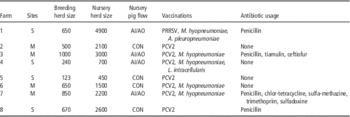
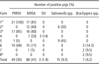
Variation in management practices, herd size and therapeutic interventions on the studied farms is described in Table 1. PRRSV, MRSA, SIV, Salmonella spp. and Brachyspira spp. were present on 4 (50%), 6 (75%), 1 (12.5%), 2 (25%) and 3 (37.5%) farms, respectively. L. intracellularis and ETEC were not detected on any of the farms. The prevalence of these pathogens among individual farms and the mean prevalence among pigs are displayed in Table 2. None of the pigs in this study exhibited clinical signs of infection. Serotyping of Salmonella isolates revealed that all isolates from farm 2 were Salmonella infantis and all isolates from farm 5 were Salmonella worthington. All isolates of Brachyspira spp. were determined to be Brachyspira murdochii. Serum titres of PRRSV on farms 1, 3, 5 and 6 were 3.4×10⁶ TCID50/ml, 4.5×10⁵ TCID50/ml, 1.4×10³ TCID50/ml and 2.6×10⁷ TCID50/ml, respectively. In addition, it was observed that farms 1, 3 and 6, which had the highest viral titres and prevalence of PRRSV, were also the farms that had the highest prevalence of MRSA. The positive correlation between farm-level prevalence of PRRSV and MRSA was significant (R 2=0.635; P=0.018). Farm-level prevalence of PRRSV was also positively correlated with nursery herd size (R 2=0.520; P=0.044). Interestingly, antibiotic usage was not associated with MRSA colonization of swine herds (P=0.98).
Descriptive characteristics of swine operations and management practices on eight farms

M=multiple sites; S=single site; AI/AO=all in/all out; CON=continuous; PRRSV=porcine reproductive and respiratory syndrome virus; PCV2=porcine circovirus type 2.
Prevalence of viral and bacterial pathogen colonization in nursery pigs from eight farms

PRRSV=porcine reproductive and respiratory syndrome virus; MRSA=methicillin-resistant Staphylococcus aureus; SIV=swine influenza virus.
a Gene expression analysis was completed on liver tissue samples from piglets originating from these farms.
Factors associated with IGF-1 expression in the liver
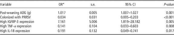
Multivariable analysis for factors associated with hepatic expression of IGF-1 is shown in Table 3. Pigs with a higher post-weaning ADG had significantly increased odds of elevated IGF-1 expression in the liver. The pig with the highest ADG (556 g/day) was 1073 (95% CI: 16.02–71 892) times more likely to have higher IGF-1 expression than the pig with the lowest ADG (140 g/day; P<0.01). Pigs that had higher expression of IGFBP-3 were more likely to have increased IGF-1 expression, and pigs that had higher expression of the cytokines TNF-α or IL-18 were less likely to have increased hepatic expression of IGF-1. Further, pigs colonized with PRRSV were less likely to have increased IGF-1 expression in the liver. None of the extraneous variables behaved as a confounder and all two-way interaction terms were not statistically significant. The ICC was not significantly different from zero (ICC=1.0×10⁻⁷; P>0.05).
Analysis of gene expression of IGF-1 in liver tissue from 73 pigs from 4 farms; multivariable logistic regression model

*Odds of increased IGF-1 expression in the liver.
OR=odds ratio; CI=confidence interval; ADG=average daily gain; PRRSV=porcine reproductive and respiratory syndrome virus; IGFBP=IGF-binding protein; TNF=tumour necrosis factor-α; IL=interleukin.
Factors associated with IGFBP-3 expression in the liver
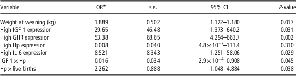
The multivariable analysis for factors associated with hepatic expression of IGFBP-1 is described in Table 4. Pigs with a higher weight at weaning were more likely to have increased hepatic expression of IGFBP-3 (P<0.05). The pig with the highest weight at weaning (14.0 kg) was 1027 (95% CI: 3.52–299 908) times more likely to have increased IGFBP-3 expression than the pig with the lowest weight at weaning (3.1 kg; P<0.05). Pigs with increased hepatic expression of IGF-1, GHR or IL-6 were more likely to have increased hepatic expression of IGFBP-3 (P<0.05). The extraneous variables showed no confounding effect on the model. However, two statistically significant interactions were detected and retained in the model; an interaction between Hp expression and IGF-1 expression, and an interaction between Hp expression and the number of live births per litter. At low Hp expression, increased IGF-1 mRNA expression was associated with increased IGFBP-3 expression (OR=29.65, P<0.05). However, IGF-1 expression was not associated with IGFBP-3 expression (P=0.54) when Hp expression was high. Live births per litter was not significantly associated with IGFBP-3 expression (P=0.60) when Hp expression was low, but it was significantly associated with IGFBP-3 expression (OR=2.57, P<0.01) when Hp expression increased. The ICC was not significantly different from zero (ICC=6.5×10⁻⁸; P>0.05).
Analysis of gene expression of IGFBP-3 in liver tissue from 73 pigs from 4 farms; multivariable logistic regression model

*Odds of increased IGFBP-3 expression in the liver.
IGFBP=IGF-binding protein; OR=odds ratio; CI=confidence interval; GHR=growth hormone receptors; Hp=haptoglobin; IL=interleukin.
Factors associated with GHR expression in the liver
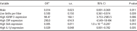
Multivariable analysis for factors associated with hepatic expression of GHR is shown in Table 5. Pigs with increased expression of IGFBP-3 or CRP were more likely to have increased expression of GHR (P<0.05). Barrows, pigs from litters with more live births, and pigs with increased expression of SAA or IL-1β were less likely to have increased GHR expression (P<0.05). It was also observed that older pigs and increased TNF-α expression had a tendency to be associated with increased expression of GHR (P<0.10). The odds ratios for these variables are listed in Table 5. None of the extraneous variables had a confounding effect and there were no statistically significant two-way interaction terms. Data clustering at the farm level was controlled for in the model as the ICC was significantly different from zero (ICC=0.57, P<0.01).
Analysis of gene expression of GHR in liver tissue from 73 pigs from 4 farms; multivariable random-effect logistic regression model

*Odds of increased GHR expression in the liver.
GHR=growth hormone receptors; OR=odds ratio; CI=confidence interval; IGFBP=IGF-binding protein; CRP=C-reactive protein; SAA=serum amyloid A; IL=interleukin.
Discussion
The results of this study demonstrate an overall inverse relationship between the hepatic expression of the IGF system and the inflammatory immune response. The IGF system appeared to be inhibited or disrupted by several proinflammatory cytokines and APPs, including IL-1β, IL-18, TNF-α, SAA and Hp. These findings support the speculation that elevated levels of proinflammatory cytokines and APPs, as a result of disease or stress, may hinder growth and development owing to unfavourable interaction with the IGF system or affiliated pathways (Roberts and Almond, Reference Roberts and Almond2003; Savage, Reference Savage2013).
The association between IGF-1 and post-weaning ADG, and between IGFBP-3 and weaning weight was expected and is consistent with previous research, which has demonstrated that these proteins are indicative of growth performance and carcass traits in pigs (Clutter et al., Reference Clutter, Spicer, Woltmann, Grimes, Hammond and Buchanan1995; Owens et al., Reference Owens, Gatford, Walton, Morley and Campbell1999; Li et al., Reference Li, Wu, Ren, Zhao and Liu2013).
Increased IGF-1 expression was inversely associated with increased expression of two potent proinflammatory cytokines; TNF-α and IL-18. The inverse relationship with TNF-α is consistent with studies which demonstrate that TNF-α can inhibit IGF-1 production of in vitro hepatocytes, even in the presence of GH stimulation (Wolf et al., Reference Wolf, Böhm, Brand and Kreymann1996; Thissen and Vernier, Reference Thissen and Verniers1997). Although limited research exists that examines the association between IL-18 and the IGF system, previous findings do indicate that IL-18 is negatively associated with IGF-1 and growth (Skopiński et al., Reference Skopiński, Rogala, Duda-Król, Lipińska, Sommer, Chorostowska-Wynimko, Szaflik, Partyka and Skopińska-Rózewska2005; Netea et al., Reference Netea, Joosten, Lewis, Jensen, Voshol, Kullberg, Tack, van Krieken, Kim, Stalenhoef, van de Loo, Verschueren, Pulawa, Akira, Eckel, Dinarello, van den Berg and van der Meer2006). Future research should explore the mechanisms by which TNF-α and IL-18 may interact with hepatic expression of IGF-1.
Hepatic expression of IGF-1 and IGFBP-3 were associated when Hp expression was low, but this association was not significant when Hp expression increased. This finding is consistent with experiments where administration of recombinant IGF-1 and IGFBP-3 to rats was associated with decreased plasma concentrations of Hp (Jeschke et al., Reference Jeschke, Herndon and Barrow2000). It appears that increased levels of IGF-1 and IGFBP-3 may inhibit Hp production in the liver. The reason for the observed interaction between Hp and live births per litter is unclear. This association is questionable as sow characteristics (live births per litter and still births per litter) were not the primary targets in this investigation and therefore sow-level clustering of data was not controlled for in the analysis. However, larger litter size is a contributor of compromised pig welfare (Rutherford et al., Reference Rutherford, Baxter, D’Eath, Turner, Arnott, Roehe, Ask, Sandøe, Moustsen, Thorup, Edwards, Berg and Lawrence2013) and previous studies have demonstrated that high Hp levels are a strong indicator of decreased welfare on pig farms (Piñeiro et al., Reference Piñeiro, Piñeiro, Morales, Carpintero, Campbell, Eckersall, Toussaint, Alava and Lampreave2007a and Reference Piñeiro, Piñeiro, Carpintero, Morales, Campbell, Eckersall, Toussaint and Lampreave2007b).
Elevated IGFBP-3 expression was associated with increased expression of IL-6, which plays an important role in the anti-inflammatory response (Tilg et al., Reference Tilg, Dinarello and Mier1997). It has previously been observed that IL-6 does stimulate production of IGF-1 in hepatocytes, which is likely caused by a cross-reaction with a known receptor of the same superfamily and through stimulation of a transduction pathway factor shared between IL-6 and the IGF system (Thissen and Verniers, Reference Thissen and Verniers1997). This may explain the positive association between IL-6 and IGFBP-3 that was observed in this study.
Expression of GHR in the liver was inversely associated with expression of SAA and IL-1β. These associations may be explained by other studies, which have demonstrated that GH reduces production of inflammatory agents, including IL-1β and SAA (Jeschke et al., Reference Jeschke, Herndon and Barrow2000; Lu et al., Reference Lu, Kumar, Fan, Sperling and Menon2010). It is likely that GH concurrently stimulates GHR expression and inhibits inflammatory cytokine expression in the liver leading to this observed association between GHR and decreased expression of IL-1β and SAA. It was also observed that male pigs were less likely to have increased GHR expression than female pigs. Similar results were reported with rats wherein the males had significantly reduced hepatic expression of GHR when compared with females, and this difference appears unrelated to castration and sex hormones (Ahlgren et al., Reference Ahlgren, Norstedt, Baumbach and Mode1995; Baumbach and Bingham, Reference Baumbach and Bingham1995). The reason for this differential hepatic expression of GHR between the sexes is unknown and further research is needed to address this discrepancy. Pigs from larger litters were also less likely to have elevated GHR expression, although this association is questionable because of the potential sow-level clustering of data.
It is unclear whether pigs that tested seropositive for PRRSV in the current study had been infected with PRRSV, vaccinated with an attenuated strain or both. However, research has demonstrated that the change in circulating GH is very similar in vaccinated and unvaccinated pigs exposed to PRRSV (Borghetti et al., Reference Borghetti, Saleri, Ferrari, Morganti, De Angelis, Franceschi, Bottarelli and Martelli2011). In this investigation, pigs seropositive for PRRSV were less likely to have increased IGF-1 expression compared with pigs that were seronegative for PRRSV. Previous research has demonstrated that pigs co-infected with PRRSV and M. hyopneumoniae have significantly reduced serum concentrations of IGF-1 despite similar feed intake when compared with a control group (Roberts and Almond, Reference Roberts and Almond2003). This finding suggests that non-nutritional factors, such as the inflammatory response, may contribute to the regulation of the IGF system in pigs (Robert and Almond, Reference Roberts and Almond2003). It is possible that early retardation of the IGF system because of nutritional and non-nutritional factors is responsible for the reduced BW observed in pigs that are seropositive for PRRSV (Regula et al., Reference Regula, Lichtensteiger, Mateus-Pinilla, Scherba, Miller and Weigel2000). Furthermore, the prevalence of PRRSV in nursery pigs was positively correlated with nursery herd size, which may indicate that larger swine operations have a higher risk of contracting PRRSV.
The current investigation also suggests that MRSA colonization does not elicit a demonstrable immune response or interact with the IGF system in pigs. This was expected as many swine herds carry this bacterium without incident and it appears to behave as a commensal bacterium (Khanna et al., Reference Khanna, Friendship, Dewey and Weese2008). However, further research should explore whether certain types of porcine MRSA are associated with a subclinical immune response. It was also observed that farm-level prevalence of MRSA is positively correlated with PRRSV. This may be the result of poor farm biosecurity or an indication of co-colonization as it has previously been shown that PRRSV reduces the bactericidal activity of porcine macrophages (Thanawongnuwech et al., Reference Thanawongnuwech, Thacker and Halbur1997). In addition, MRSA was present on farms, regardless of antibiotic usage, which suggests that other factors are contributing to its persistence on pig farms.
S. infantis was not associated with hepatic expression of the IGF system despite previous studies that identified reduced serum IGF-1 and IGFBP-3 in pigs colonized with Salmonella (Jenkins et al., Reference Jenkins, Turner, Dritz, Durham and Minton2004). The lack of association observed in the present investigation may be because of the limited number of liver samples used for RT-qPCR from pigs colonized with Salmonella (n=6). RT-qPCR was not performed on liver samples from pigs colonized with S. worthington. Both S. infantis and S. worthington recovered from pigs in this study are rarely reported from clinical salmonellosis in pigs, and it is possible that the effect of Salmonella on the IGF system in pigs is serotype specific.
One of the limitations of this study is that transportation of the pigs to the postmortem facilities may have affected expression of some immune-related genes, particularly the APPs. However, transportation is unlikely to be a confounding variable as there is no evidence to suggest that it independently affects growth factor expression. Transportation likely acted as an intervening variable that introduced variation, although all pigs were transported under relatively similar conditions.
Another limitation of this study emerged from the qPCR method used for quantifying relative gene expression; although most of the genes had >95% efficiency, those genes that performed with <90% efficiency are questionable and the results may be useful for hypothesis generation. In addition, dichotomization of ΔC q values could create undue bias, although it was necessary for multivariable analysis and coherent interpretation of the data. Further research should explore the magnitude to which growth factor gene expression is affected by immunomodulators using RNA copy numbers.
Overall, hepatic expression of the IGF system was inversely associated with hepatic expression of several cytokines and APPs, which suggests that an inflammatory response, perhaps as a result of disease, vaccination or stress, can disrupt the balance of the IGF system. However, future investigations need to assess whether prolonged dysregulation of the IGF system because of sustained increase in cytokines and APPs causes significant changes in production outcomes. Differential expression of the IGF system may result in changes to growth performance, carcass quality or increase the risk of health ailments such as lameness. Lastly, the genetic composition of pigs may partly contribute to the transcriptional expressivity of the IGF system and immune response, which was not investigated in the current study, and should be investigated thoroughly.
Acknowledgements
We appreciate the assistance and support of Jing Zhang, Angela Holliss and Joyce Rousseau who helped with the laboratory work in this study.