INTRODUCTION
The 4 major human gastrointestinal (GI) nematodes, Ascaris lumbricoides, Trichuris trichiura, Ancylostoma duodenale and Necator americanus, infect millions of people throughout the world. Although these parasites rarely cause acute infections, the resulting chronic infections are serious and produce a greater burden on life (39·0 million DALYs; DALYs=disability-adjusted life years) than, for example, malaria (35·6 million DALYs) (Chan, Reference Chan1997; Molyneux et al. Reference Molyneux, Hotez and Fenwick2005). GI nematode infections are also recognized as being the cause of, economically, the most important diseases of livestock, particularly sheep and goats (Nieuwhof and Bishop, Reference Nieuwhof and Bishop2005).
At present, the most popular means for controlling GI nematodes is by the use of anthelmintic drugs (Albonico et al. Reference Albonico, Crompton and Savioli1999). However, widespread anthelmintic resistance has developed amongst the GI nematodes of livestock, especially in Australia, New Zealand, South America and South Africa, where it is increasing to the extent that sheep farming is now becoming impossible in some areas due to the occurrence of triple resistance to all 3 classes of anthelmintic (van Wyk et al. Reference van Wyk, Malan and Randles1997), particularly on farms where both sheep and goats are grazed (Waller, Reference Waller1986; Varady et al. Reference Varady, Praslicka, Corba and Vesely1993; Mwamachi et al. Reference Mwamachi, Audho, Thorpe and Baker1995; Coles et al. Reference Coles, Warner and Best1996; Hong et al. Reference Hong, Hunt and Coles1996; Gill and Lacey, Reference Gill and Lacey1998). Resistance is currently less of a problem in the control of GI nematodes of humans, but similar problems will soon be faced if lessons are not learnt from the veterinary experience (Coles, Reference Coles1995; Geerts and Gryseels, Reference Geerts and Gryseels2000); falling efficacies of the drugs normally used against human intestinal nematodes are already being seen in Africa and Australia (de Clercq et al. Reference de Clercq, Sacko, Behnke, Gilbert, Dorny and Vercruysse1997; Reynoldson et al. Reference Reynoldson, Behnke, Pallant, MacNish, Gilbert, Giles, Spargo and Thompson1997; Albonico et al. Reference Albonico, Bickle, Ramsan, Montresor, Savioli and Taylor2003). Hence, there is an urgent need for the development of new anthelmintics with novel mechanisms of action.
For centuries, plant extracts have been used in traditional medicine for the treatment of nematode infections in tropical communities (Waller et al. Reference Waller, Bernes, Thamsborg, Sukura, Richter, Ingebrigtsen and Höglund2001). Of these, perhaps the best studied are extracts from the fruit and stem of pineapple (Ananas comosus) (Berger and Asenjo, Reference Berger and Asenjo1939) and the latex from unripe fruits such as papaya (Carica papaya) (Berger and Asenjo, Reference Berger and Asenjo1940) and fig (Ficus species) (Robbins, Reference Robbins1930), which have been found to have anthelmintic activity when administered to pigs infected with Ascaris suum (Satrija et al. Reference Satrija, Nansen, Bjorn, Murtini and He1994) and humans infected with various GI nematodes (Hansson et al. Reference Hansson, Veliz, Naquira, Amren, Arroyo and Arevalo1986). These plants all contain proteolytic enzymes, cysteine proteinases, of the papain family [Sub-family C1A in the Merops database (http://merops.sanger.ac.uk/)] in their latex or fruit (refs in Stepek et al. Reference Stepek, Behnke, Buttle and Duce2004). If the cysteine proteinases from plants are to be developed as new treatments against intestinal nematode infections, their efficacy needs to be assessed reliably and their modes of action need to be understood comprehensively. Early work by Robbins (Reference Robbins1930) indicated that the mechanism of action of the latex from the fig plant, Ficus glabrata, was to digest the cuticle, although the active constituent of the latex was not fully determined. Subsequent work in vitro established that the specific mechanism of action of the plant-derived cysteine proteinases, particularly the pure and crude enzymes from papaya, fig and pineapple, but not from kiwi fruit, against the rodent GI nematode, Heligmosomoides polygyrus, was to rapidly digest the cuticle and thereby kill the nematode (Stepek et al. Reference Stepek, Buttle, Duce, Lowe and Behnke2005). However, whether the same mechanism of action is responsible for loss of worms in vivo has not been demonstrated.
In this study, again exploiting H. polygyrus as our model system, we assessed the in vivo efficacy of papaya latex and related cysteine proteinases, with the specific aims of determining (a) the rate at which the worms are lost following sustained treatment; (b) the effective dose required for anthelmintic activity in terms of the concentration of active enzyme; (c) whether the cysteine proteinases in the plant latex are indeed the active constituents; (d) whether the cysteine proteinases in the plant latex prevent nematode development in the intestinal mucosa; (e) whether the mechanism of action of these enzymes is similar to that observed in vitro; (f) whether treatment with the cysteine proteinases results in adverse side-effects to the host; and (g) whether the pure enzymes have the same in vivo efficacy as the crude latex. This information is essential to evaluate the potential of these enzymes if they are to be developed further as alternatives to the currently available synthetic anthelmintic drugs, and to provide the necessary details for improvements in delivery and formulation.
MATERIALS AND METHODS
Animals
Male C3H mice were purchased from Charles River UK Ltd, at 5 weeks of age and infected at 6 weeks of age. The animals were provided with food and water ad libitum. All animal procedures were carried out under UK Home Office licence number 40/2242 and under the regulations of the Animals (Scientific Procedures) Act 1986.
Parasites
Mice were infected with a suspension of 200 H. polygyrus L3 in 0·2 ml of distilled water on day 0. At days 14, 16, 18, 19, 21, 23 and 25 post-infection, faecal samples were collected from each mouse and the number of eggs present was counted using the McMaster technique modified from Behnke and Parish (Reference Behnke and Parish1979). Briefly, faeces were collected from individual mice, weighed and then 10 ml of saturated NaCl was added. The pellets were broken up for 1 h using a rotary mixer, and then washed through a sieve with 50 ml of saturated NaCl. The eggs were counted using a 2-chamber McMaster slide, each chamber holding a volume of 0·15 ml. The number of eggs per gram of faeces was calculated from the following equation:

From day 18 to day 24 post-infection, each mouse was treated orally once daily with 0·2 ml of the relevant treatment. All mice were killed by exposure to CO2 on day 25 post-infection and the intestines were removed in their entirety and opened longitudinally with a pair of blunt-ended dissecting scissors. Each intestine was placed into a net in a beaker with 50 ml of pre-warmed (37°C) Hanks' Balanced Salt Solution (HBSS), in a 37°C water bath, for approximately 4 h. The adult male and female worms that had collected in the bottom of the beaker were then counted separately under a dissecting microscope.
Enzymes
The crude latex from papaya (Carica papaya) (Sigma), which contains at least 4 cysteine proteinases (Dekeyser et al. Reference Dekeyser, Buttle, Devreese, van Beeumen, Demeester and Lauwers1995), was used throughout this study (5 g dispersed in 8 ml of sterile distilled water, prior to filtration). Purified papain (Merops identifier C01.001; Sigma), the crude latex from Ficus carica (freshly collected from plants maintained in the University of Sheffield Experimental Gardens) and fruit bromelain [from acetone precipitation of the fruit of fresh pineapples (Merops identifier C01.028; Rowan et al. Reference Rowan, Buttle and Barrett1990)] were also examined in vivo. To determine the operational molar concentration of active cysteine proteinase, the samples were titrated with increasing concentrations of the cysteine proteinase-specific inhibitor, L-trans-epoxysuccinyl-leucylamido(4-guanidino) butane (E-64) (Sigma) (Zucker et al. Reference Zucker, Buttle, Nicklin and Barrett1985), as described previously (Stepek et al. Reference Stepek, Buttle, Duce, Lowe and Behnke2005).
Scanning electron microscopy
Following the commencement of treatment on day 18 post-infection, representative mice were killed and dissected, and male and female worms were removed from the entire intestinal tract every hour for 4 h after 3 consecutive days of treatment (on day 20). They were washed in HBSS prior to immediate fixation for 1 h in 2·5% glutaraldehyde in 0·15 m phosphate buffer, pH 7·2. These worms were then prepared for scanning electron microscopy, as described previously (Stepek et al. Reference Stepek, Buttle, Duce, Lowe and Behnke2005).
Histological analysis
In order to examine the effects of papaya latex on the host, mice were killed and the small intestine was removed. One cm sections were cut 10 cm from the stomach (the first 10 cm were used for the worm counts, as described above) and were fixed with either Carnoy's fixative (for mast cells) or 10% formalin buffered saline (for Goblet cells and villi measurements). These fixed samples (with the exception of the sections intended for villi measurements) were processed by dehydration through 70% to 100% ethanol, followed by xylene and paraffin polymer (wax), in preparation for embedding in paraffin wax. The embedded samples were mounted onto small wooden blocks and 5 μm sections were cut using a microtome. These sections were deparaffinized by heating to 50°C before being brought to water through xylene washes and 100% to 70% ethanol. Different stains were then used for the different cell populations. Alcian blue (1% in 3% acetic acid), pH 2·5 for 10 min, 1% periodic acid for 5 min and finally Schiff's Reagent for 10 min were used to stain for Goblet cells. Mast cells were stained with pre-warmed (50°C) Alcian blue (1% in 0·7 m HCl), pH 0·3 for 45 min, rinsed with 0·7 m HCl for 30 sec, before counterstaining with pre-warmed (50°C) 0·5% Safranin O (in 0·125 m HCl), pH 1·0 for 5–10 min. The stained sections were dehydrated through 70% to 100% ethanol and cleared with xylene, prior to mounting in DPX mounting solution. The numbers of mast cells and Goblet cells were counted using a Weible graticule under ×20 magnification.
The sections for measurements of the villi were placed into 50% ethanol for 10 min, followed by tap water for 10 min. Pre-heated (60°C) 1 m HCl was added for 7·5 min before tap water for a further 10 min. The intestinal sections were then stained with Schiff's Reagent for 30 min. Sections of tissue (with villi) were cut using a long cataract knife and placed onto a slide with 45% acetic acid. The lengths of 20 random villi from each section were measured under ×4 magnification using a calibrated eyepiece-measuring graticule.
Statistical analysis
Faecal egg counts were analysed by repeated measures ANOVA (rmANOVA) in SPSS (version 12.0.1) on log10 (EPG+25) transformed data, with time after infection as the within-subject factor, and treatment (papaya latex, water, papain, fruit bromelain, or Ficus carica latex) as the between-subject factor. When the data did not meet the requirements of sphericity (Mauchley's Test of Sphericity), we used the Huynh-Feldt adjustment to the degrees of freedom to interpret significance on the side of caution. Cell counts and villus height were also analysed by rmANOVA with replicate counts as the within-subject factor, and infection and treatment as the between-subject factors. Worm burdens were analysed by 1- or 2-way ANOVAs, as relevant and, whilst we explored models based on transformed data and with negative binomial error structures, the best fit for the adult worm data were models based on the raw worm counts. Where only two groups required comparison, we used the Mann-Whitney U test in SPSS. All parametric models were assessed for goodness of fit by R 2, and residuals were checked for normal or negative binomial distribution, as relevant. The final statistical models fitted to the data are explained more comprehensively in the legends. For Fig. 2, a sigmoidal dose-response curve was fitted with an upper limit of 100 and a lower limit of zero in GraphPad Prism (version 4), and the log10 EC50 and log10 EC95 were calculated together with 95% confidence limits (CL). The model fit was assessed by R 2.
RESULTS
Papaya latex shows substantial anthelmintic activity in vivo
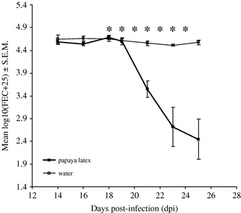
In order to determine the efficacy of crude papaya latex against intestinal nematodes in vivo, we administered papaya latex daily for 7 days (133 nmol active cysteine proteinase/mouse at each treatment), from days 18–24 post-infection, to 5 male C3H mice which had been infected with 200 L3 of H. polygyrus on day 0. The parasite faecal egg output (FEC) from each mouse was counted on days 14, 16, 18, 19, 21, 23 and 25 post-infection. Fig. 1 shows that already by day 4 of consecutive treatment (day 21 of infection), there was a marked decline in FEC (87%) of the treated mice compared to the untreated mice, and this divergence between the groups was significant (2-way interaction between time∗treatment in rmANOVA (within-subject measure), F 3·3,26·2=15·8, P<0·001 and main effect of treatment (between-subject measure), F 1,8=27·9, P=0·001). This decline in FEC in the treated group continued until the end of the experiment (97%) and was accompanied by a significant reduction in worm burden (92%) of the treated mice compared to those only administered water (mean worm recovery from control group=173·2±4·2; treated group=13·6±1·9; Mann-Whitney U test: z=2·611, P=0·009). These results clearly demonstrate the anthelmintic efficacy of papaya latex, and show that worms are lost over several days as treatment is sustained daily.
Papaya latex has substantial anthelmintic activity against the GI nematode, Heligmosomoides polygyrus, in vivo. Male C3H mice were infected with 200 H. polygyrus L3 on day 0 and then treated with papaya latex (133 nmol active cysteine proteinase/mouse) from days 18 to 24 post-infection, inclusively. The egg output from each mouse was counted on days 14, 16, 18, 19, 21, 23, and 25 post-infection. The anthelmintic efficacy of papaya latex was expressed in terms of the percentage reduction in egg output of the treated group compared to the untreated animals, and showed a significant decline in egg output with increasing numbers of doses. Error bars represent the standard error of the mean (n=5 per group). *Indicates day of treatment.

The anthelmintic effect of papaya latex is dose-dependent
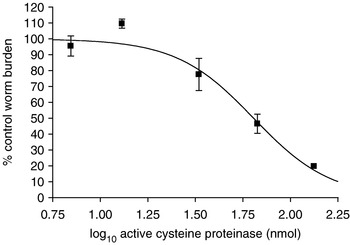
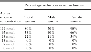
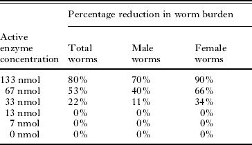
To determine whether the in vivo effect was dose-dependent, a range of doses of papaya latex [expressed as nmol of active cysteine proteinase, determined by active-site titration with E-64 (Zucker et al. Reference Zucker, Buttle, Nicklin and Barrett1985)] were used. Six groups of 3 male C3H mice were infected with 200 H. polygyrus L3 on day 0 and then treated with increasing concentrations of papaya latex (0 nmol, 7 nmol, 13 nmol, 33 nmol, 67 nmol and 133 nmol active cysteine proteinase/day) from days 18 to 24 post-infection, inclusively. The number of worms remaining in the intestinal tract was counted on day 25 post-infection and FEC were quantified throughout. As Fig. 2 shows, the in vivo activity of papaya latex was dose-dependent with respect to the worm burdens (log10 EC50=1·8 (95% CL=1·72–1·89) (67 nmol) and log10 EC95=2·4 (95% CL=2·14–2·67) (133 nmol); model R 2=0·901 for 13 degrees of freedom), and egg counts were reduced to a similar extent (data not shown). Doses above 33 nmol/treatment (log10=1·50 nmol), which equates to 1·15 g/kg for the preparation of papaya latex used in this study, successfully reduced parasite burdens. Additionally, at all effective doses, the reduction in the number of female worms was relatively greater than that of male worms (e.g. with 133 nmol papaya latex/treatment, there was a 90% reduction in the number of female worms but only a 71% reduction of male worms; Table 1).
Papaya latex produces a dose-dependent quantitative reduction in worm number of the GI nematode, Heligmosomoides polygyrus, in vivo. Six groups of 3 male C3H mice were infected with 200 H. polygyrus L3 on day 0 and then treated with increasing concentrations of papaya latex (0 nmol, 7 nmol, 13 nmol, 33 nmol, 67 nmol and 133 nmol active cysteine proteinase) from days 18 to 24 post-infection, inclusively. The number of worms remaining in the intestinal tract was counted on day 25 post-infection. The anthelmintic efficacy of papaya latex was expressed in terms of the percentage reduction in total worm burden of the treated groups compared to the untreated animals, and showed a dose-dependent reduction. A sigmoidal dose-response curve was fitted with an upper limit of 100 and a lower limit of zero in GraphPad Prism (version 4), and the log10 EC50 and log10 EC95 were calculated together with 95% confidence limits (CL). Error bars represent the standard error of the mean (n=3 per concentration).

Comparison of reductions in Heligmosomoides polygyrus male and female worms from C3H mice treated with papaya latex in a dose-dependent manner
(Male C3H mice were infected with 200 H. polygyrus L3 on day 0 and then treated with various concentrations of papaya latex from days 18 to 24 post-infection, inclusively. The number of worms remaining in the intestinal tract was counted on day 25 post-infection and the anthelmintic efficacy of papaya latex was expressed in terms of the percentage reduction in worm burden of the treated groups compared to the untreated animals, n=3 per concentration.)
The anthelmintic effect of papaya latex is dependent upon the activity of cysteine proteinases
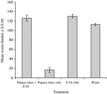
To determine whether this anthelmintic effect was due solely to the cysteine proteinases contained in the crude latex of papaya, we administered papaya latex (135 nmol active enzyme/treatment dose) with and without prior incubation with the cysteine proteinase-specific inhibitor, E-64 (pre-incubation for 15 min with 0·64 mm E-64; Barrett et al. Reference Barrett, Kembhavi, Brown, Kirschke, Knight, Tamai and Hanada1982), daily for 7 days (from day 18 to day 24 post-infection) to groups of male C3H mice (n=5/group) infected with H. polygyrus. Mice were killed on day 25 post-infection and the number of worms was counted. Statistical analysis of worm counts was by 2-way ANOVA (treatment with papaya latex and/or E-64) on the raw worm counts. The only mice to show a significant reduction in faecal egg number (96%) over the 7-day treatment period were those animals administered crude papaya latex alone, and this was accompanied by a significant reduction in worm burden of 85% (for main effect of papaya latex, F 1,16=125·4; for main effect of E-64, F 1,16=201·3; for 2-way interaction, F 1,16=104·4; P<0·001 in all cases. Model adjusted R 2=0·958) (Fig. 3). The results clearly demonstrate the anthelmintic efficacy of papaya latex (the only treatment group to show a reduction in worm burden), and the lack of effect of papaya latex pre-treated with E-64 shows that cysteine proteinase activity is an absolute requirement for efficacy.
Active cysteine proteinases in papaya latex are required for efficacy. The number of mature adult worms of Heligmosomoides polygyrus present in the intestinal tract of infected mice treated for 7 days with oral administration of either papaya latex (135 nmol active enzyme) pre-incubated for 15 min with 0·64 mm of the cysteine proteinase-specific inhibitor, E-64, papaya latex (135 nmol active enzyme) alone, E-64 alone, or sterile distilled water was determined. Papaya latex alone was the only treatment that caused a major reduction in worm burden. Hence, E-64 blocked the worm loss that would have resulted from papaya latex treatment given alone. Error bars represent the standard error of the mean (n=5 per group).

Papaya latex does not affect the development of the post-infective larval stages in the intestinal mucosa
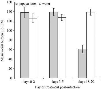
Although treatment with papaya latex produced a marked reduction in adult GI nematodes that reside in the gut lumen, its efficacy against earlier nematode developmental stages (L3 and L4), which are found in the mucosa and are thus not in direct contact with the contents of the gut lumen, was still not known. Therefore, we treated groups (n=5) of H. polygyrus-infected mice with papaya latex or water for 3 days, either from days 0–2 post-infection (270 nmol active enzyme/treatment dose; against L3), days 3–5 post-infection (335 nmol active enzyme/treatment dose; against L4) or days 18–20 post-infection (316 nmol active enzyme/treatment dose; against adult worms). As can be seen in Fig. 4, the only mice to show a significant reduction in worm burden (56%) were those treated between days 18 and 20 post-infection (i.e. against the adult stage) (for main effect of treatment: F 1,23=10·4, P=0·04; for main effect of stage of infection: F 2,23=13·2, P<0·001; for the 2-way interaction: F 2,23=20·9, P<0·001. Model adjusted R 2=0·727). Thus, the cysteine proteinases in papaya latex do not prevent the development of the mucosal-dwelling post-infective larval stages of GI nematodes.
Papaya latex is only efficacious against the adult worm stage of Heligmosomoides polygyrus. Male C3H mice were infected with 200 H. polygyrus L3 on day 0 and then treated with papaya latex or water for 3 days, either from days 0–2 post-infection (270 nmol active enzyme/treatment; against L3), days 3–5 post-infection (335 nmol active enzyme/treatment; against L4) or days 18–20 post-infection (316 nmol active enzyme/treatment; against adult worms). The number of worms remaining in the intestinal tract was counted on day 25 post-infection. The anthelmintic efficacy of papaya latex was expressed in terms of the percentage reduction in total worm burden of the treated groups compared to the untreated animals, and showed that only the adult worms were significantly reduced in number. Error bars represent the standard error of the mean (n=5 per group).

Plant cysteine proteinases attack the cuticle of GI nematodes in vivo
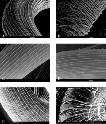
We investigated the mechanism of action of papaya latex in vivo, by removing and examining adult worms of H. polygyrus every hour for 4 h after the third daily dose of papaya latex (133 nmol active enzyme/treatment dose). The results showed that the appearance of the worms was indistinguishable from that recorded in vitro (Fig. 5). The worms retrieved 1–2 h post-treatment had a similar appearance to worms incubated with plant cysteine proteinases in vitro in that their smooth longitudinal ridges had become transverse wrinkles and folds. These worms were located in the lower third of the small intestine after 1 h (Fig. 5A) and in the large intestine after 2 h (Fig. 5B), sites which are not optimal for their survival and in which they are never normally found unless they are in the process of being expelled from the host, as H. polygyrus usually resides in the upper two-thirds of the small intestine. This, therefore, suggests that worms damaged by the action of cysteine proteinases are eliminated from the host. The worms obtained 3–4 h post-treatment (Fig. 5C and D) were only found in the usual upper two-thirds of the small intestine and appeared to be undamaged, indicating that they had escaped the effects of treatment; at this point, there were no worms, damaged or otherwise, found in the lower third of the small intestine or in the large intestine. Thus, the damaged worms had been rapidly expelled within 4 h of treatment.
Plant cysteine proteinases disrupt the structure of the cuticle of GI nematodes in vivo. Papaya latex (133 nmol active enzyme) was administered to C3H mice infected with Heligmosomoides polygyrus for 3 consecutive days from day 18 post-infection. One mouse was killed every hour post-treatment for 4 h on day 20. The worms were then collected from the whole intestinal tract and processed for scanning electron microscopy, but only representative worms are shown. The smooth longitudinal ridges of the cuticle of H. polygyrus adult worms became transverse wrinkles and folds. These worms were found in the lower third of the small intestine 1 h post-treatment (A) and the large intestine 2 h post-treatment (B), where H. polygyrus does not usually reside. H. polygyrus adult worms, demonstrating the smooth longitudinal ridges of the cuticle, were retrieved from the usual upper two-thirds of the small intestine 3 (C) and 4 (D) h post-treatment. Adult worms incubated before (E) and after (F) 60 min of incubation with 6·25 μM (active enzyme) papaya latex in vitro, for comparison with the worms retrieved in vivo after 1–2 h. Note cuticle digestion of the worm shown in (F). Scale bar=10 μm.

Histology of the intestinal mucosa after treatment with papaya latex
An important consideration for potential anthelmintic candidates is whether any adverse reactions occur to the host. To begin to address this issue, we examined the intestine of mice which had been (a) infected with H. polygyrus and treated with papaya latex, (b) infected but not treated, (c) uninfected but treated with papaya latex, or (d) uninfected and untreated, by histological examination of the intestine for size of the intestinal villi and numbers of mast cells and Goblet cells. There were no significant differences in the numbers of either type of immune cells examined between the different groups of mice (Table 2), although the group which was infected but not treated (group B) had slightly reduced numbers of mast cells compared to the remaining 3 groups. Mean Goblet cell counts were also higher in both infected groups and in the group treated with papaya latex, relative to the non-infected mice given treatment with water, but overall there was no significant difference between treatment groups and no significant interaction. The intestinal villi were much smaller in the infected but untreated mice (group B) compared to the mice in the other 3 groups, with a significant 2-way interaction (Table 2). Infections with H. polygyrus adult worms cause erosion of the intestinal villi as a result of both the attachment and feeding of the worms on the intestine (Bansemir and Sukhdeo, Reference Bansemir and Sukhdeo1994) and from the accompanying intestinal immune response. This demonstrates that the patho-physiological changes in the mice, associated with the presence of the worms, were reversed by treatment with papaya latex, and hence pathology was reduced rather than exacerbated.
Comparison of the numbers of mast cells and goblet cells, and the size of the intestinal villi of C3H mice infected and uninfected with Heligmosomoides polygyrus and treated with either papaya latex or water
(Male C3H mice were infected with 200 H. polygyrus L3 on day 0 and then treated with 316 nmol (active enzyme/treatment) papaya latex from days 18 to 24 post-infection, inclusively. The lengths of the intestinal villi were measured and the numbers of mast cells and Goblet cells were counted on day 25 post-infection, and expressed as the number of cells/mm2, before comparison between the treated and untreated groups to determine the histological effect of papaya latex, n=5.)

Statistical analysis of results:
a For the main effect of infection: F 1,15=9·6, P=0·007; main effect of papaya latex treatment: F 1,15=9·0, P=0·009; 2-way interaction of infection∗papaya latex treatment: F 1,15=10·8, P=0·005.
b For the main effect of infection: F 1,15=1·6; main effect of papaya latex treatment: F 1,15=2·3; 2-way interaction of infection∗papaya latex treatment: F 1,15=0·15. For all, P>0·1.
c For the main effect of infection: F 1,15=0·759; main effect of papaya latex treatment: F 1,15=2·6; 2-way interaction of infection∗papaya latex treatment: F 1,15=2·9. For all, P>0·1.
Cysteine proteinases from other plants are also efficacious against H. polygyrus adult worms in vivo
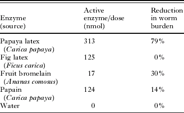
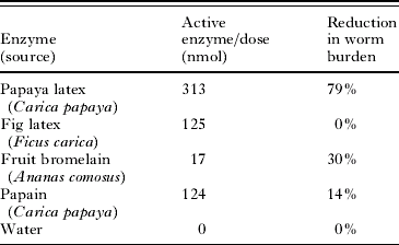
Since papaya latex proved highly efficacious against adult worms, we determined whether other plant extracts containing cysteine proteinases also shared this property in vivo. Mice (n=5/group) infected with 200 L3 of H. polygyrus were treated for 7 days (from day 18) with papaya latex (313 nmol active enzyme per dose), Ficus carica (fig) latex (125 nmol active enzyme/dose), fruit bromelain (isolated from pineapple fruit by acetone precipitation; 17 nmol active enzyme/dose) or pure papain (from papaya latex; 124 nmol active enzyme/dose). As Table 3 shows, papaya latex, at a daily dose of 313 nmol, provided considerable anthelmintic activity, with 79% reduction in worm burden. Fruit bromelain, at a daily dose of only 17 nmol, also produced a statistically significant reduction in worm burden (30%), despite the relatively lower dose (analysis of the raw data by 1-way ANOVA (with 5 levels to the treatment factor) gave F 4,20=36·7, P<0·001; model adjusted R 2=0·856). Post hoc analysis by Tukey's HSD test indicated that only papaya latex (P<0·001) and fruit bromelain (P=0·04) differed significantly from the control group. This suggests that the anthelmintic efficacy in vivo is not restricted to cysteine proteinases in the latex of papaya.
Comparison of the in vivo anthelmintic efficacy of cysteine proteinases from different plants against Heligmosomoides polygyrus in C3H mice
(Male C3H mice were infected with 200 H. polygyrus L3 on day 0 and then treated with either papaya latex, fig latex, fruit bromelain or pure papain from days 18 to 24 post-infection, inclusively. The number of worms remaining in the intestinal tract was counted on day 25 post-infection and the anthelmintic efficacy of papaya latex was expressed in terms of the percentage reduction in worm burden of the treated groups compared to the untreated animals, n=5.)
DISCUSSION
With the rapid appearance of anthelmintic resistance, and the lack of new drugs and vaccines under current development, interest is now being shown in the use of extracts from traditional medicinal plants as novel anthelmintics (Tagboto and Townson, Reference Tagboto and Townson2001; Waller et al. Reference Waller, Bernes, Thamsborg, Sukura, Richter, Ingebrigtsen and Höglund2001; Githiori et al. Reference Githiori, Höglund, Waller and Baker2004). Because extracts of plants, such as papaya and fig, have been used traditionally for centuries in some developing countries, studies have been carried out to examine the anthelmintic properties of these and related plants, both in vitro (Robbins, Reference Robbins1930; Berger and Asenjo, Reference Berger and Asenjo1939, Reference Berger and Asenjo1940; Hansson et al. Reference Hansson, Veliz, Naquira, Amren, Arroyo and Arevalo1986; Stepek et al. Reference Stepek, Buttle, Duce, Lowe and Behnke2005, Reference Stepek, Lowe, Buttle, Duce and Behnke2006, Reference Stepek, Lowe, Buttle, Duce and Behnke2007) and in vivo (Hansson et al. Reference Hansson, Veliz, Naquira, Amren, Arroyo and Arevalo1986; Satrija et al. Reference Satrija, Nansen, Bjorn, Murtini and He1994, Reference Satrija, Nansen, Murtini and He1995; Stepek et al. Reference Stepek, Lowe, Buttle, Duce and Behnke2006, Reference Stepek, Lowe, Buttle, Duce and Behnke2007). Although several earlier studies showed that plants, such as papaya, pineapple and fig, were active against gastrointestinal nematodes in vitro, and a basic mechanism was described (Robbins, Reference Robbins1930), the actual mechanism of action in vitro, based on ultrastructural observations on the cuticle at the scanning electron microscope level, has only recently been described in detail (Stepek et al. Reference Stepek, Buttle, Duce, Lowe and Behnke2005) and attributed to the cysteine proteinases present in the latex or fruit (Stepek et al. Reference Stepek, Buttle, Duce, Lowe and Behnke2005). Few studies have been carried out to assess the in vivo anthelmintic effect of these plant extracts, with Satrija et al. (Reference Satrija, Nansen, Murtini and He1995) being the first to demonstrate that papaya latex did indeed have anthelmintic properties. However, to our knowledge, the current study is the first (i) to establish, unequivocally, that it is upon the cysteine proteinases present in the latex that the anthelmintic efficacy of papaya latex depends, and (ii) to provide evidence that the mechanisms of action in vivo and in vitro are essentially indistinguishable.
In vitro studies (Stepek et al. Reference Stepek, Buttle, Duce, Lowe and Behnke2005, Reference Stepek, Lowe, Buttle, Duce and Behnke2006, Reference Stepek, Lowe, Buttle, Duce and Behnke2007) have demonstrated that cysteine proteinases attack the protective cuticle of the nematode, rapidly leading to complete dissolution of the parasite, a mechanism of action that differs from all the current anthelmintic drugs (Sangster and Gill, Reference Sangster and Gill1999; Martin and Robertson, Reference Martin and Robertson2000). This effect was similar to that described previously for 3 other gastrointestinal nematodes, H. polygyrus (Stepek et al. Reference Stepek, Buttle, Duce, Lowe and Behnke2005), T. muris (Stepek et al. Reference Stepek, Lowe, Buttle, Duce and Behnke2006) and Ascaris suum (Robbins, Reference Robbins1930; Berger and Asenjo, Reference Berger and Asenjo1939), indicating that plant cysteine proteinases have a broad spectrum of activity against GI nematodes in vitro. The same appearance of the nematode cuticle has now been seen in vivo.
In contrast to adult GI nematodes, papaya latex was not effective in vivo against parasitic larval stages (L3 and L4). When we treated H. polygyrus-infected mice with papaya latex for 3 days, either against L3, L4 or adult worms, the only mice to show reductions in worm burden were those treated between days 18 and 20 post-infection, when the mature worms had developed. Similar observations were reported for the nematode of the large intestine, Trichuris muris (Stepek et al. Reference Stepek, Lowe, Buttle, Duce and Behnke2006), and this was attributable to the location of the different developmental stages of the parasite within the tissues of the intestine because, for both parasite species, the L3 and L4 are located within the intestinal mucosa, hidden from direct contact with the papaya latex as it passes through the gut lumen after oral administration. In contrast, adult worms are exposed to papaya latex in the lumen of the small intestine. However, our data do not necessarily imply that efficacy begins with 18-day-old worms because H. polygyrus emerge from their site of development in the mucosa on days 8–9 post-infection and, thereafter, live in the intestinal lumen, where they are likely to be susceptible to treatment as with the 18-day-old worms in our current experiments (Bryant, Reference Bryant1973). Therefore, cysteine proteinases from plants will only be effective anthelmintics against lumen-dwelling, not mucosal-dwelling, species and stages. Since this will essentially leave a proportion of worms in refugia, it will help to delay any onset of resistance (Coles, Reference Coles2005).
We have shown that the cysteine proteinase activity of papaya latex passes very rapidly (within 20 min) through the upper small intestine (Stepek et al. Reference Stepek, Lowe, Buttle, Duce and Behnke2007), perhaps explaining why multiple treatments (daily doses for 7 consecutive days) are required for greater anthelmintic efficacy. A few worms do remain after the final treatment, which will allow the natural immunity to infection to develop in species that are controlled by the host's immune response (Waller, Reference Waller1997). Cysteine proteinases of the papain family are stable between pH 4 and 9, with optimal activity at neutral pH, but their activity decreases greatly below pH 4 and above pH 9, due largely to the pKas of the active-site thiolate and imidazolium ions (Salih et al. Reference Salih, Malthouse, Kowlessur, Jarvis, O'Driscoll and Brocklehurst1987). Thus, the enzymes become temporarily inactive when passing through parts of the gastrointestinal tract with an acidic pH, such as the stomach, but regain their activity on entering the small intestine with a pH of 7–8. Hale (Reference Hale2004) found that bromelain, when administered with antacid, retained its activity throughout the intestinal tract of mice, and it is possible that, by neutralizing stomach acidity prior to treatment, the enzyme activity of papaya latex may be enhanced. Indeed, we have already reported that anthelmintic activity against the stomach nematode, Protospirura muricola, is substantially increased when treatment is combined with antacids (Stepek et al. Reference Stepek, Lowe, Buttle, Duce and Behnke2007), but whether the same will be the case for worms in the small intestine is not yet known.
Athanasiadou et al. (Reference Athanasiadou, Kyriazakis, Jackson and Coop2001) and Paolini et al. (Reference Paolini, Frayssines, de la Farge, Dorchies and Hoste2003) reported that an extract of condensed tannins from plants reduced worm burdens of nematode species in the small intestine, but not of those in the abomasum, of small ruminants. A possible reason for this, as suggested by Athanasiadou et al. (Reference Athanasiadou, Kyriazakis, Jackson and Coop2001), could be the pH of the abomasum and small intestine. These authors stated that condensed tannins are stable between pH 5 and 7, and that infections in sheep of the abomasal nematodes, Haemonchus contortus and Teladorsagia circumcincta, were probably not affected due to the abomasum having a pH of 2·5, in contrast to that of the small intestine (pH of 7–8), explaining why Trichostrongylus colubriformis worm burdens were reduced. Perhaps, in this situation, pre-treatment with an antacid with the capacity to neutralize the pH of the abomasum would have facilitated tannin-mediated reduction in worm burden of nematode species found in the abomasum, as we found with papaya latex against P. muricola infections in an earlier study (Stepek et al. Reference Stepek, Lowe, Buttle, Duce and Behnke2007). Another problem that is likely to be faced when attempts are made to evaluate cysteine proteinases in ruminants is possible degradation of the enzymes by rumen microbes but, currently, this aspect has not been investigated. However, we believe that current advances in delivery methods and formulation of drugs (Vandamme and Ellis, Reference Vandamme and Ellis2004) should eventually enable a formulation to be devised that will help to preserve activity during passage through the rumen and also through the acid environment of the abomasum. Equally, with further refinement of the methods of formulation and delivery, it should be possible to reduce multiple drug administration, as here in our study, to single dose treatments that are much more likely to be attractive to livestock owners, but these are challenges for future research.
We found that adult female H. polygyrus were more susceptible to treatment with papaya latex than the male worms in vivo. Previous studies have reported a similar sex bias with T. muris (Stepek et al. Reference Stepek, Lowe, Buttle, Duce and Behnke2006) and P. muricola (Stepek et al. Reference Stepek, Lowe, Buttle, Duce and Behnke2007) although, especially in the case of H. polygyrus and T. muris, these species were substantially reduced in terms of worm burdens and egg outputs. This sex bias does pose interesting questions about possible differences in cuticular structure between the sexes. However, of more immediate practical importance, is the fact that the susceptibility of female worms makes this a potentially attractive mechanism for reduction of nematode egg production and associated contamination of the environment with parasite transmission stages, and hence control of life-cycle and transmission.
The pure forms of these plant cysteine proteinases are already available for medicinal use against other ailments: for example, chymopapain is a licensed drug for the treatment of intervertebral disc disease. Many of these plant cysteine proteinases have also been used as meat tenderisers and in other aspects of food preparation, and so would appear to be safe for human and animal use. Our preliminary histological analysis showed that treatment of H. polygyrus-infected mice with papaya latex caused no detectable adverse side-effects. Whilst this suggests that this treatment would not damage the internal structures, more in-depth immunological analyses are required because papaya latex is known to cause allergies. However, these usually arise following intake through the airways, as is commonly the case with IgE-mediated allergy (Soto-Mera et al. Reference Soto-Mera, López-Rico, Filgueira, Villamil and Cidrás2000; Nelde et al. Reference Nelde, Teufel, Hahn, Duschl, Sebald, Bröcker and Grunewald2001), and serious allergic reactions following oral ingestion would be expected to be extremely rare. Nevertheless, Hale (Reference Hale2004) found anti-bromelain antibodies in the serum of mice, but only following prolonged (18 weeks) daily oral administration of the enzyme, and this contrasts with the considerably briefer period of daily treatment (1 week) required to clear the majority of the worms from the intestinal tract in the current study.
Now that the anthelmintic efficacy and mechanism of action of papaya latex have been clearly demonstrated, both in vitro and in vivo, and shown to be absolutely dependent on the presence of cysteine proteinases, the way is open for the development of plant cysteine proteinases as drugs to treat a range of GI nematode infections of humans and livestock, where resistance to current synthetic drugs is becoming a serious problem. It is doubtful that resistance to cuticle hydrolysis would arise quickly as it is likely to be polygenic and would entail major structural changes to the cuticle. Therefore, these naturally-occurring plant extracts should be ideal candidates for novel anthelmintics. To our knowledge, only one human trial has been conducted to date (Hansson et al. Reference Hansson, Veliz, Naquira, Amren, Arroyo and Arevalo1986), and this did not detect adverse effects on the subjects. Undoubtedly, further clinical trials and field evaluations will be required and further refinement of the methods of delivery and formulation should increase efficacy against nematodes residing in various parts of the GI tract.
Cysteine proteinases from plants, such as papaya, pineapple or fig, may provide a relatively inexpensive alternative to the currently available chemotherapeutic drugs for the treatment of GI nematode infections of humans and livestock in the areas of the world most requiring alternative treatments. In so far as they are derived from naturally-occurring plant sources and the active principles are proteins, they can be classified as natural products with little chance of long-lived harmful residues. Moreover, they may spawn novel agricultural industries, particularly in the tropics, in promoting the growth of anthelmintic plants as sources of active ingredients for future medicines.
This work was funded by a research grant from The Leverhulme Trust held by J. M. B., D. J. B. and I. R. D. We thank Steve Ellin and the late Ken Cartmell (Tapton Experimental Gardens, University of Sheffield) for their help in the maintenance of the fig trees and in the collection of the latex, and Ian Mellor (University of Nottingham) for his help in producing the sigmoidal dose-response curve in Fig. 2. Finally, we are sincerely grateful to Simon Clifford and Tim Smith for excellent technical assistance in parasite maintenance and electron microscopy.