Introduction
As an important Asian pollinator, the Chinese honeybee (Apis cerana cerana Fabricius, A. c. cerana) plays a significant role in maintaining the balance between biodiversity and agricultural economic development (Yang, Reference Yang2005; Weinstock et al., Reference Weinstock, Robinson, Gibbs, Worley, Evans and Maleszka2006). Compared with Apis mellifera Linnaeus (A. mellifera), A. c. cerana offers some irreplaceable advantages such as strong resistance to mites, an acute sense of smell, and ability to forage a wide range of nectars and pollens (Ratnieks, Reference Ratnieks2006). However, the population of honeybees has declined severely due to various abiotic stresses such as viral diseases, indiscriminate use of insecticides, ultraviolet radiation (UV), and global warming, all of which have been reported to produce reactive oxygen species (ROS) damage (Balaban et al., Reference Balaban, Nemoto and Finkel2005; Narendra et al., Reference Narendra, Bhatracharyulu, Padmavathi and Varadacharyulu2007; Xu et al., Reference Xu, Shi and Chen2009).
Organisms have developed elaborate antioxidant mechanisms to avoid the ROS damage caused by abiotic stress. Organisms protect themselves by using a battery of antioxidant enzymes and detoxifying enzymes, including peroxidase (POD), superoxide dismutase (SOD), catalase (CAT), glutathione-S-transferase (GST), glutathione peroxidase (GPX), and Cytochrome P450 monooxygenases (P450s) enzymes (Wang et al., Reference Wang, Oberley and Murhammer2001; Li et al., Reference Li, Schuler and Berenbaum2007; Dubovskiy et al., Reference Dubovskiy, Martemyanov, Vorontsova, Rantala, Gryzanova and Glupov2008; Feyereisen, Reference Feyereisen2011; Matteis et al., Reference Matteis, Ballou, Coon, Estabrook and Haines2012). Furthermore, P450s play an important role in the metabolism of xenobiotics (drugs, pesticides, and plant toxins) by catalyzing oxidation reactions (Scott, Reference Scott1999; Anzenbacher and Anzenbacherova, Reference Anzenbacher and Anzenbacherova2001; Nebert and Russell, Reference Nebert and Russell2002; Thomas Reference Thomas2007). In insects, most P450s genes are divided into four clades: mitochondrial P450s, CYP2, CYP3 (including CYP6 and CYP9), and CYP4. The CYP4 clade is thought to be largely responsible for detoxifying functions and other abiotic stress responses and has been implicated in both pesticide metabolism and chemical communication (Claudianos et al., Reference Claudianos, Ranson, Johnson, Biswas, Schuler, Berenbaum, Feyereisen and Oakeshott2006; Feyereisen, Reference Feyereisen2011). Early studies by Shi et al. (Reference Shi, Sun, Xu and Li2013) provided evidence that the CYP4G11 gene was involved in protecting honeybee workers from abiotic stress damage. Several CYP4 genes are overexpressed in the pesticide-resistant Culex pipiens, Diabrotica virgifera, and Helicoverpa armigera species (Pittendrigh et al., Reference Pittendrigh, Aronstein, Zinkovsky, Andreev, Campbell, Daly, Trowell and Ffrench-Constant1997; Scharf et al., Reference Scharf, Parimi, Meinke, Chandler and Siegfried2001; Shen et al., Reference Shen, Dong, Tian, Ma, Li, Wu and Zhu2003). In 2015, the publication of the A. c. cerana genomic sequence provided plenty of information for a better understanding of the natural biology and complex behavior of the Asian honeybee (Park et al., Reference Park, Jung, Choi, Jayakodi, Lee and Lim2015). There are 46 P450s genes, four of which are CYP4 genes in honeybee (Park et al., Reference Park, Jung, Choi, Jayakodi, Lee and Lim2015). However, it remains essential to identify specific genes and their corresponding proteins and to characterize their expression profiles and related biological functions during the response to abiotic stresses.
There are four CYP4 genes (CYP4G11, CYP4AZ1, CYP4AV1, and CYP4AA1) in the honeybee. Previous research has suggested that AccCYP4G11 and AccCYP4AZ1 play a crucial role in response to abiotic stress and development in A. c. cerana (Shi et al., Reference Shi, Sun, Xu and Li2013; Zhang et al., Reference Zhang, Chen, Li, Ma, Yu, W, Liu and Xu2018). However, the functions of AccCYP4AV1 during the response to abiotic stress in A. c. cerana have not been well understood, yet. Thus, it is necessary to gain insight into the functions of AccCYP4AV1 and to elucidate the defense mechanisms in A. c. cerana. To identify the role of the AccCYP4AV1, we characterized this gene and investigated its transcription levels in different tissues and at different developmental stages. We also simulated common abiotic stress conditions [low- and high-temperatures (4°C and 44°C, respectively), cadmium chloride (CdCl2), mercury (II) chloride (HgCl2), deltamethrin, paraquat, dichlorovos], which are commonly encountered in unfavorable environments by A. c. cerana during the collection of pollen and nectar outside the hives, to examine the expression profiles of AccCYP4AV1 mRNA and protein. Thus, this study provides a better understanding of the role of CYP4 in the defense against abiotic stresses in A. c. cerana and contributes to our knowledge of the supergene family.
Materials and methods
Insects and treatments
Honeybee workers were collected from three healthy colonies provided by the Shandong Agricultural University (Tai'an, Shandong, China). Whole bodies of single larvae (L1-L5), pupae [prepupae (PP), white eyes (Pw), brown eyes (Pp), and dark eyes (Pd)], newly emerged honeybee workers (<12 h of age, NE), and adults were collected and staged according to Michelette and Soares (Reference Michelette and Soares1993). Newly emerged honeybee workers were marked with paint and collected at 15 days of age. Tissues of the brain (BR), epidermis (EP), venom gland (VG), and midgut (MG) were isolated from 15-day-old bees (20 individuals pooled together) to examine tissue-specific expression. Adult workers were housed in wooden cages (22 × 9 × 5 cm3, external dimensions). Each cage was placed in a climate chamber maintained at 31 ± 1°C and 65 ± 5% relative humidity, and the honeybees were fed 50% powdered sugar (1:1 sugar-water) and water. Cages were divided into ten groups (50 honeybees per cage, six cages per group): groups 1 and 2 were placed at low (4°C) and high (44°C) temperatures, respectively; groups 3 and 4 were fed sucrose solutions containing CdCl2 and HgCl2, respectively; groups 5–7 were fed three different pesticides, deltamethrin, paraquat, or dichlorovos, respectively, diluted in 50% powdered sugar (online Supplementary Table 1; honeybees were starved for 6 h before feeding); group 8 was exposed to UV light; group 9 served as the untreated control (CK). Honeybees were collected at an appropriate time for each condition, frozen in liquid nitrogen, and stored at −80°C. All experiments were carried out in triplicate.
RNA extraction and cDNA synthesis
E.Z.N.A.® Total RNA Kit II (OMEGA, USA) was used to extract total RNA from samples according to the manufacturer's protocol. TransScript ® All-in-One First-Strand cDNA Synthesis SuperMix for qPCR (One-Step gDNA Removal; TransGen Biotech, China) was used to generate the first-strand cDNA, according to the manufacturer's instructions.
Cloning of the cDNA sequence of AccCYP4AV1
The synthesized cDNA of newly emerged workers was used as the PCR template for cloning. All the primers used in this study (shown in online Supplementary Table 2) were designed by Primer 5 (Premier Biosoft International, USA). PCR products were visualized in 1% agarose gels and purified using a Gel Extraction Kit (Solarbio, China). The purified products were cloned into PMD18-T vectors (TaRaKa, China) and transformed into competent Escherichia coli DH5α (E. coli DH5α) cells for sequencing by BioSune Biotech (China).
Bioinformatics analysis
National Centre for Biotechnology Information (NCBI) bioinformatics tools (http://blast.ncbi.nlm.nih.gov/Blast.cgi) were used to detect conserved domains in AccCYP4AV1. The DNAMAN version 5.2.2 (Lynnon Biosoft, Quebec, Canada) was used to search for the ORF of AccCYP4AV1. The theoretical isoelectric point and molecular weight were predicted using PeptideMass (http://us.expasy.org/tools/peptidemass.html). Homologous AccCYP4AV1 sequences in other species were retrieved from the NCBI server and aligned using the DNAMAN software. Phylogenetic analysis was performed with the Molecular Evolutionary Genetics Analysis software (MEGA version 7.0.26) by applying the neighbor-joining method. The signal peptide was analyzed on the SignalP 4.0 website (http://web.expasy.org/protscale/). The transmembrane segments were analyzed using the TMHMM website (http://www.cbs.dtu.dk/services/TMHMM/).
Real-time quantitative polymerase chain reaction (RT-qPCR) analysis
The stably expressed housekeeping gene β-actin (XM_017065464) was selected as a reference gene to normalize RT-qPCR experiments (Lourenço et al., Reference Lourenço, Mackert, Dos and Simões2008; Scharlaken et al., Reference Scharlaken, de Graaf, Goossens, Peelman and Jacobs2008; Umasuthan et al., Reference Umasuthan, Revathy, Lee, Whang, Choi and Lee2012). The AccCYP4AV1 primers 4AV1-F/4AV1-R and the β-actin primers β-actin-F/β-actin-R were used for RT-qPCR with the SYBR® PrimeScriptTM RT-PCR Kit (Roch, Germany), in a 7500 Real-Time System (ABI, USA). The following RT-qPCR protocol was performed: pre-denaturation at 95°C for 10 min, 40 cycles of amplification at 95°C for 15 s and 60°C for 60 s, and a melt cycle from 65°C to 95°C. Data were analyzed with the ABI SDS software, using the 2−ΔΔCt method (Livak and Schmittgen, Reference Livak and Schmittgen2001). Three independent biological replicates were processed for all experimental conditions, and all the RT-qPCR reactions were analyzed in three technical replicates.
Antibody production
Custom-made polyclonal antibodies were used. An epitope was predicted using the DNASTAR 7.1 Optimum Antigen design tool (predicted AccCYP4AV1 peptide antigen: EWKDRKDTNFNEKS), and the corresponding peptide antigen was, then, synthesized by Sangon Biotech (Shanghai, China). After the coupling reaction and mixing with complete adjuvant (Sigma, USA), the coupled antigen was used for injection into five 6-week-old pathogen-free male ICR mice (Taibang, China) for the first time. Next, the coupled antigen was mixed with incomplete adjuvant (Sigma, USA) and injected into male ICR mice, three times at 3-week intervals. Mice had free access to food (standard pellet diet) and tap water and were equally housed in conventional steel cages, with a 12 h light and dark cycle at room temperature. Subsequently, blood was collected by eyeball puncture and incubated at 37°C for 1 h, followed by centrifugation at 3000 × g for 10 min. Finally, the antiserum was prepared. The collected antibody was hybridized to a blot containing total proteins to detect the specificity of the antiserum.
Western blotting analysis
Protein expression was measured by western blot. Samples were homogenized in RIPA lysis buffer containing 10 mM protease inhibitor (CWBIO, Beijing, China). The supernatants were recovered by centrifugation at 10,000 g at 4°C for 10 min. Protein concentrations were quantified by a BCA Protein Assay Kit (Beyotime, Beijing, China). The total protein samples were quantified using a bicinchoninic acid (BCA) Protein Assay Kit (CWBIO, China). Equal amounts of protein were loaded onto a 12% SDS-PAGE gel, separated, and subsequently transferred to a polyvinylidene fluoride membrane (Millipore, Bedford, MA). After blocking for 1.5 h at 25°C, the membranes were incubated with the anti-AccCYP4AV1 polyclonal antibody (primary antibody) (1:500), at 4°C overnight. After incubation for 4 h with horseradish peroxidase-conjugated IgG secondary monoclonal antibody (1:1000) at 4°C, the membranes were immersed in enhanced chemiluminescence reagent (Beyotime, China) and visualized on a Fusion FX imaging system (Vilber Lourmat, China). The optical density of each band was quantified using the Fusion Capt Advance FX7 software (Jiangsu, China), with tubulin (antibody acquired from Beyotime, China) as a reference protein.
Silencing of the AccCYP4AV1 gene and assessment of the transcriptional levels of other genes after AccCYP4AV1 knockdown
RNA interference (RNAi) was performed to knockdown the AccCYP4AV1 transcript and was carried out as previously described by Amdam et al. (Reference Amdam, Simões and Guidugli2003). The non-conserved region of the AccCYP4AV1 ORF was chosen for targeting by specific primers containing a T7 polymerase promoter region at both forward and reverse orientations. The RiboMAXTM Large Scale RNA Production System T7 (Promega, USA) was used to produce AccCYP4AV1 double-stranded RNA (dsRNA), according to the manufacturer's instructions (Elias-Neto et al., Reference Elias-Neto, Soares, Simões, Hartfelder and Bitondi2010). The gene encoding the green fluorescent protein (GFP, GenBank ID: U87974) was used as a control (Elias-Neto et al., Reference Elias-Neto, Soares, Simões, Hartfelder and Bitondi2010). A. c. cerana genes do not share homology with GFP; thus, an RNAi response should not be triggered by dsGFP in the body of A. c. cerana. Newly emerged adult worker bees were randomly divided into four groups (n = 50 group−1) and used for RNAi experiments. Two groups were injected with 0.5 μl bee−1 (9 μg) of either dsAccCYP4AV1 or dsGFP. The third group was injected with 0.5 μl of double distilled water per honeybee worker. The fourth group served as the untreated control. The aforementioned groups were maintained in an incubator (31 ± 1°C, 65 ± 5% relative humidity). Healthy adult honeybee workers were sampled each day and stored at −80°C until use. The expression profiles of AccsHsp22.6 (KF150016), AccSOD2 (JN637476), AccTpx4 (KJ551847), AccTpx1 (HQ387025), AccGSTO1 (KF496073), and AccTrx1 (JX844652) were examined at 2 and 3 days post injection. Catalase and peroxidase activities were then tested at 2 and 3 days post injection by a catalase and peroxidase assay kits (Jiancheng, China), respectively. All experiments were carried out in triplicate.
Immunohistochemistry (IHC)
The MGs of the honeybee, which had been injected with H2O (H2O group), dsGFP (dsGFP group), or dsAccCYP4AV1 (dsAccCYP4AV1 group), were collected and fixed using 4% paraformaldehyde, for at least 24 h. All samples were dehydrated, infiltrated, and embedded in paraffin, following a common histological routine. Blocks were sectioned at 5-μm thickness and stained with antiserum (anti-AccCYP4AV1; 1:150 dilution), for 1 h at room temperature. After several washes with phosphate buffer saline (pH 7.2), samples were reacted with HRP-conjugated secondary antibody (Solarbio, China), for 1 h at room temperature. Samples were visualized with a DAB Color Development Kit (Solarbio, China), counterstained with hematoxylin, and mounted in neutral gum. For MG morphology, one slide per worker was observed at 200 × magnification under an optical microscope (Nikon Eclipse 80i, Nikon, Japan). The methodology was adapted from Britten et al. (Reference Britten, Howarth and Roche1993).
Mortality rate
A total of 720 honeybee workers were randomly collected and divided into 12 cages, each cage being randomly assigned to one of four groups: two treatment groups and two control groups. Honeybee workers of groups I and II were fed a 50% powdered sucrose diet (1:1 sugar-water) containing dichlorovos, and those of groups III and IV were fed a 50% powdered sucrose diet (1:1 sugar-water) containing paraquat (the concentrations of paraquat and dichlorovos are shown in online Supplementary Table 1). The honeybee workers of groups I and III were injected with 0.5 μl bee−1 (9 μg) of dsAccCYP4AV1, constituting the treatment groups; honeybee workers of groups II and IV were injected with dsGFP and constituted the control groups (CK). Sucrose diet and water were available ad libitum. RNAi-treated bees were compared with untreated bees. The mortality was scored for 4 days. Dead bees were discarded, and their number was carefully recorded in every group (Malone and Burgess, Reference Malone and Burgess2001).
Primers
All the specific primers and PCR amplification conditions are listed in online Supplementary Tables 2 and 3, respectively. All the RT-qPCR primers complied with the RT-qPCR requirements (Bustin et al., Reference Bustin, Benes, Garson, Hellemans, Huggett and Kubista2009; Yang et al., Reference Yang, Kemps-Mols, Spruyt-Gerritse, Anholts, Claas and Eikmans2016). The R2 of each RT-qPCR primer pair are shown in online Supplementary Table 2. All the primers used in the study were synthesized by BioSune Biotech (China).
Statistical analysis
All data are presented as mean ± standard error of the means (SEM). Data were analyzed by one-way ANOVA, followed by the Tukey's honestly significant difference test (Tukey's HSD). ANOVA was performed with the Statistical Analysis System version 9.1 software (SAS 9.1, USA). Equivalence of variance among groups was evaluated using the Levene's test for homogeneity of variance. Different letters above the bars designate significant differences (P < 0.05).
Results
ORF cloning and characterization of AccCYP4AV1
The full-length AccCYP4AV1 ORF sequence (MN930745) is 1506 base pairs long and encodes a deduced polypeptide composed of 501 amino acids (AA) (online Supplementary Fig. 1) with a theoretical molecular mass and isoelectric point of 57.84 kDa and 8.67, respectively. The predicted AccCYP4AV1 protein shares a number of characteristics with other members of the P450s, such as a charge pair consensus (ExxR) in Helix-K, a consensus sequence (WxxxR) within Helix-C, and the consensus sequence (A/G/E) GxxT (Grahamlorence et al., Reference Grahamlorence, Amarneh, White, Peterson and Simpson1995; Nelson, Reference Nelson1998) (online Supplementary Fig. 1). In addition, a partial AA sequence alignment of P450s revealed a highly conserved AA sequence consisting of PERK (online Supplementary Fig. 1). The phylogenetic tree analysis was performed to analyze the evolutionary relationship among CYP4AV1 of different species (online Supplementary Fig. 2). In this analysis, the homologous proteins AccCYP4AV1 of A. c. cerana and AmCYP4AV1 of A. mellifera were found to be more closely related to each other than to other homologues. Based on this homology search, the cDNA sequence was identified as cDNA encoding AccCYP4AV1. A potential signal peptide was identified in the N-terminal sequence, from AA 1 to 10 (online Supplementary Fig. 3a). The analysis of the AccCYP4AV1 potential transmembrane segments, which are strong hydrophobic areas, identified one such segment in the N-terminus (online Supplementary Fig. 3b).
Developmental and tissue-specific expression patterns of AccCYP4AV1
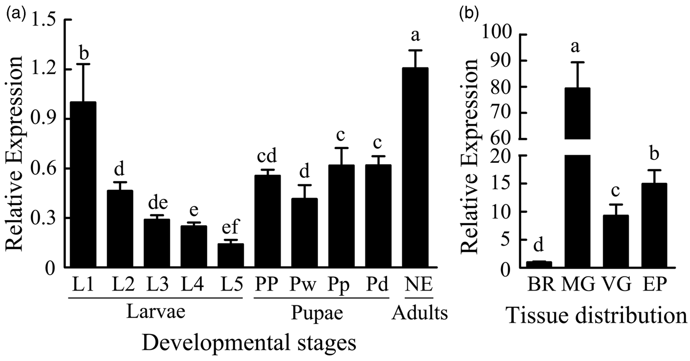
To gain insight into the function of AccCYP4AV1, its relative mRNA expression levels were examined during different development stages and in several different tissues. On average, AccCYP4AV1 expression levels were the highest during the adult stage and the lowest during the larval stage (fig. 1a). During the larval stage, the highest level of expression appeared at L1; during the pupal stage, the expression levels in Pw pupae were lower than those during other stages. As shown in fig. 1b, the spatial expression patterns revealed that AccCYP4AV1 transcripts were the highest in the MG, followed by the EP and VG, and the lowest transcription levels were observed in the BR.
Tissue-specific expression of AccCYP4AV1 in different tissues and developmental stages determined by RT-qPCR. (a) The AccCYP4AV1 expression levels at diverse developmental stages including larvae (L1–L5, from the first to fifth instars), pupae (PP prepupae, Pw white-eyed, pupae, Pp brown eyes, and Pd dark-eyed pupae), and newly emerged workers (NE). (b) AccCYP4AV1 transcription levels in the brain (BR), venom gland (VG), epidermis (EP), midgut (MG). The different tissues were isolated from 15-day-old bees. The mRNA level in different tissues and developmental stages were normalized using β-actin as a reference gene. The error bars indicate standard errors of the mean (SEM) (n = 6). Different letters above the bars indicate significant difference as determined by one-way ANOVA analysis (P < 0.05).

Expression patterns of AccCYP4AV1 exposed to different abiotic stresses
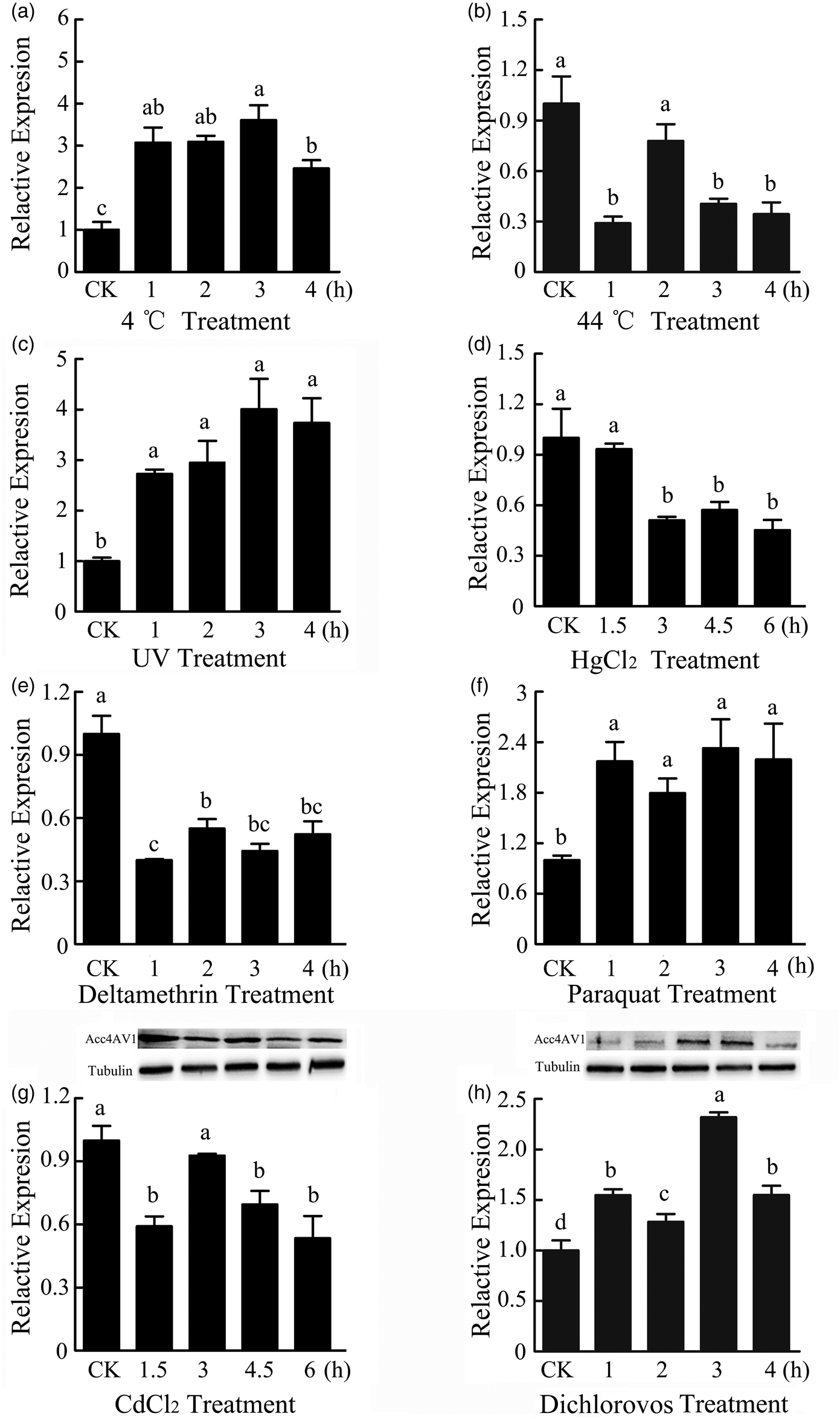
RT-qPCR was used to assess the mRNA expression profiles of AccCYP4AV1 after exposure to severe abiotic stresses. There are no significant differences in the levels of AccCYP4AV1 transcript at different time points in the untreated control group. Surprisingly, AccCYP4AV1 was induced by most of the treatments tested (fig. 2). AccCYP4AV1 responded robustly when honeybee workers were exposed at 4°C, which dramatically induced the AccCYP4AV1 transcript to the highest level in 3 h (fig. 2a). In contrast, after treatment at 44°C, the AccCYP4AV1 level was reduced, compared with that of the control, reaching a minimum at 1 h (fig. 2b). We also found that AccCYP4AV1 transcript level was increased by 4.1-fold after 3 h of exposure to UV (fig. 2c). However, The HgCl2 treatment caused a nearly 55% decrease in the relative expression of AccCYP4AV1 at 6 h (fig. 2d). As shown in fig. 2e, AccCYP4AV1 levels were downregulated after treatment with deltamethrin. Regarding paraquat, the relative expression levels of AccCYP4AV1 increased by 2.4-fold at 3 h after paraquat treatments (fig. 2f). All the results indicated that AccCYP4AV1 might be involved in the process of response to various ROS, and the response is stressor-dependent. As shown in fig. 2g, the AccCYP4AV1 transcript level was significantly reduced to a nearly 40% after 1.5 h of exposure to CdCl2, being equal to that of the control at 3 h but reducing again after more than 4.5 and 6 h of treatment. In general, the translation of AccCYP4AV1 was gradually decreased when compared with that of the control group (fig. 2g). After dichlorovos treatment, the relative expression levels of AccCYP4AV1 increased by 2.3-fold at 3 h (fig. 2h). The AccCYP4AV1 expression clearly increased and then gradually decreased (fig. 2h). The protein expression levels of AccCYP4AV1 were consistent with the RT-qPCR data (fig. 2g, h).
Expression profiles of AccCYP4AV1 in adult worker bees under various abiotic stresses. RT-qPCR was performed on total RNA extracted from 15 days post-emergence adult bees. These stresses are as follows: (a) 4 °C, (b) 44 °C, (c) UV, (d) HgCl2, (e) deltamethrin, (f) paraquat, (g) CdCl2, (h) dichlorovos. Western blot analysis of AccCYP4AV1 changes after CdCl2 (g) and dichlorovos (h) treatment. The data are given as the means ± SEM of six replicates. The letters above the bar indicate significant differences (P < 0.05) as determined by Tukey's HSD tests using SAS software version 9.1.

AccCYP4AV1 is required for defense against abiotic stress damage
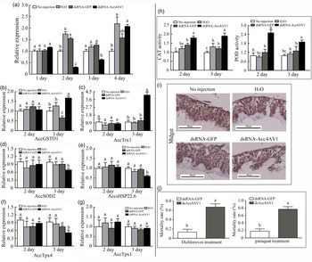
RNAi was performed to analyze the response of several stress response-related genes, which have been reported to be involved in abiotic stress responses, after AccCYP4AV1 knockdown. The AccCYP4AV1 gene was successfully silenced in RNAi-treated groups compared with that in control groups. The lowest expression levels of AccCYP4AV1 were observed at 2 and 3 days post injection of dsRNA-AccCYP4AV1 (fig. 3a). RT-qPCR results showed that the transcription of AccGSTO1 and AccTrx1 were induced when AccCYP4AV1 was knocked down, suggesting that the induced genes are involved in compensating for AccCYP4AV1 knockdown in A. c. cerana (fig. 3b, c). Furthermore, the transcription levels of AccHSP22.6, AccSOD2, and AccTpx4 decreased when AccCYP4AV1 was knocked down (fig. 3d, e, f), and the transcription levels of AccTpx1 did not show a significant effect (fig. 3g). The CAT and POD capacities of the honeybees following AccCYP4AV1 silencing were increased compared to those of the control groups (fig. 3h).
RNA interference on AccCYP4AV1 transcript levels in adults and the effect of AccCYP4AV1 silencing on other genes in A. c. cerana. (a) The mRNA levels of AccCYP4AV1; (b–g) expression profiles of other genes when AccCYP4AV1 was knocked down. (h) Effects of AccCYP4AV1 knockdown on antioxidant enzymatic activities of CAT and POD. Each value is given as the means ± SEM. Different letters above the bars indicate significant differences (P < 0.05), according to SAS software. (i) Immunolabeling of A. c. cerana midgut using our anti-AccCYP4AV1 antisera (200X). Scale bar, 100 μm; n = 4 biological replicates. (j) Mortality rate of AccCYP4AV1-knocked-down workers exposed to dichlorovos and paraquat. Means capped with different letters are significantly different (Tukey's HSD, P < 0.05).

To further determine the effect of AccCYP4AV1 knockdown in honeybee workers, IHC was also used. In this study, fig. 3i shows the MG of workers at 2 day after injection with dsAccCYP4AV1. It is possible to observe that the MG sections from AccCYP4AV1-knockdown workers were more lightly stained than those from the controls, although no visible morphological alterations were observed in the MG, which plays crucial roles in the process of detoxification.
The workers in the treatment groups had a significantly higher mortality rate than that of bees in the control group (fig. 3j). When exposed to dichlorovos, the control group had significantly higher survival rate compared with the bees subjected to AccCYP4AV1 knockdown (fig. 3j). When honeybee workers exposed to paraquat, the mortality is higher in the AccCYP4AV1-silenced group than in the control group (15% vs. 77%).
Discussion
In many insects, P450s play important roles in detoxification, including endogenous and exogenous agents (Shen et al., Reference Shen, Dong, Tian, Ma, Li, Wu and Zhu2003; Sasabe et al., Reference Sasabe, Wen, Berenbaum and Schuler2004; Kirischian and Wilson, Reference Kirischian and Wilson2012). However, only a few P450s genes from A. c. cerana have been reported (Shi et al., Reference Shi, Sun, Xu and Li2013; Zhu et al., Reference Zhu, Zhang, Liu, Chen, Li and Xu2016; Zhang et al., Reference Zhang, Chen, Li, Ma, Yu, W, Liu and Xu2018). Therefore, the identification and characterization of new P450s genes have become very attractive research areas. In this study, we isolated a novel P450s gene (AccCYP4AV1) from A. c. cerana.
As shown in online Supplementary Fig. 1, four conserved sequences and functionally important motifs, Helix-C, Helix-K, AGXE/DTT/S and a conserved PERF sequence are present in AccCYP4AV1. Helix C, helix I, helix K, PERF and heme-binding motifs are universal among P450s and are often considered the ‘signature’ of P450s (Feyereisen, Reference Feyereisen2011). Overall, these results suggest that AccCYP4AV1 is a member of the P450s. As shown in the online Supplementary Fig. 2, the phylogenetic analysis suggested that AccCYP4AV1 orthologues are present in many organisms and present a higher degree of homology with the AmCYP4AV1 sequence of A. mellifera than with other sequences.
The diversification of these P450s genes reveals that each of the identified P450s might play a different physiological role or might exhibit distinct substrate specificity (Zhu et al., Reference Zhu, Zhang, Liu, Chen, Li and Xu2016). Consistent with this hypothesis, different P450s genes are differentially expressed during the developmental stages of A. c. cerana. AccCYP4AV1 was specifically expressed in NE rather than in the other developmental stages. We speculated that AccCYP4AV1 may play a vital role in preventing abiotic stresses mediated damage during the adult stage and would be essential for the honeybee's development.
The highest level of AccCYP4AV1 was found in the MG, consistent with its role in against oxidative stimulation from various abiotic stresses. When AccCYP4AV1 was silenced, the translation level of AccCYP4AV1 was significantly reduced in MG as the IHC analysis. The MG is important for digestion and absorption and is known to function in the defense against xenobiotics in insects (Ahmad et al., Reference Ahmad, Duval, Weinhold and Pardini1991; Snyder et al., Reference Snyder, Stevens, Andersen and Feyereisen1995; Yu et al., Reference Yu, Lu, Li, Xiang and Zhang2009). Lower level of AccCYP4AV1 transcript was found in the BR and VG, indicating that the expression of AccCYP4AV1 is tissue-specific. Taken together, the variation in the transcription levels of the AccCYP4AV1 gene among different developmental stages and different tissues reveals that gene plays a special role in either stage- or tissue-specific metabolism (Chung et al., Reference Chung, Sztal, Pasricha, Sridhar, Batterham and Daborn2009).
Temperature is an abiotic environmental factor that causes physiological changes and oxidative stress in organisms (An and Choi, Reference An and Choi2010; Lushchak, 2010; Mi and Choi, Reference Mi and Choi2010). Honeybees face extreme temperatures during their lifespan due to climate changes. Here, we found that AccCYP4AV1 might play crucial roles in the process that avoids cold lesions, thus protecting honeybees against ROS damage. Previous studies showed that heat-shock stress can generate O2− and result in polyamine oxidation and that cold temperature can induce cells to undergo apoptosis due to ROS damage (Rauen et al., Reference Rauen, Polzar, Stephan, Mannherz and De Groot1999). The discrepancy in AccCYP4AV1 responses to 4°C and 44°C may be attributed to the differences in the regulation mechanism. Heavy metals may influence the behavior and normal development of insects (Park and Park, Reference Park and Park2007; Fujimura et al., Reference Fujimura, Usuki, Sawada and Takashima2009; Tang et al., Reference Tang, Huang, Zhou, Li, Xie and Liu2012). Heavy metals Cd and Hg are well-known to be the most poisonous ones and can directly bind to enzyme metal ion sites, thus inactivating the function of proteins (Rashed, Reference Rashed2001). As a pollinator, forager honeybees may encounter heavy metals when they forage for pollen and nectar outside the hive in the environment. We found that CdCl2 affected the expression of AccCYP4AV1 in a time-dependent manner, which could be due to more pronounced toxic effects of CdCl2 at longer times. Cd2+ can cause toxicity by displacing metal ions such as Cu2+ and Fe2+ (Kozlowski et al., Reference Kozlowski, Kolkowska, Watly, Krzywoszynska and Potocki2014). Furthermore, western blot analysis showed that the translation level of AccCYP4AV1 was correlated with its mRNA expression. However, the transcription levels of AccCYP4AV1 were reduced by HgCl2 treatment. Different heavy metals have different mechanisms of toxicity.
Pesticides are important abiotic factors that could lead to environmental pollution and influence insect behavior and lifestyle (Asghari et al., Reference Asghari, Moloudizargari, Bahadar and Abdollahi2016). P450s may be included in the metabolism and resistance to various pesticides in insects (Scott, Reference Scott1999; Karatolos et al., Reference Karatolos, Williamson, Denholm, Gorman, Ffench-Constant and Bass2012; David et al., Reference David, Ismail, Chandor-Proust and Paine2013; Edi et al., Reference Edi, Djogbénou, Jenkins, Regna, Muskavitch and Poupardin2014). The transcription levels of AccCYP4AV1 were induced by paraquat and dichlorovos treatments, suggesting a role of AccCYP4AV1 in the protection of honeybees against the damage induced by pesticides. Protein expression was consistent with the transcriptional level at dichlorovos treatments. Nevertheless, different abiotic stresses could lead to different expression patterns of these genes. Indeed, the expression level of AccCYP4AV1 was repressed under deltamethrin treatment. This phenomenon suggests it might have some other effect on this gene's expression by an unknown mechanism. Furthermore, there is also evidence that UV radiation is a harmful environmental stressor that can produce oxidative damage in honeybees, which damages membrane lipids and proteins (Ravanat et al., Reference Ravanat, Douki and Cadet2001; Cadet et al., Reference Cadet, Sage and Douki2005; Schauen et al., Reference Schauen, Hornig-Do and Schomberg2007; Nguyen et al., Reference Nguyen, Michaud and Cloutier2009). In this study, the RT-qPCR results revealed that AccCYP4AV1 can be induced during UV radiation exposure, which indicated that AccCYP4AV1 may play important roles in UV resistance.
Previous researches have proven that gene silencing is crucial for studying the function of genes in A. c. cerana (Huvenne and Smagghe, Reference Huvenne and Smagghe2010; Goic et al., Reference Goic, Vodovar, Mondotte, Monot, Frangeul and Blanc2013; Wilson et al., Reference Wilson, Kenny and Dearden2014). In this study, we found that the transcription levels of AccGSTO1 and AccTrx1 were upregulated when AccCYP4AV1 was silenced. Studies have proved that these genes are involved in the process of abiotic stress defense (Yu et al., Reference Yu, Kang, Meng, Guo and Xu2011; Yao et al., Reference Yao, Chen, Yan, Feng, Zhang and Guo2014), which indicates that AccCYP4AV1 may participate in the antioxidant process. When AccCYP4AV1 silenced, AccsHSP22.6, AccSOD2, and AccTpx4 were reduced, which have been proved that AccsHSP22.6, AccSOD2, and AccTpx4 could play important roles not only in stresses defense but also in the development in A. c. cerana (Jia et al., Reference Jia, Sun, Shi, Yan, Han and Guo2014; Zhang et al., Reference Zhang, Liu, Guo, Li, Gao, Guo and Xu2014; Huaxia et al., Reference Huaxia, Fang, Yan, Feng, Wang and Guo2015).
In organisms, CAT and POD are antioxidant enzymes that play crucial roles in the response to ROS scavenging (Corona and Robinson, Reference Corona and Robinson2006). When AccCYP4AV1 was silenced, the increased activities of CAT and POD indicate that AccCYP4AV1 may be involved in antioxidation. When AccCYP4AV1 was knocked-down, the dichlorovos and paraquat treatments significantly increased the mortality rate. These results further confirmed that AccCYP4AV1 may play essential role in the survival of insects in response to pesticide stress.
In this study, we isolated and identified AccCYP4AV1 from A. c. cerana. Our results further demonstrated that AccCYP4AV1 might play a crucial role in the development and was involved in honeybee resistance to abiotic stresses. Our study may help to further investigate the functions of the insect P450s genes in response to abiotic stress at the molecular level and provide information for researchers to clarify the potential consequences of abiotic stress exposure in insects and other organisms.
Supplementary material
The supplementary material for this article can be found at https://doi.org/10.1017/S0007485320000644
Acknowledgements
This work was financially supported by Shandong agricultural fine varieties breeding Projects (2017LZN006), the National Natural Science Foundation of China (No. 31572470), the earmarked fund for the China Agriculture Research System (No. CARS-44).
Conflict of interest
The authors declare that they have no conflicts of interest with the contents of this article.
Ethical standards
Animal housing facilities and handling protocols were approved by the Animal Welfare and Health Committee of Shandong Agricultural University.
Ethical approval
All applicable international, national, and/or institutional guidelines for the care and use of animals were followed.