Since first performed in 1989, and reported in 1991,1 stenting of both native and recurrent coarctation of the aorta has become widely practiced and widely reported, with nearly 20 series published since the initial description.2–20 This literature demonstrates that the technique is generally safe and effective in the short term. Despite this extensive literature, there are several unresolved concerns. First, there is limited information on intermediate outcomes. Second, the frequency, significance, and types of complications related to stenting are not well characterized. Third, placement of stents may result in catastrophic complications. We are aware of at least 10 instances of aortic rupture, apart from the isolated cases reported in the literature.21 All these episodes are life-threatening, and many did, indeed, prove fatal.
Placement of stents for treatment of coarctation involves angioplasty, which by definition injures the aortic wall. Among the most common adverse events occurring after transcatheter therapy for coarctation are formation of aneurysms, and aortic rupture, with some form of traumatic aortic injury reported in up to one-third of patients.22–24 The understanding of the nature of the injury to the aortic wall, however, is limited. There is no standardized nomenclature for characterizing the mural injury. Risk factors for, and mechanisms of, injury have not been defined. Data regarding the natural history of acute injuries is lacking, and the incidence of new mural injuries over time is unknown. The purpose of this study, therefore, was to review a large series of stenting procedures performed at a single institution for treatment of aortic coarctation, to assess their acute and intermediate efficacy, to evaluate associated injuries to the aortic wall, and to determine the rates of stent-related complications.
Methods
Patients
Transcatheter treatment of aortic coarctation may include simple angioplasty, angioplasty followed by stenting, or primary stenting without prior angioplasty. Lacking evidence-based criterions for stenting obstruction within the aortic arch, operators at our institution rarely state an explicit “intent to place a stent” prior to a procedure. Rather, in most cases, decisions about stenting are made after initial angioplasty is performed and assessed. The decision to place a stent typically depends on a combination of factors, including the age and size of the patient, the proximity of the obstructive lesion to the carotid and subclavian arteries, the severity and nature of the obstruction, presence of associated haemodynamic abnormalities, and the response to the initial angioplasty. As a result of this practice, an intention-to-treat analysis is not practical in patients referred for endovascular treatment of aortic coarctation at our centre. Accordingly, this study was designed as a retrospective analysis of patients who had undergone placement of stents for the treatment of aortic coarctation.
The database relating to our cardiovascular programme was queried for patients who underwent implantation of an aortic stent between December 1989 and July 2005. In this way, we identified a total of 172 patients. We excluded 17 patients who underwent stenting for aortic obstructions associated with a systemic arteriopathy, and/or abdominal coarctation.20 Also excluded were 2 patients who did not have aortic obstruction but underwent placement of an aortic stent to stabilize an embolized fragment of a subaortic stent, or to tack down a traumatic aortic dissection. Thus, our cohort was comprised eventually of 153 patients.
Catheterization and placement of stents
The techniques of implantation have been described previously,12 but our practice has evolved somewhat, particularly with regard to pre-dilation and multi-view angiography (see Aortic imaging below). Pre-dilation, that is inflation of a balloon without a stent, is now performed prior to placement of the stent in nearly all cases, using 1 or more low-pressure balloons inflated to pressures of less than 8 atmospheres. The diameter of the initial balloon typically ranges from 150 to 300% of the diameter of the coarctation, and is no larger than 150% of the diameter of the adjacent aorta. Pre-dilation serves a number of purposes. It can provide therapeutic benefit by initiating a tear that can be stabilized and supported with a stent, creating a larger final aortic diameter. In some instances, the tear associated with “pre”-dilation provides sufficient relief of obstruction that stenting is not necessary. These cases, however, are not part of our study group. When stenting is indicated, pre-dilation allows localization of the waist or waists in the balloon, the identification of multiple waists, an estimation of aortic compliance, and anticipation of any migration of the balloon that may occur during placement of the stent. Occasionally, pre-dilation will identify an unexpectedly tight waist, less than 75 to 80% of the diameter of the balloon. In these cases, a smaller balloon is used for deploying the stent.
Whenever reasonable, we use a stent that can be expanded to an adult size. Occasionally, in small patients, low-profile pre-mounted stents may be used despite the limited expansion diameter. The stent is either front-loaded into a long sheath, or advanced through a sheath previously positioned above the coarctation. A side-arm adapter on the end of the long sheath allows for local injection of contrast during the procedure, which enables fine adjustments of position. In some instances of difficult anatomy within the arch, additional angiography is performed through a second catheter introduced via the radial artery, or into the ascending aorta by a transseptal approach. After deployment of the stent, expansion with a larger balloon may be performed, depending on a variety of factors, including the residual gradient, the presence of a waist in the stent, and the appearance of the aorta.
Aortic imaging
Two authors (AMQ, ACM) reviewed pre-intervention and follow-up angiograms and computed tomography scans, when applicable, for all patients. Echocardiographic imaging of the arch was not evaluated or analyzed for this study.
Pre-intervention aortograms were performed to define the anatomy of the coarctation, the adjacent aorta, and brachiocephalic branches, and to identify existing abnormalities involving the aortic wall. Angiograms performed after angioplasty and stenting were typically profiled in 4 different projections, which varied depending on the anatomy of the arch and the coarctation. Imaging in multiple views facilitated visualization of unanticipated vascular tears occurring at or adjacent to the coarcted segment. Repeated images also helped distinguish small or irregular vascular injuries from the adjacent aorta or collateral vessels, and allowed for more complete and accurate assessment of these three-dimensional findings.
Jailed vessels and stent fractures.
Coverage of brachiocephalic arterial branches by a stent was noted, and events, symptoms, or signs suggestive of neurovascular compromise or embolism were recorded. The degree of coverage by the stent was recorded. Arteries that were covered completely were considered “jailed”, while those with more than 50%, but less than 100%, of the orifice covered by the stent were considered “partially jailed”. Patients who underwent follow-up catheterization or chest fluoroscopy were assessed for stent fractures. Because stent fractures may only be evident with dynamic imaging or direct inspection, simple chest radiography was not considered sufficient to exclude the possibility of fracture.
Injury to the aortic wall
Lacking an established nomenclature for categorizing aortic mural injuries in patients undergoing transcatheter treatment for aortic coarctation, we developed a set of definitions designed to reflect the spectrum of injuries encountered in this population. Injuries were characterized angiographically and categorized as therapeutic tears, dissections, aneurysms, or ruptures. A therapeutic tear, necessary for successful angioplasty, is a tear in the intima and part of the media, with contrast confined to the aortic wall in the radial dimension, and to the coarcted segment in the axial dimension. A dissection is a tear in the intima and media that extends beyond the coarcted segment in the axial dimension, permitting extraluminal contrast to track proximally or distally from the dilated segment. An aneurysm, or area of loss of muscular integrity of the vessel wall, is defined angiographically as a defect in the aortic wall that extends more than 3 millimetres beyond the presumed adventitial plane in the radial dimension, has an angle of incidence of greater than 30 degrees relative to the adjacent aorta, and hence has a discrete length. In our series, we have distinguished aneurysms from “aneurysmal dilatation”, the latter feature often being seen in patients with native or repaired coarctation as pre- and post-stenotic dilation. An aortic rupture is a frank disruption of the aortic wall, which appears angiographically as extravasation of contrast beyond the confines of the aorta into the mediastinum or pleural space.
Analysis of data
Outcomes included the acute reduction in gradient, the increase in diameter at the site of coarctation, survival, freedom from reintervention, stent fracture, and adverse events. Independent variables analyzed for association with outcomes included age, prior aortic intervention, associated anomalies, pre-existing aortic mural abnormalities, the location of the coarctation, and the gradient across it. Gradients as reported are those measured from peak-to-peak during catheterization, unless otherwise stated. We also recorded that type of stent, the sizes of balloons and their ratio to the diameter at the site of coarctation, and the magnitude of residual aortic obstruction. Survival and freedom from reintervention were assessed with Kaplan-Meier analysis. Association between independent variables and time-dependent outcomes was assessed using Cox regression, including multivariable analysis with forward stepwise entry if appropriate. For comparison of continuous variables before and after intervention, we used paired t-testing or the Wilcoxon signed-rank test. For analysis of factors associated with acute outcomes, and to make comparisons between groups of means and proportions, we employed independent-samples t-test and Fisher's exact test, respectively. Data is presented as mean with standard deviation, or median with range. Our study was approved by the Children's Hospital Committee for Clinical Investigation.
Results
Patients
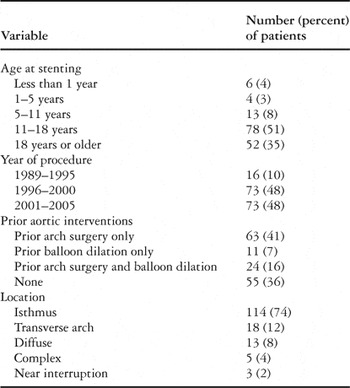
We identified 153 patients who underwent implantation of stents for treatment of aortic coarctation between December 1989 and July 2005 and who met our criterions for inclusion in the study. The median age at stenting in the 153 study patients was 15.8 years, with a range from 6 days to 60 years. Demographic, anatomic, and clinical data are summarized in Table 1.
Table 1. Baseline demographic and clinical details (n = 153).

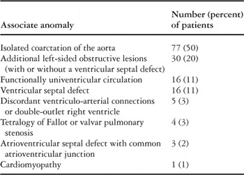
Coarctation was an isolated anomaly in 77 patients (50%). Among the remaining 76 patients in whom additional cardiovascular defects were present, 16 had a functionally single ventricle (Table 2).
Table 2. Associated cardiovascular anomalies (excluding bicuspid aortic valve) (n = 153).

A total of 142 prior interventions had been performed in 98 patients (65%). The median time from the most recent arch intervention in these patients was 8.3 years, with a range from 1 day to 44 years.
Catheterization and placement of stents
At the initial catheterization, 171 stents were implanted in 153 patients, with multiple stents in 16. Implanted stents included 123 Palmaz®™ large or XL stents (Cordis Endovascular, Warren, NJ), 41 Palmaz® Genesis™ XD stents (Cordis Endovascular, Miami, FL), 1 of which was covered with expanded polytetrafluoroethylene membrane, and 7 premounted stents (1 Palmaz® Genesis™ and 6 coronary stents). Palmaz® Genesis™ stents were incorporated into our practice in July 2002, and gradually replaced Palmaz large and XL stents as the latter were removed from the Cordis product line. The 7 premounted stents were implanted in 6 neonates or infants, 2 stents being implanted in 1 patient, including 2 who were acutely ill after palliative surgery for functionally single ventricles or repair of interrupted aortic arch, 1 who developed an aortic aneurysm after balloon dilation alone (see below), and 3 who were not considered good surgical candidates due to extremely low birth weight, very low birth weight with associated anomalies, or multiple associated medical problems.
The coarctation was predilated immediately preceding implantation of the stent in 113 patients (75%), and in 12 additional patients a test dilation was performed as part of a prior catheterization procedure. The size of the balloon used to place the stent was the same as that of the balloon used for predilation in 45 cases. In 67 cases, the balloon was larger, by a median of 3 millimetres, with a range from 1 to 10 millimetres, and in 1 case it was smaller. In 111 patients (73%), the stent or stents were expanded with at least 1 additional balloon after implantation. The ratio of the maximum diameter of the balloon to the narrowest diameter of the coarctation was high in patients with a diameter at the site of coartation of 5 millimetres or less, with ratios up to 6 to 1 and between 1.4 to 1 and 3 to 1 in patients with a diameter at the coarctation larger than 5 millimetres. Cutting balloons were not used in any patient in this series.
Acute outcomes
Haemodynamics and relief of obstruction.
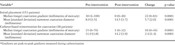
The haemodynamics and diameters of the coarctation prior to dilation are summarized in Table 3. Factors associated with a lower gradient across the coarctation prior to intervention included presence of a functionally univentricular circulation (p less than 0.001), associated cardiovascular anomalies (p equal to 0.001), and prior intervention to the aortic arch (p equal to 0.003).
Table 3. Haemodynamic data at initial placement and follow-up reinterventions.

The haemodynamics and diameters at the site of coarctation after stenting are summarized in Table 3. Residual obstruction of at least 20 millimetres of mercury measured as a peak-to-peak gradient was present in 7 patients (5%), and residual obstruction of 15 millimetres of mercury or more was present in 16 (10%). The only factors associated with increased probability of residual obstruction of 15 millimetres of mercury or more were a higher gradient prior to stenting, at 50.2 plus or minus 21.6, versus 29.2 plus or minus 15.4 millimetres of mercury (p less than 0.001), and diffuse or complex obstruction of the transverse aortic arch, found in 7 of 16 versus 29 of 136 patients with other types of obstruction (p = 0.05).
Aortic mural injury.
Angiography demonstrated pre-existing aneurysms in 19 patients (12%), 16 of whom had undergone prior interventions to the aortic arch (15% of 103). These comprised 6 of 63 (10%) patients who underwent prior surgery alone, 5 of 11 (45%) who underwent prior balloon dilation alone, and 5 of 24 (21%) who underwent prior surgery and dilation. These aneurysms had been previously diagnosed in 5 of the 10 patients who underwent prior balloon dilation, and in 1 of the 6 who had prior surgery only.
After dilation or stenting of the aortic coarctation, acute mural injuries were observed in 3 patients (2%), including 1 rupture, 1 dissection, and 2 aneurysms in a single patient. The rupture occurred in a 6-week-old patient with a functionally univentricular circulation who underwent balloon dilation 1 month after the first stage of the Norwood sequence, and who was treated successfully with placement of a stent and coil embolization. The dissection occurred in a 20-year-old patient with a native coarctation and near occlusion of the aortic arch. This patient developed an anterior intimal flap at the site of the anterior coarctation, with subsequent dissection 4 centimetres down the descending aorta (Fig. 1). The entry point of the dissection was stented and the flap tacked down. The aneurysms occurred in an 11-year-old patient with a recurrent coarctation, who developed 2 aneurysms around and through the superior portion of the stent. A covered stent was placed and successfully excluded the larger aneurysm. In the first 2 patients, the dissection or rupture occurred during initial dilation, and upon immediate recognition of the potentially catastrophic injury, a stent was placed. In the third patient, the aneurysms were noted after placement of the stent. Thus, overall, aortic mural injuries other than therapeutic tears occurred in 2% of patients, and stent-induced acute aortic injury occurred in 0.7%, that is in only 1 of 153 patients. No patient died as a result of, or required surgical management for, an aortic injury.

Figure 1. Aortic angiograms in a 20-year-old patient who developed a dissection of the descending aorta after balloon dilation of a severe, nearly occluded, native coarctation, which was subsequently stented. (a) An angiogram in the aortic arch demonstrates a very tight coarctation (arrow), with post-stenotic dilation of the descending aorta. The left subclavian artery, from which major collaterals originated, is notably enlarged. (b) After dilation of the coarctation, there was initially a tear at the site of narrowing. (c) Shortly thereafter, a dissection was seen extending caudally along the medial aspect of the aorta (black arrows). The intimal tissue plane between the true and false lumens is indicated with black arrowheads, and the balloon-mounted stent prior to deployment is indicated with white arrows. (d) Three months after stenting, angiography demonstrated no significant change in the appearance of the dissection, the mouth of which is covered by the distal aspect of the stent (white arrows indicate the proximal and distal edges of the stent). At the time of follow-up catheterization, 2 additional stents were placed across the proximal and distal mouths of the dissection, and flow through the false lumen was decreased but not eliminated.
None of the patients with pre-existing aneurysms, or with pre-existing therapeutic tears at the site of prior balloon angioplasty, developed larger aneurysms or more advanced aortic mural injuries after placement of a stent (Fig. 2).

Figure 2. (a) A therapeutic tear (arrow) was present at the site of coarctation in a patient who had previously undergone arch repair followed by balloon dilation. There is also significant poststenotic dilation of the descending aorta. (b) After pre-dilation of the coarctation, the tear is slightly extended (arrows). (c) After stent placement, the tear is no longer seen, and there is no evidence of aneurysm or other aortic wall injury (arrows mark the proximal and distal edges of the stent).
Jailed arteries.
We identified complete jailing in 13 patients, and partial jailing in 11, of either 21 subclavian arteries, or 4 common carotid arteries, along with separate jailing of the vertebral artery in 1 patient. Jailing, therefore, occurred in 24 patients (16%). In 10 of these cases, stents were modified after implantation either by dilation through stent cells into the jailed artery in 3 patients, or overdilation and flaring of the end of the stent into the affected artery in 7. In 25 other patients (16%), the stent extended less than 50% over the origin of a subclavian artery in 21, the common carotid artery in 3, or the common brachiocephalic trunk in 1, of whom 13 had the overhanging stent flared by dilation into the covered branch. Jailing of arteries did not result in clinically apparent thromboembolic events, neurologic symptoms, reduced flow to the affected artery, or haemodynamic compromise.
Acute adverse events.
Major adverse events occurred acutely in 3 patients. Wire-related dissection of the ascending aorta, with compromise of flow to the left coronary artery, occurred in 1 patient early in our experience, as reported previously.12 Neurologic injuries diagnosed as vertebrobasilar strokes occurred in 2 patients, both of whom had persistent neurologic deficits. Less serious adverse events included injury to the femoral or iliac arteries in 3 patients, and production of arteriovenous fistulas in the vessels of access in 2, with surgical treatment required in 2 cases. There were 6 cases of embolization or malposition of the stent, all of which were managed percutaneously, by deployment of an embolized or malpositioned stent in a non-therapeutic distal position in 5, and removal of an embolized stent through a large sheath in 1. We were unable to identify any risk factors for embolization.
Early mortality.
Of our cohort, 2 patients died from complications of preexisting cardiac disease. Both patients were acutely ill, with low cardiac output and obstruction to the aortic arch after surgical procedures for functionally univentricular palliation, and were being supported on extracorporeal membrane oxygenation. Despite successful placement of stents producing relief of the obstruction within the aortic arch, both patients failed to recover and were removed from support 2 weeks later.
Intermediate outcomes
Patients.
Cross-sectional follow-up was obtained in 140 of the 151 surviving patients (93%), a median of 2.5 years, with a range from 2 months to 12.5 years, after placement of the stents. Follow-up of 5 years or longer was available in 30 patients. Among 121 patients with available medication data, 69 (57%) were prescribed antihypertensive medications.
Survival.
There were 2 late deaths, neither being related to the stent or catheterization. Both patients dying were adolescents with failing physiology of the Fontan circulation, who died 3.5 and 4 years after placement of the aortic stent. There were no deaths after aortic reinterventions. Estimated survival by Kaplan-Meier analysis was 99% at 1 year, and 94% at 5 and 10 years after placement of the stents. All 4 deaths occurred in patients with functionally univentricular physiology. No patients with a biventricular circulation died, and none of the patient-related or procedural variables analyzed were significantly associated with decreased survival over time.
Aortic reinterventions.
Aortic reinterventions were performed in 51 patients, these being surgical in 3 patients, and 1 catheter reintervention in 41, with more than one catheter-based reintervention in the other 7, including redilation of the existing stent or stents in all, and placement of additional stents in 2. The 3 patients who underwent surgical reintervention included the patient with the highest residual gradient after stenting, measured at 40 millimetres of mercury, this patient having diffuse hypoplasia of the aortic arch. The other 2 patients included one who underwent stenting as an extremely low-weight neonate, then after 3 months of haemodynamic stability and growth underwent closure of a ventricular septal defect and arch repair; and another whose original surgical repair included insertion of an ascending-to-descending aortic tube graft that was causing obstruction to the superior caval vein and the tracheobronchial tree, and was thus resected along with augmentation of the arch. The peak gradient prior to reintervention was 10 millimetres of mercury or less in 19 of the 48 patients who underwent repeated transcatheter intervention (40%), reflecting our practices of staged therapy for severe obstructions and elective redilation of stents to account for somatic growth in children and adolescents.
Acute results of the catheter-based reinterventions are summarized in Table 3. An increased diameter was achieved in almost all patients, with increases typically 1 to 3 millimetres, and by as much as 6 millimetres. By Kaplan-Meier analysis, freedom from reintervention was 83% at 1 year, 48% at 5 years, and 45% at 10 years (Fig. 3). The only factors associated with shorter freedom from aortic reintervention by Cox regression were a higher gradient prior to stenting, and higher gradients after stenting (both p = 0.01). By multivariable Cox regression, only the higher gradient prior to stenting remained as a significant independent predictor of shorter freedom from aortic reintervention.

Figure 3. Kaplan-Meier curve demonstrating estimated freedom from reinter-vention after aortic stenting. Patients were censored at the time of the first aortic reintervention. The numbers of patients at risk are listed below the curve.
Adverse events.
Adverse events related to aortic reintervention occurred in 3 patients. In 1 patient with functionally univentricular physiology, multiple dilations were needed prior to completion of the Fontan circulation. At surgery, a stented portion of the aorta was exposed. During catheterization in the early postoperative period, redilation of the stent resulted in aortic rupture. The rupture was excluded with a covered stent and the patient survived with temporary circulatory support. The other 2 adverse events consisted of a wire-related mediastinal haematoma that resolved with observation, and embolization of an additional stent that was placed at the follow-up catheterization, which was addressed by deploying the embolized stent in the femoral artery.
Aortic mural injury.
Follow-up aortic imaging was performed in 82 patients (54% of surviving patients) at a median of 2.8 years, with a range from 1 day to 11.7 years, after placing the stents, including angiography in 52 patients, computed tomography in 15, and both in 15. This imaging revealed new aneurysms in 5 of 82 patients (6%), 0.7 to 4.9 years after stenting. In 1 patient, an aneurysm formed on the posterior aspect of the transverse arch, remote from the site of implantation, apparently due to an injury to the transverse arch resulting from wire position and movement of the nose of the balloon. In 3 patients, small aneurysms originating within the stented segment of aorta, in the region of the coarctation, were seen on follow-up angiography (Fig. 4). It could not be determined whether the aneurysms developed from progression of a therapeutic tear. One of these aneurysms was obliterated at follow-up catheterization after the stent was re-expanded, and another was diagnosed at the second reintervention, and was presumably a result of the first reintervention, this being redilation of the stent. In the other patient, a new aneurysm at the margin of the stent was observed. None of the independent variables analyzed were associated with new aneurysmal formation.

Figure 4. A small circumferential aneurysm originating from within the stent is seen in this patient one year after stent implantation (white arrow). The black arrows mark the proximal and distal edges of the stent. This aneurysm was not seen on straight anteroposterior and lateral projections. Also note the pre-stenotic dilation of the arch.
All 3 patients in whom aortic mural injuries were diagnosed acutely after placement of stents had follow-up imaging. In the patient who suffered an aortic rupture, follow-up angiography demonstrated no evidence of aneurysm or extravasation. Similarly, in the patient with 2 aneurysms, the aneurysm that had been excluded by the covered stent was not evident on follow-up angiography, and the other aneurysm was decreased in size. In the patient with a dissection, follow-up catheterization was performed 3 months after the initial placement, at which time the dissection was in communication with the aortic lumen both proximally and distally, and had not grown. After placement of 2 additional stents during the follow-up catheterization, covering both the entry and exit of the dissection, there was reduced flow into the false lumen.
Jailed vessels and stent fractures.
Among the 49 patients with jailed, partially jailed, or partially covered brachiocephalic arteries, 20 underwent follow-up catheterization. None of these patients had obstruction of the jailed vessel, evident thrombus, or clinical embolic events associated with the overhanging portion of the stent.
Fractures (Fig. 5) were diagnosed a median 3.5 years after stenting in 12 of 67 patients (18%) who underwent follow-up angiography. Nine of the fractured stents were Palmaz stents and 3 were Palmaz Genesis stents (p = not significant). Of the 12 fractures, 6 were complete, 3 being longitudinal, 2 transverse, and 1 compound, and the other 6 were partial. In 2 cases, fracture was observed only after redilation. All of the fractured stents were well apposed to the aortic wall, with no discernible embolization of fragments. None of the patient-related or procedural variables analyzed were associated with fracture.

Figure 5. Fluoroscopic images in 2 patients who developed stent fractures that were diagnosed approximately 3 years after placement of Palmaz Genesis stents for (a) native coarctation or (b) postoperative transverse arch obstruction demonstrate complete circumferential fracture of the stents (arrow in image (a)).
Discussion
Acute and intermediate efficacy of stenting
In this large retrospective series, placement of stents was highly effective at acutely relieving various types of aortic obstruction. This finding supports prior reports of smaller cohorts in which it was demonstrated that stenting consistently reduces directly measured gradients to less than 5 millimetres of mercury in both simple and complex anatomies.2–20
During a median follow-up of 2.5 years, with 30 patients followed for 5 years or longer, aortic reintervention was common. Freedom from reintervention was just under 50% at 5 years, notably higher than in most surgical series. This rate of reintervention is attributable in part to a deliberate multi-stage approach to the transcatheter treatment of aortic coarctation in some patients. In our population, predominantly made up of children and adolescents, reintervention was undertaken “electively” in some patients to reexpand the stent to account for somatic growth, and not necessarily to treat residual or recurrent gradients. In other patients, those with angiographically severe coarctation, stents were initially placed at submaximal diameter, with the intention of redilating the stent at a second procedure. This staged approach theoretically allows time for the initial therapeutic tear to heal, and may reduce the likelihood of catastrophic aortic injury. The relatively high rate of reintervention may also reflect our practice of aggressively treating even mild obstruction, with two-fifths of reinterventions performed in patients with a gradient of 10 millimetres of mercury or less. We adopted this policy after finding that relief of even mild obstruction can reduce left ventricular end-diastolic pressure.12 Regardless of the indication, redilation with or without placement of additional stents was effective, both at relieving any gradient and increasing the size of the stent, by 2 to 3 millimetres in most cases.
Aortic mural injury
The therapeutic mechanism of balloon angioplasty requires disruption of the intimal and medial layers of the stenotic segment of vessel. Accordingly, vascular tears are almost universally present after effective balloon angioplasty for aortic coarctation. More extensive aortic mural injuries can take a variety of forms.8, 22–24 One of the most common acute adverse events described in patients undergoing angioplasty or stenting for aortic coarctation is the formation of aneurysms and other non-therapeutic mural injuries, which have been reported in up to one-third of patients undergoing angioplasty22–24 and one-sixth of patients undergoing stenting.8 The most extreme form of aortic mural injury due to dilation or stenting of an aortic coarctation is frank aortic rupture, which is uncommon but frequently catastrophic.2, 21, 22
Despite the reported frequency and potential severity of aortic mural injuries in patients undergoing treatment of coarctation, the risk factors for and implications of such injuries are not well understood, in part because there is no standardized nomenclature for discussing them. In fact, most reports of stenting or angioplasty for coarctation fail to define the criterions by which aneurysms or other aortic injuries are diagnosed. Where definitions are specified,25 such as aortic dimension greater than 150% of the diameter of the aorta at the level of the diaphragm, they are typically derived from the adult surgical literature.26 In patients with native or repaired coarctation of the aorta, in whom non-aneurysmal dilation of the aorta in pre- or post-stenotic position is common, the relevance of these criterions is arguable, and underscores the need for a specific, standardized, definition for use in children or patients with congenital lesions. In this study, we have used a simple angiographically based categorization of aortic mural injuries that recognizes both pathologic and therapeutic tears, reflecting both the potential adverse implications of aortic injury and the necessity of controlled tears for the effective treatment of aortic coarctation.
Overall, pathologic aortic mural injuries such as aneurysm, dissection, or rupture occurred at the time of dilation or stenting in 3 of 153 patients (2%) in our series, with no catastrophic events and no injuries requiring surgical intervention. At follow-up reintervention, aortic rupture after redilation of an existing stent occurred in 1 of 47 cases. Due to the low incidence of non-therapeutic injuries in our series, we were unable to identify risk factors, and our ability to comment on the natural history of these lesions is limited. In 2 of the 3 patients with non-therapeutic acute aortic injuries, the angiographic abnormalities were improved or no longer evident on follow-up imaging, suggesting that such injuries generally remain stable.
It is not known how often new aneurysms form in the intermediate- or late-term after aortic stenting. Of the 82 patients in this series who underwent follow-up imaging, 5 (6%) developed new aneurysms, 1 of which occurred only after a second intervention at which the stent was redilated. The locations of the aneurysms may provide some insight as to their aetiology. The origins of the aneurysms were around the stent in 1 patient, implying injury from the margins of the stent, and within the stent in 3 patients, implying balloon-induced injury to the aortic wall in the region of the coarctation. Importantly, 1 of the aneurysms detected at follow-up was remote from the coarctation and the stent, and appeared to be due to a tear induced by the nose of the pre-dilation balloon. Of note, none of these were at the site of angiographically apparent therapeutic tears seen after pre-dilation, which supports our impression that pre-dilation does not increase the risk of pathologic aortic mural injury after placement of the stents. In some cases, stenting may help to stabilize aortic mural injuries that occur after angioplasty by redistributing mural stress in the region of vascular injury.
Technical considerations
Recognizing that an important element of effective endovascular treatment of aortic coarctation involves a controlled tearing of the intima and media, we have increasingly incorporated pre-dilation into the stenting procedure. Although opinions differ about the utility and safety of pre-dilation before stenting, we believe that pre-dilation may help to avoid the rare catastrophic aortic rupture by revealing the location and compliance of stenoses, as well as the compliance of the surrounding aorta. Aside from the potential to prevent catastrophic injury, the assessment of compliance and response afforded by pre-dilation may allow the operator safely to implant stents at a larger diameter than would be chosen for primary stenting, when waists in the balloon may not be apparent. Moreover, in some cases, there may be a favourable response to dilation alone, such that implantation of a stent may not be necessary. Despite our bias in favour of pre-dilation, we recognize that the effect of pre-dilation on safety is difficult to demonstrate due to the relative infrequency of major aortic complications. Another technical factor that may help avoid major aortic mural injuries is multi-projection angiography following dilation and prior to placement of the stents, which allows for optimal visualization and definition of therapeutic tears.
Other adverse events, jailed vessels, and stent fractures
Major adverse events occurred in 3% of patients, including stroke in 2, wire-related aortic dissection in 1, and arterial complications requiring surgery in 2. These events, along with the aneurysm caused by the nose of the pre-dilation balloon, highlight the importance of attention to technical details such as stable and appropriate distal positioning of the wire, minimally traumatic vascular access and cannulation, and fastidious maintenance of procedural anticoagulation.
Coverage of an arterial branch arising from the aortic arch can be difficult to avoid in certain situations, such as transverse arch obstruction or stenosis immediately adjacent to the left subclavian artery. Complete coverage or coverage of more than 50% of an artery occurred in 16% of patients, and coverage of less than 50% occurred in another 16%. In most cases, the subclavian artery was covered, but an artery directly supplying the cerebral circulation was affected in 9 patients. In none of these patients did coverage of the artery lead to clinically evident neurovascular events or apparent compromise of flow. We frequently attempted to modify the overhanging stent by dilation through cells of the stent or flaring the overhanging ends. The relative risks and benefits of such modifications are not clear, but did not lead to any acute or follow-up complications, neurovascular symptoms, or haemodynamic compromise.
Among 67 patients undergoing follow-up angiography or fluoroscopy, fractures were identified in 12 (18%), in 2 cases after redilation. The fractured fragments did not embolize or cause noticeable haemodynamic compromise. Breakage of stents used to treat aortic coarctation has been described previously.3, 27, 28 The cause of such fractures is unknown, and we did not identify any patient-related or procedural risk factors. Other sites in which vascular stents commonly fracture, such as conduits placed in the right ventricular outflow tract, femoropopliteal arteries, and subclavian vessels, are vulnerable to repetitive external compression,29–31 which is not the case with aortic stents. Ultimately, the importance and mechanisms of fracture will require further observation and evaluation.
Limitations
There are several limitations of this study. The small number of non-therapeutic aortic mural injuries precluded determination of risk factors, and did not permit clear demonstration of the benefits or shortcomings of technical measures such as predilation. Also, the lack of follow-up aortic imaging in 46% of patients may introduce ascertainment bias with respect to estimating the frequency of new aneurysms developing after aortic stenting. Another factor that may limit the importance of our findings is that our practice does not lend itself to an intention-to-treat analytic strategy, and we did not design the study as a comparison of stenting and simple angioplasty, so our data do not speak to the question of which of these treatments is most appropriate for any given patient.
Acknowledgement
This study was supported in part by the John F. Keane Operating Fund.