INTRODUCTION
Antischistosomal chemotherapy is still restricted to the use of praziquantel. The antimalarial drug mefloquine, a synthetic analogue of quinine has promising antischistosomal activity in vitro and in vivo (Keiser et al. Reference Keiser, Chollet, Xiao, Mei, Jiao, Utzinger and Tanner2009, Reference Keiser, Vargas and Doenhoff2010; Xiao et al. Reference Xiao, Mei and Jiao2011). The mode of action and the target of mefloquine in Schistosoma spp. are, however, not known. Knowledge of the drug target might, however, aid in the design and discovery of novel antischistosomal drugs (Renslo and McKerrow, Reference Renslo and McKerrow2006; Sleno and Emili, Reference Sleno and Emili2008).
There is good evidence that in Plasmodium spp. mefloquine interferes with haemoglobin degradation. Toxic haem is released when the parasite feeds on haemoglobin. The haem is rapidly oxidized to haemin and subsequently converted to haemozoin. In the presence of mefloquine the conversion of free haem to haemozoin is inhibited. As a consequence, toxic haem and haemin complexes accumulate and kill the parasite (Dorn et al. Reference Dorn, Vippagunta, Matile, Jaquet, Vennerstrom and Ridley1998; Zhang et al. Reference Zhang, Krugliak and Ginsburg1999; Pasternack et al. Reference Pasternack, Munda, Bickford, Gibbs and Scolaro2010).
Schistosomes feed on blood and haem is detoxified into large insoluble crystals identical to Plasmodium haemozoin (Oliveira et al. Reference Oliveira, D'avila, Torres, Oliveira, Tempone, Rumjanek, Braga, Silva, Dansa-Petretski, Oliveira, De Souza and Ferreira2000). Therefore, a similar mode of action of mefloquine in both parasites is probable. A recently conducted in vitro study suggests that mefloquine interferes with the haem detoxification pathway in Schistosoma mansoni, since a more pronounced activity of the drug was observed against S. mansoni in the presence of haemin (Manneck et al. Reference Manneck, Braissant, Ellis and Keiser2011a). However, mefloquine also shows high in vitro activities on 1-day-old S. mansoni schistosomula and on adult schistosomes without addition of haemin or red blood cells. In fact, RNAi studies have shown that schistosomula show an extensive uptake of exogenous material into the gut immediately after transformation of cercariae into schistosomula. Haemoglobin-derived pigment is present in the gut of 2-day-old schistosomula, but there is no evidence for blood uptake at earlier time-points (Stefanic et al. Reference Stefanic, Dvorak, Horn, Braschi, Sojka, Ruelas, Suzuki, Lim, Hopkins, Mckerrow and Caffrey2010). This suggests that the drug may have targets independent of haemoglobin digestion.
The purpose of the present work was to identify targets for mefloquine in 1-day-old, most likely non-haem feeding S. mansoni schistosomula. Schistosomula can easily be produced by mechanical transformation of cercariae obtained from infected Biomphalaria glabrata snails (Keiser, Reference Keiser2010).
Here, we present a mefloquine affinity chromatography study (Müller et al. Reference Müller, Sidler, Nachbur, Wastling, Brunner and Hemphill2008) in order to identify mefloquine-binding proteins in schistosomula and report on the identification of S. mansoni enolase (EC 4.2.1.11; Q27877) as a major mefloquine-binding protein. Enolase is the enzyme catalysing the conversion of phosphoglycerate to phosphoenol-pyruvate in the Embden-Meyerhoff-Parnas pathway of glycolysis. These investigations were complemented by functional enolase assays and by isothermal microcalorimetry studies (Manneck et al. Reference Manneck, Braissant, Haggenmüller and Keiser2011b).
MATERIALS AND METHODS
Parasite
The S. mansoni life cycle is maintained at the Swiss Tropical and Public Health Institute in Basel, Switzerland. The intermediate host snails (Biomphalaria glabrata) were exposed to light for approximately 3 h around noon and subsequently cercariae of S. mansoni were harvested.
Chemicals and drugs
Mefloquine hydrochloride (414·81 g/mol) was kindly provided by Mepha Pharma AG (Aesch, Switzerland). Penicillin/streptomycin solution and M199 culture medium are products from Gibco/ Invitrogen (Carlsbad, USA) obtained by Lubio Science GmbH (Lucerne, Switzerland). If not otherwise stated, all chemical reagents were from Sigma-Aldrich Chemie GmbH (Buchs, Switzerland). Drug stock solutions were prepared in DMSO or water and stored at −20°C.
Preparation of S. mansoni schistosomula
Cercariae of S. mansoni were mechanically transformed into schistosomula by repeat vortexing as described previously (Keiser, Reference Keiser2010; Manneck et al. Reference Manneck, Haggenmüller and Keiser2010). The schistosomula were then kept in M199 culture medium supplemented with 5% heat-inactivated foetal calf serum and 100 U/ml penicillin and 100 μg/ml streptomycin at 37°C in an atmosphere of 5% CO2 for a minimum of 3–12 h, to assure that transformation into the schistosomular stage was completed. Afterwards the parasite suspension was centrifuged (8000 g, 5 min, room temperature), washed twice with phosphate-buffered saline (PBS), and followed by another centrifugation step. The pellet was stored at −20°C.
Isothermal microcalorimetry
A 48-channel isothermal microcalorimeter (Model TAM 48, TA Instruments, New Castle, Delaware, USA) was used to measure the production of heat by S. mansoni schistosomula incubated with 1 of 3 different glycolytic inhibitors, namely fluoride (1 and 20 mM), menadione (50 μM) and 3-bromopyruvate (0·5 mM) or mefloquine (24 and 48 μM; 10 and 20 mg/l). Untreated schistosomula and dead parasites served as controls. Parasites were killed with ethanol and death was confirmed by microscopical evaluation as described previously (Manneck et al. Reference Manneck, Braissant, Ellis and Keiser2011a). Pure medium represented the background control. The calorimeter and the injection system were described in detail in a previous publication (Manneck et al. Reference Manneck, Braissant, Haggenmüller and Keiser2011b). Briefly, glass ampoules were filled with 1800 μl of culture medium M199 with or without glucose supplementation (20 mM). Then 100 μl of schistosomula suspension containing approximately 600 schistosomula were added. Ampoules were placed into the measuring channels and when a stable signal was obtained (approximately after 12 h), 100 μl of medium containing different volumes of drug stock solution or of DMSO (as a solvent control) were injected into the ampoules. Heat-flows with 1 data point per 10 min were recorded for 96 h at maximum. Heat-flow curves of treated and untreated parasites were examined before and after addition of drug. Heat-production of schistosomula treated with the compound alone or in the presence of glucose were compared 24 h after drug addition. All samples were analysed in triplicate.
Preparation of schistosomula crude extracts
For protein extraction, frozen schistosomula pellets were re-suspended in ice-cold extraction buffer consisting of PBS, 1% Triton-X-100, and a protease inhibitor cocktail (Halt, Thermo Scientific, Rockland, IL, USA). Suspensions were vortexed thoroughly following repeated freezing on dry ice-isopropanol and thawing. After 3 cycles of freezing and thawing, the suspension was centrifuged (15 200 g, 10 min, 4°C). Supernatants were collected, and extraction of pellets was repeated twice. The supernatants were combined and subjected to enolase assays or to mefloquine-agarose affinity chromatography. For affinity chromatography, approximately 8·5×105 parasites (10 transformation procedures with 70–90 intermediate host snails each) were extracted in 3×1 ml of extraction buffer. For enolase assays less parasites (about 4×10⁵ schistosomula) and accordingly modified extraction volumes were used.
Affinity chromatography with S. mansoni schistosomula crude extracts on mefloquine-epoxy agarose
In order to produce mefloquine-agarose, 0·3 g lyophilized epoxy-agarose with a C-12-spacer was suspended in 15 ml of H2O and centrifuged at 300 g for 5 min. Washes in water were repeated twice, and the suspension was washed once in coupling buffer (NaHCO3 0·1 M, pH 9·5). After the last wash, epoxy-agarose was suspended in 2 ml of coupling buffer. Twenty mg mefloquine were dissolved in 2 ml of dimethyl-formamide and added to the agarose. The mixture was incubated for 3 days at 37°C under slow but continuous shaking in order to allow coupling of the epoxy group to mefloquine via the OH- group of the piperidine-2-yl-methanol part of the molecule. For the negative control a mock column was prepared by blocking a column with ethanol-amine only (Müller et al. Reference Müller, Sidler, Nachbur, Wastling, Brunner and Hemphill2008). Prior to the runs, both columns (bed volumes ca. 1 ml each) were combined in a tandem (mock column first, then mefloquine column) and washed with 50 ml of PBS equilibrated at 20°C. Then 3 ml of schistosomula crude extract was loaded onto the column tandem at a flow rate of 0·25 ml/min. The columns were washed with PBS until a flat baseline was detected (more than 10 column volumes). The columns were separated, and proteins binding to the columns were eluted with 1 mM mefloquine in PBS followed by elution with a pH shift (glycine Cl− 100 mM, pH 2·9) in order to remove non-specifically bound proteins. Moreover, fractions were taken before elution with mefloquine (pre-mefloquine) and following a pH shift (pre-pH shift). Sizes of these fractions ranged between 3 and 5 ml. Aliquots from each fraction (200 μl) and from the crude extract (50 μl) were taken and analysed by SDS-PAGE. SDS-PAGE and silver staining were performed as summarized in a recent publication (Müller et al. Reference Müller, Sidler, Nachbur, Wastling, Brunner and Hemphill2008).
The protein identified in the mefloquine eluate only was then further analysed by mass spectrometry. For protein sequencing by mass spectrometry, protein was prepared (Müller et al. Reference Müller, Sidler, Nachbur, Wastling, Brunner and Hemphill2008) and sequenced by the Proteomics Core Facility, Faculty of Medicine, University of Geneva, Switzerland.
Cloning and heterologous expression of recombinant S. mansoni enolase
Cloning, heterologous expression and His-tag purification of recombinant enolase were carried out as described (Müller et al. Reference Müller, Sidler, Nachbur, Wastling, Brunner and Hemphill2008). Briefly, in order to clone enolase into the His-tag-expression vector pET151 directional TOPO (Invitrogen, Carlsbad, CA), the primers pENOfor (CACCATGTCCATTTTAACGATCCAC) and pENOrev (TTATACTTTGGGATGGCGGAAG) were created for the amplification of a 1260 base pair product encoding the enolase (Q27877) polypeptide with 4 additional bases at the 5′end allowing directional cloning (MWG Biotech, Ebersberg, Germany). cDNA was created from adult S. mansoni following a protocol by Lochmatter et al. (Reference Lochmatter, Schifferli and Martin2009) and PCR was performed using Phusion Polymerase and the appropriate protocol (Phusion TM DNA Polymerase, High Fidelity PCR KIT, Finnzymes). The resulting product was cloned into pET151 TOPO vector and transformed into E. coli TOP 10 cells (Invitrogen). Heterologous expression of enolase in positive transformants of Escherichia coli (BL21 Star) and subsequent his-tag purification of recombinant enolase was performed as previously described (Müller et al. Reference Müller, Sidler, Nachbur, Wastling, Brunner and Hemphill2008). Purified protein was stored in 50% glycerol at −20°C.
Enolase assay
Enolase activity was determined using an enzymatic assay coupled to pyruvate kinase and L-lactate dehydrogenase by measuring the conversion of NADH to NAD by the latter enzyme according to a protocol of Sigma-Aldrich. The assay was performed in a 96-well-plate containing a total volume of 250 μl in each well and 80 mM triethanolamine buffer (pH 7·4), 0·12 mM ß-NADH, 25 mM magnesium sulfate, 100 mM potassium chloride solution, 1·3 mM ADP, 7 U/ml pyruvate kinase, 10 U/ml of lactate dehydrogenase and enolase (crude extracts or recombinant, 2–5 μl). The reaction was initiated by adding the enolase substrate 2-phosphoglycerate (0–1·8 mM). Blanks without enolase and blanks without substrate served as controls.
For inhibition studies, mefloquine and sodium fluoride were added as indicated in Fig. 3 of the results section (24–240 μM). All samples were assessed in triplicate. The decrease of absorbance of NADH was recorded for 20 min at 340 nm by continuous photometric rate determination (VersaMax™ Absorbance Microplate Reader, Molecular Devices, Sunnyvale, California, USA). Effects of mefloquine on pyruvate kinase and lactate dehydrogenase were monitored by adding phosphoenolpyruvate (final concentration of 1·8 mM) instead of enolase and 2-phosphoglycerate to the reaction mix.
Statistics
Pairwise t-tests were performed using the StatsDirect statistical software package (version 2.7.2., StatsDirect Ltd; Cheshire, UK). Km and Vmax values were determined after regression analysis by the corresponding software tool contained in the Excel software package (Microsoft, Seattle, Washington, USA).
RESULTS
Enolase is a major mefloquine-binding protein in S. mansoni schistosomula
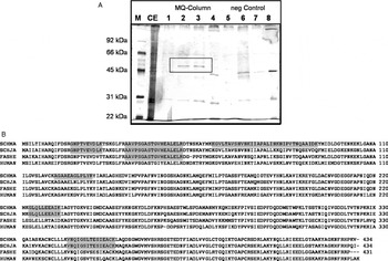
Since mefloquine was found to affect the non-haematophagous schistosomula (Manneck et al. Reference Manneck, Haggenmüller and Keiser2010) we aimed to identify the cellular target of this drug in schistosomula. Mefloquine was coupled to epoxy-agarose, a mock-column was prepared, and affinity chromatography was performed with schistosomula crude extracts on both columns mounted in tandem. Both columns were washed and separately eluted with mefloquine or a low-pH buffer. The elution with low pH of both columns yielded bands of ca. 40 and 30 kDa. These bands were also present to various extents in mefloquine elutions. In addition, only elution with mefloquine of the mefloquine-column yielded a band of ca. 50 kDa (Fig. 1A, rectangle). This band was subjected to mass spectrometry analysis. Eight peptides aligned with 100% protein probability to S. mansoni enolase Q27877 (identical to AAC46884, AAC46886, CBN61518.1) and to S. mansoni enolase C4Q3S7 (identical to XP_002573848.1, CAZ30081.1) with 23% coverage of the sequence (Fig. 1B). C4Q3S7 differed in 2 amino acids only from Q27877, namely an isoleucine instead of a threonine in position 52 and a phenylalanine instead of a tyrosine in position 93. Q27877 was identified through an analysis of trans-spliced mRNAs (Davis et al. Reference Davis, Hardwick, Tavernier, Hodgson and Singh1995), C4Q3S7 via the genomic sequencing effort (Berriman et al. Reference Berriman, Haas, Loverde, Wilson, Dillon, Cerqueira, Mashiyama, Al-Lazikani, Andrade, Ashton, Aslett, Bartholomeu, Blandin, Caffrey, Coghlan, Coulson, Day, Delcher, Demarco, Djikeng, Eyre, Gamble, Ghedin, Gu, Hertz-Fowler, Hirai, Hirai, Houston, Ivens, Johnston, Lacerda, Macedo, Mcveigh, Ning, Oliveira, Overington, Parkhill, Pertea, Pierce, Protasio, Quail, Rajandream, Rogers, Sajid, Salzberg, Stanke, Tivey, White, Williams, Wortman, Wu, Zamanian, Zerlotini, Fraser-Liggett, Barrell and El-Sayed2009). Analysis by BLAST (Altschul et al. Reference Altschul, Madden, Schaffer, Zhang, Zhang, Miller and Lipman1997) revealed that the coding sequences for both proteins were identical (data not shown). One peptide aligned also to enolases from S. japonicum (P33676) and F. hepatica (Q27655), 3 aligned with the S. japonicum enolase only, 4 were unique to S. mansoni. The protein with the highest similarity in humans is alpha-enolase (P06733; Fig. 1B).
(A) SDS-PAGE of affinity chromatography eluates of Schistosoma mansoni schistosomula extracts. Eluates were obtained by mefloquine-agarose affinity chromatography (MQ-column; lanes 1–4) and by an ethanolamine coupled mock column (negative control; lanes 5–8). Marker (M), crude extract (CE), Wash plus pre-mefloquine (1 and 5), eluates with 1 mM mefloquine (2 and 6 and 3 and 7), pH-shift with 0·1 M glycine at pH 2·9 (4 and 8). Bands were visualized by silver staining. (B) Protein sequence of S. mansoni enolase Q27877 (SCHMA) and alignment with other enolases, namely P33676 from S. japonicum (SCHJA), Q27655 from Fasciola hepatica (FASHE) and P06733 from human (HUMAN). Peptides identified by mass spectrometry are highlighted in grey.

Enolase activity from schistosomula crude extract is inhibited by mefloquine
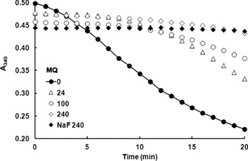
Enolase activity in parasite crude extracts was inhibited by mefloquine in a concentration-dependent manner at 24 μM and above. At 240 μM, both mefloquine and sodium fluoride, a well-known enolase inhibitor (Warburg and Christian, Reference Warburg and Christian1941), nearly completely inhibited enolase activity in crude extracts (Fig. 2). The inhibition by mefloquine was not due to inhibition effects on the coupled reactions catalysed by pyruvate kinase and lactate dehydrogenase. This was shown by control assays where the product of the reaction catalysed by enolase, namely phosphoenol-pyruvate, was added to the reaction mix instead of enolase and the substrate 2-phosphoglycerate. Activities of the coupled reactions performed without mefloquine (Control, 60·6 ΔmA340/min ±3·5) or with 240 μM mefloquine (59·3 ΔmA340/min ±4·6) were identical.
Mefloquine inhibits enolase activity in Schistosoma mansoni crude extracts. Enolase assays were performed with S. mansoni schistosomula crude extracts in the presence of different concentrations of mefloquine (MQ 0–240 μM) or sodium fluoride as a positive control for inhibition (NaF 240 μM). Data are expressed as the decrease of absorption at 340 nm over time.

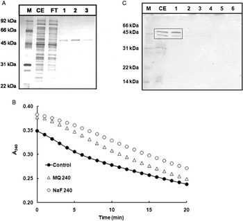
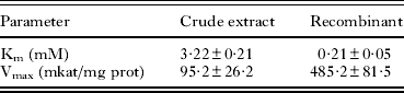
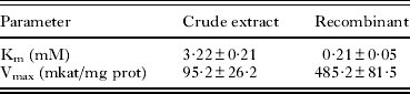
Subsequently, Q27877 was over-expressed as a His-Tag protein in E. coli and affinity purified under non-denaturing conditions (Fig. 3A). The recombinant protein had enolase activity, but was not inhibited, either by fluoride or by mefloquine at 240 μM (Fig. 3B). In order to see whether the recombinant enzyme had the same catalytic properties as the enolase activity in crude extracts, we performed enolase assays with both enzymes at various substrate concentrations and determined Km and Vmax values according to Lineweaver-Burke. Enolase from crude extracts had nearly 1 magnitude higher Km and about 5 times lower Vmax values than the recombinant enzyme (Table 1). Moreover, we performed a pull-down assay on mefloquine agarose with the recombinant enolase. The protein was detected quantitatively in the flow-through, hence the recombinant protein did not bind to the column (Fig. 3C).
Recombinant enolase Q27877 is not inhibited either by fluoride or by mefloquine. (A) SDS-PAGE of his-tag purified recombinant enolase. Marker (M), crude extract (CE), flow through (FT), eluates (1–3). (B) Enolase activity of recombinant Q27877 in the presence of 240 μM mefloquine (MQ 240) or 240 μM sodium fluoride (NaF 240). (C) SDS-PAGE of mefloquine agarose affinity chromatography of recombinant enolase Q27877. Crude extract (CE), flow-through (1), wash (2), pre-mefloquine (3), mefloquine (4), mefloquine pre pH-shift (5), pH-shift (0·1 M glycine, pH 2·9) (6). Bands were visualized by silver staining.

Kinetic parameters of enolase activity from Schistosoma mansoni schistosomula crude extracts and of recombinant enolase Q27877
Glucose interferes with the antischistosomal activity of mefloquine
Based on previously published results on metabolic inhibition of schistosomula by mefloquine, we compared the effect of mefloquine to compounds well known for interfering with glycolysis, namely sodium fluoride (Warburg and Christian, Reference Warburg and Christian1941) and 3-bromopyruvate (Pelicano et al. Reference Pelicano, Martin, Xu and Huang2006). We hypothesized that a supplementation of the medium with glucose may counteract inhibition by these compounds. Additional glucose would offer more substrate for each reaction during glycolysis, increase the rate of glycolysis and might decrease the inhibitory effect of the tested glycolytic inhibitors and of mefloquine. As a control, we included menadione (Bueding, Reference Bueding1950) interfering with glycolysis via induction of oxidative stress (Verrax et al. Reference Verrax, Stockis, Tison, Taper and Calderon2006).
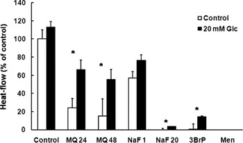
Figure 4 illustrates the relative heat production (metabolic activity of untreated control worms in the absence of glucose was set to 100%) of schistosomula incubated with the 4 tested compounds in the presence or absence of glucose (0 and 20 mM) 24 h post-incubation. The addition of glucose caused no significant increase as compared to the control group. After incubation with 24 μM mefloquine in glucose-free medium, metabolic activity of schistosomula dropped to nearly one fifth of the control values. Schistosomula incubated with 20 mM glucose showed a significantly higher metabolic activity after exposure to mefloquine as compared to schistosomula incubated in plain medium. The same effect was seen for schistosomula incubated with 48 μM mefloquine. By 24 h post-incubation, metabolic activity of parasites incubated in glucose-free medium had decreased to less than 20% compared to control values, whereas worms exposed to mefloquine incubated in medium containing glucose had a significantly higher activity. When schistosomula were incubated with a lower concentration of 10 mM glucose (data not shown) again a significantly higher heat-flow after exposure to mefloquine was seen, when compared to schistosomula incubated in medium without glucose. The effects of higher mefloquine concentrations could not be evaluated since these doses resulted in a rapid decrease of metabolic activity of schistosomula, which was too fast to be recordable by our system (Manneck et al. Reference Manneck, Braissant, Haggenmüller and Keiser2011b).
Heat-flow of Schistosoma mansoni schistosomula 24 h post-incubation with 24 μM (MQ 24) and 48 μM mefloquine (MQ 48), 1 mM or 20 mM sodium fluoride (NaF 1, NaF 20), 0·5 mM 3-bromopyruvate (3-BP), or with 50 μM menadione (Men) in the absence of glucose (Control) or with glucose (20 mM Glc). Mean values±s.e. are indicated for 4 replicates. Values with significant differences (t-tests; P< 0·05) between samples with and without additional glucose are labelled with asterisks.

The addition of sodium fluoride caused a concentration-dependent decrease of the heat-flow. Incubation with 20 mM sodium fluoride resulted in death of all schistosomula. In the presence of 20 mM sodium fluoride plus glucose metabolic activity was still detectable. Both parasites incubated with and without additional glucose had similar metabolic activities 24 h after incubation with 50 μM 3-bromopyruvate (data not shown). In the presence of 0·5 mM 3-bromopyruvate metabolic activity was nearly at background levels. In the presence of glucose and 0·5 mM 3-bromopyruvate metabolic activity was detectable at nearly 10% of the control levels. Finally, menadione (50 μM) killed schistosomula 24 h post-incubation regardless of glucose supplementation.
DISCUSSION
Isothermal microcalorimetry, which measures the heat produced by microorganisms has proven to be a suitable method to evaluate the activity of antischistosomal compounds (Manneck et al. Reference Manneck, Braissant, Haggenmüller and Keiser2011b). Energy acquisition in schistosomes is provided mainly via glycolysis (Bueding, Reference Bueding1950; Van den Bossche, Reference Van den Bossche1985; Yang et al. Reference Yang, Qiu, Xia, Yao, Fu, Yuan, Feng and Lin2010). Compounds interfering with glycolysis thus rapidly block muscular contraction and heat development. We have investigated a series of compounds directly interfering with glycolysis, namely sodium fluoride, 3-bromopyruvate, and menadione on schistosomula using microcalorimetry. Sodium fluoride is a well-characterized inhibitor of enolase, the enzyme catalysing the conversion of phosphoglycerate to phosphopyruvate (Warburg and Christian, Reference Warburg and Christian1941). However, sodium fluoride is not selective to enolase and inhibits a variety of other enzymes such as phosphatases or ATPases. Therefore, the reduction of glucose consumption in schistosomula may also be due to effects of sodium fluoride on other metabolic pathways. In adult schistosomes, sodium fluoride reduces the consumption of glucose and the production of lactate (Bueding, Reference Bueding1950). Menadione is a naphthoquinone with structural similarities to mefloquine. Similar to sodium fluoride, it reduces glucose consumption and lactate production in adult schistosomes (Bueding, Reference Bueding1950), but most likely not by an inhibition of glycolysis but instead by an induction of oxidative stress (Verrax et al. Reference Verrax, Stockis, Tison, Taper and Calderon2006). 3-bromopyruvate inhibits hexokinase in the glycolytic pathway and decreases the ATP content in cells by inhibition of ATP production and depletion of cellular ATP. Due to its cytotoxic activity against cancer cells, pre-clinical trials have been launched to confirm its activity for anticancer treatment (Pelicano et al. Reference Pelicano, Martin, Xu and Huang2006). To our knowledge, the antischistosomal activity of 3-bromopyruvate has not been described so far. Our microcalorimetric studies reveal that simultaneous addition of glucose attenuates the metabolic inhibition by sodium fluoride, 3-bromopyruvate and mefloquine, but not by menadione. Note that, in the case of sodium fluoride, the effective concentration is very high (above 1 mM), which is most likely due to a reduced uptake of this compound by schistosomula. This phenomenon has also been described for adult schistosomes with at least 20 mM sodium fluoride being necessary to inhibit glucose utilization in the parasites by 50% (Bueding, Reference Bueding1950). Our findings suggest that mefloquine directly interferes with glycolysis similar to the two inhibitors of glycolytic enzymes rather than acting indirectly as menadione. This assumption is corroborated by biochemical data.
Using affinity chromatography followed by mass spectrometry, we have identified the glycolytic enzyme enolase as a major mefloquine-binding protein in S. mansoni schistosomula. Enolase activity in schistosomula crude extracts is inhibited by mefloquine in a similar concentration range as by sodium fluoride. The recombinant enolase Q27877 is, however, not inhibited by either mefloquine or sodium fluoride. Moreover, the recombinant enolase has different kinetic parameters from the activity in crude extracts.
At first glance, these results suggest that mefloquine may inhibit an enolase isoform other than our recombinant Q27877. Mammals have 3 enolase isoforms, alpha, beta and gamma with multiple functions beside glycolysis (Pancholi, Reference Pancholi2001) and for S. mansoni to date 2 isoforms of enolase, namely Q27877 and C4Q3S7, are listed in genomic databases. However, although these 2 isoforms are provided in genomic databases, it is likely that enolases other than Q27877 do not exist in S. mansoni. The small differences between Q27877 and C4Q3S7, the protein derived from the genomic sequencing effort, are most likely due to errors, because the corresponding coding sequences are identical. A BLAST analysis showed that Q27877 has 75% identity to human alpha-enolase, 72% to beta-enolase, and 71% to gamma-enolase and may thus fulfil – to a certain extent – the functional requirements for all classes in mammals.
More likely, enolase from crude extracts has been subjected to post-translational modifications and/or interacted with other proteins present in its natural cellular environment resulting in catalytic properties differing from the recombinant enzyme. For example, in mammalian cells such as human muscle fibres, enolases may be phosphorylated (Hojlund et al. Reference Hojlund, Bowen, Hwang, Flynn, Madireddy, Geetha, Langlais, Meyer, Mandarino and Yi2009). The functional implication of this phosphorylation is unknown so far. In yeast, enolase is bound to vacuoles (Decker and Wickner, Reference Decker and Wickner2006) and may be part of a large macromolecular complex associated with mitochondria (Brandina et al. Reference Brandina, Graham, Lemaitre-Guillier, Entelis, Krasheninnikov, Sweetlove, Tarassov and Martin2006). In Plasmodium, enolase has been described to have undergone post-translational modifications that are associated to different subcellular fractions (Pal-Bhowmick et al. Reference Pal-Bhowmick, Vora and Jarori2007). Taken together, these results indicate that a native enolase may be different from the recombinant enolase produced in E. coli. Results obtained with the latter may thus not reflect the original situation.
Moreover, there is increasing evidence that in S. bovis (Ramajo-Hernandez et al. Reference Ramajo-Hernandez, Perez-Sanchez, Ramajo-Martin and Oleaga2007) and S. japonicum (Yang et al. Reference Yang, Qiu, Xia, Yao, Fu, Yuan, Feng and Lin2010) enolase is localized not only intracellularly but also at the cell surface. The role of this localization is unclear, but extracellular enolase may be involved in the interaction with host proteins such as plasminogen or the immune system (Pancholi, Reference Pancholi2001). It is possible that an extracellular, extensively modified enolase is the target for mefloquine. In S. japonicum, enolase is expressed in all developmental stages, but has higher expression levels in schistosomula than in adult worms suggesting that enolase is critical for growth, migration and adaption of the young schistosomes (Yang et al. Reference Yang, Qiu, Xia, Yao, Fu, Yuan, Feng and Lin2010).
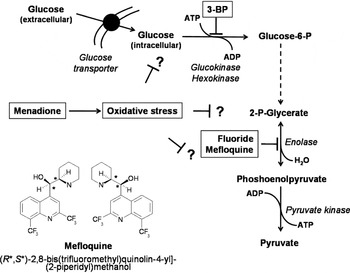
Overall, our results suggest, that in S. mansoni schistosomula, mefloquine inhibits enolase resulting in an interference with glycolysis and/or in debilitating the parasite via an unknown mechanism in the case of extracellular enolase. Since mefloquine has 3 fluorine residues, it is possible that it blocks enolase at the same site as fluoride ions as suggested in the scheme presented in Fig. 5. Fluoride forms a complex with magnesium and phosphate at the active site (Bunick and Kashket, Reference Bunick and Kashket1982; Qin et al. Reference Qin, Chai, Brewer, Lovelace and Lebioda2006). There is, however, a non-deniable discrepancy between the concentrations of mefloquine inhibiting enolase activity in crude extracts and the nearly 1 magnitude lower concentrations needed for metabolic inhibition measured by microcalorimetry and by drug sensitivity assays (Manneck et al. Reference Manneck, Braissant, Haggenmüller and Keiser2011b). This suggests the existence of other molecular targets for mefloquine. Further work should focus on this aspect as well as on the role of enolase in the mode of action of mefloquine in Schistosoma spp.
Scheme summarizing glycolysis in Schistosoma and targets for inhibitors (in rectangles) as used in the present study. A structural formula of mefloquine is shown.

ACKNOWLEDGEMENTS
We would like to thank Olivier Braissant for helpful discussions and his support concerning microcalorimetry and we are grateful to Corinne Lochmatter, Cristian Köpfli, Dania Müller and Yvette Endress for laboratory support. Furthermore, we would like to thank Professor Dr Andrew Hemphill (University of Berne) for his support.
FINANCIAL SUPPORT
T. M. and J. K. are grateful to the Swiss National Science Foundation (project no. PPOOA–114941) for financial support. J. M. is a recipient of a research fellowship provided by Novartis Animal Health.