Introduction
Post-auricular cutaneous mastoid fistula is a rare condition. It can occur following radical mastoid surgery, chronic suppurative otitis mediaReference Bhat, Udayashankar, Venkatesha and Kumar1 or spontaneous exteriorisation of a cholesteatoma from the mastoid through the post-auricular skin surface.Reference Wadhera, Gulati, Kalra, Ghai and Garg2 Necrosis, breakdown and fistula development may also occur after closure of a post-auricular incision with poor skin edges.Reference Luetje3 Primary closure of the defect is usually unsuccessful because of the necrotic skin edges. Secondary wound closure may be achieved with a dressing regimen of antibiotic ointment covered with dry gauze. However, this often needs a long time to heal, with consequent discomfort for the patient. Several alternative types of flap reconstruction have been described.
Herein, we describe a surgical refinement for post-auricular cutaneous mastoid fistula closure, involving a fibro-muscular-periosteal flap to cover the mastoid cavity, combined with a bilobed flap from the mastoid and lateral neck regions for skin closure. We report the case of a post-auricular cutaneous mastoid fistula that developed after a revision tympanoplasty for a cholesteatoma, which was successfully treated with the presented technique.
Materials and methods
Case presentation
A 27-year-old man presented with a history of chronic cholesteatoma. Seven months earlier he had undergone a revision of a previous right open tympanoplasty for cholesteatoma recurrence. Unfortunately, four weeks after the surgery, he developed a post-auricular cutaneous mastoid fistula with a chronic discharge of clear fluid. No signs of infection were observed.
The post-auricular cutaneous mastoid fistula was initially treated with sterile dressings with iodoform gauze twice a week, and a course of oral antibiotics. However, after a month of using the dressings, only a minimal reduction in post-auricular cutaneous mastoid fistula dimension was observed.
The suprathreshold audiometry showed a right-sided, moderate-to-severe, mixed hearing loss, with normal left-sided hearing. The surgical option of a lateral petrosectomy with adipose tissue obliteration of the cavity, which is often the preferred choice for a discharging retroauricular fistula, would have definitely prevented the feasibility of an air conduction hearing aid for impaired hearing, as well as the possibility of otomicroscopic follow up. For this reason, we decided to plan a plastic closure of the fistula using a fibro-muscular-periosteal flap to fill the cavity, in combination with a bilobed flap for skin reconstruction.
Surgical technique
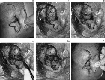
The patient is positioned supine, with the affected ear uppermost, and the face turned away from the surgeon. The post-auricular skin is cleaned, and the fistula and fistula tract are exposed. A bilobed flap is planned, harvesting skin from the mastoid and lateral neck regions, and incision lines are marked. The first lobe is transversely oriented over the mastoid and posterior neck. The dimension of this lobe corresponds to the extent of the defect. The second lobe is angled 90 degrees in order to make its longitudinal axis lie in the lateral neck. From the superior margin of the fistula, a vertical incision line is extended in the temporal-mastoid-occipital area to allow the fibro-muscular-periosteal flap harvesting (Figure 1a).
(a) Pre-operative view of post-auricular cutaneous mastoid fistulae, with incision lines marked. (b) Dissection of the bilobed flap and exposure of fibro-muscular-periosteal tissue, after excision of the fistula. (c) Preparation of the fibro-muscular-periosteal flap. The muscular-periosteal flap is anteriorly rooted (d), and is secured with stitches and an absorbable haemostatic gelatine sponge (e). (f) Bilobed flap inset and primary closure of the donor site.

The first surgical step consists of the incision of the skin around the fistula, and the identification of the fistula tract in the mastoid region. The fistula is resected to include an amount of surrounding healthy skin. The incision is then extended upward in the temporal-mastoid-occipital area to expose an adequate portion of fibro-muscular-periosteal tissue. The bilobed flap is then harvested. Dissection is performed in a subcutaneous plane, paying attention to not damage the great auricular nerve (Figure 1b).
In the second step, the fibro-muscular-periosteal flap is dissected in the mastoid-occipital surgical field, anteriorly pedicled, and rooted (Figure 1c and d). Under microscopic vision, the fistula defect is filled up with the fibro-muscular-periosteal flap, and is secured with stitches and absorbable haemostatic gelatine sponge (Figure 1e). Once it has been fixed and the mastoid obliterated, the defect is closed by transposing the two lobes of the bilobed flap (Figure 1f).
Medicated gauze is placed in the external auditory canal. The gauze is kept in place for four weeks, to protect the middle-ear cavity of the open tympanoplasty and avoid cicatricial stenosis. The post-auricular wound is treated with Steri-Strips™ and gauzes.
Results
On post-operative day 2, the patient was discharged with a short course of antibiotics and analgesics, and informed to cleanse the wound and change the dressing daily. The stitches were removed after 7 days.
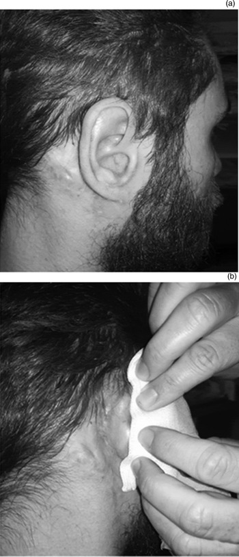
The fistula had healed completely. The patient did not report any complication. He continued to have a self-cleaning open tympanoplasty with mastoid obliteration. No post-auricular cutaneous mastoid fistula relapse has been observed to date, six months after the surgery (Figure 2a and b).
(a & b) Post-operative result, at six months’ follow up.

Discussion
Post-auricular cutaneous mastoid fistulae are rarely reported. They can be a complication of chronic ear disease or ear surgery, or both. Direct closure has extremely high failure rates associated with the necrotic skin edges, and it may result in an enlargement of the fistula.
Papers describing techniques for post-auricular cutaneous mastoid fistula closure are very exiguous, with only a few cases reported. Most of the authors agree on the difficulty in managing such fistulae because of the necrotic nature of the margins and the need to deal with an empty mastoid cavity.
In 1933, Asherson managed a post-auricular cutaneous mastoid fistula by transplanting a portion of the temporalis muscle into the cavity.Reference Asherson4 In 1980, Luetje used an anteriorly based fibro-muscular-periosteal flap covered with bone pâté and a free graft of abdominal fat, and closed the defect with a rotational post-auricular skin flap.Reference Luetje3 Choo et al. reported a case of a post-auricular cutaneous fistula in a patient with chronic suppurative otitis media, treated with a small strip of temporalis muscle flap rotated inferiorly to cover the tegmen bony defect and the mastoid cavity, followed by direct skin closure.Reference Choo, Shaw and Chong5 Lee et al. described the closure of a post-auricular cutaneous mastoid fistula using a temporalis fascia transposition flap.Reference Lee, Lee, Rhie and Ahn6 Vira and Andrews, in four patients with a post-auricular cutaneous mastoid fistula following mastoidectomy, adopted a double-layer closure technique using temporalis muscle and fascia, and conchal cartilage with skin.Reference Vira and Andrews7 Recently, Olusesi and Opaluwah, in two women with a post-auricular cutaneous mastoid fistula caused by cholesteatoma, used a post-auricular fascio-cutaneo-periosteal advancement flap with Burow's triangles, following excision of the fistula margins.Reference Olusesi and Opaluwah8
Each technique described has advantages and disadvantages. Temporalis muscle flaps cause a depression in the temporal region of the scalp, with an aesthetic defect that is frequently not accepted by younger patients. Furthermore, fat grafts for mastoid cavity obliteration, as in petrosectomy, require closure of the external auditory canal to prevent fat necrosis, with unavoidable worsening of hearing loss. The fibro-muscular-periosteal flap used to cover the mastoid cavity in the technique described here is able to solve all these issues, by filling the cavity, and maintaining the open tympanoplasty with mastoid obliteration.
The main purpose of skin closure was to limit, as much as possible, suture tension. The bilobed flap has been successfully used for the reconstruction of large central defects of the auricle,Reference Weerda, Soutar and Tiwari9 and for complex defects of the postero-medial surface of the auricle, the retroauricular sulcus and the mastoid region.Reference D'Arpa, Cordova and Moschella10 The bilobed flap from the mastoid and lateral neck regions is easy to harvest, and allows free tension wound closure with a good aesthetical result.
Conclusion
The management of post-auricular cutaneous mastoid fistulae can be challenging for the surgeon. The fibro-muscular-periosteal flap combined with a bilobed flap from the mastoid and lateral neck regions, in our view, can be considered a valid option for post-auricular cutaneous mastoid fistula closure. Avoiding a straightforward petrosectomy allows the option of hearing aids when mixed hearing loss cannot be rectified by bone conduction hearing devices. In particular, in the case of cholesteatoma, monitoring is mandatory, and by maintaining a self-cleaning open tympanoplasty, clinical follow up remains feasible.
Competing interests
None declared