1. Introduction
Xinjiangchelyidae is a family of basal eucryptodiran turtles that flourished in Asia during the Jurassic (Sukhanov, Reference Sukhanov2000; Rabi et al. Reference Rabi, Joyce and Wings2010; Tong, Reference Tong, Li and Tong2017). Originating in the Middle Jurassic, this family underwent a quick radiation and became the dominant group in the Late Jurassic freshwater ecosystems in mainland Asia. Abundant specimens have been collected from numerous localities in southern and northern China, Mongolia and other places in Central Asia (Ye, Reference Ye1986; Peng & Brinkman, Reference Peng and Brinkman1993; Nessov, Reference Nessov1995; Matzke et al. Reference Matzke, Maisch, Ge, Pfretzschner and Stöhr2004, Reference Matzke, Maisch, Sun, Pfretzschner and Stöhr2005; Tong et al. Reference Tong, Danilov, Ye, Ouyang, Peng and Li2012a, b; Wings et al. Reference Wings, Rabi, Schneider, Schwermann, Sun, Zhou and Joyce2012; Brinkman et al. Reference Brinkman, Eberth, Xu, Clark and Wu2013b; Rabi et al. Reference Rabi, Zhou, Wings, Ge and Joyce2013, Reference Rabi, Sukhanov, Egorova, Danilov and Joyce2014; Tong et al. Reference Tong, Dong and Wang2015a). In SE Asia, this group is less well documented. The first basal cryptodiran turtle from SE Asia, Siamochelys peninsularis, closely related to xinjiangchelyids, was reported from the Jurassic of the southern peninsula of Thailand (Tong et al. Reference Tong, Buffetaut and Suteethorn2002). More recently, remains of a xinjiangchelyid turtle, Phunoichelys thirakhupti Tong, Naksri, Buffetaut, Suteethorn, Suteethorn, Deesri, Sila, Chanthasit and Claude, 2015, were reported from the Upper Jurassic Phu Kradung Formation at the Phu Noi locality (Kalasin Province) in NE Thailand (Tong et al. Reference Tong, Naksri, Buffetaut, Suteethorn, Suteethorn, Deesri, Sila, Chanthasit and Claude2015b). In this paper, we describe a second xinjiangchelyid turtle taxon from Phu Noi locality. The material consists of shells and a nearly complete skull. Our study provides additional evidence about the age of the Phu Kradung Formation and new insight on the evolution of the basal eucryptodiran turtles. The material is housed in Sirindhorn Museum, Sahatsakhan, Kalasin Province, Thailand. For more geological information about Phu Noi locality, see Tong et al. (Reference Tong, Naksri, Buffetaut, Suteethorn, Suteethorn, Deesri, Sila, Chanthasit and Claude2015b).
2. Systematic palaeontology
Testudines Linnaeus, Reference Linnaeus1758
Cryptodira Cope, Reference Cope1868
Xinjiangchelyidae Nessov in Kaznyshkin etal., Reference Kaznyshkin, Nalbandyan and Nessov1990
Kalasinemys prasarttongosothi gen. and sp. nov.
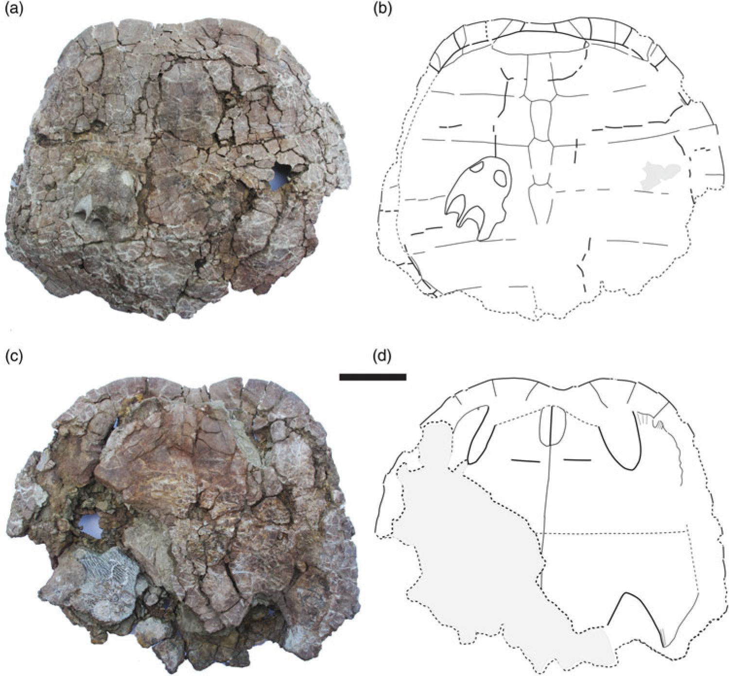
Kalasinemys prasarttongosothi n. gen. n. sp. (KS34-190, holotype) from the Phu Kradung Formation, Phu Noi locality, Kalasin Province, Thailand. Carapace and skull in dorsal view (a, b); plastron in ventral view (c, d). Scale bar = 5 cm.

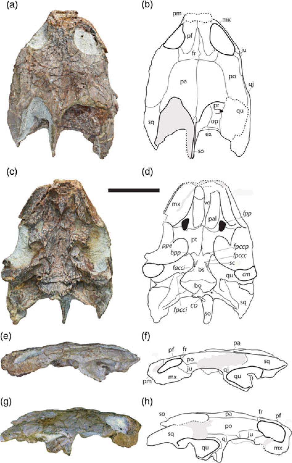
Kalasinemys prasarttongosothi n. gen. n. sp. (KS34-190, holotype) from the Phu Kradung Formation, Phu Noi locality, Kalasin Province, Thailand. Skull in dorsal (a, b), ventral (c, d), left lateral (e, f) and right lateral (g, h) views. Scale bar = 2 cm. Abbreviations: bo, basioccipital; bpp, basipterygoid process; bs, basisphenoid; cm, condylus mandibularis; co, condylus occipitalis; ex, exoccipital; facci, foramen anterius canalis carotici interni; fpccc, foramen posterius canalis carotici cerebralis; fpcci, foramen posterius canalis carotici interni; fpccp, foramen posterius canalis carotici palatinum; fr, frontal; ju, jugal; ma, maxilla; op, opisthotic; pa, parietal; pal, palatine; pf, prefrontal; pm, premaxilla; po, postorbital; ppe, processus pterygoideus externus; pr, prootic; pt, pterygoid; qj, quadratojugal; qu, quadrate; sc, sulcus caroticus; so, supraoccipital; sq, squamosal; vo, vomer.

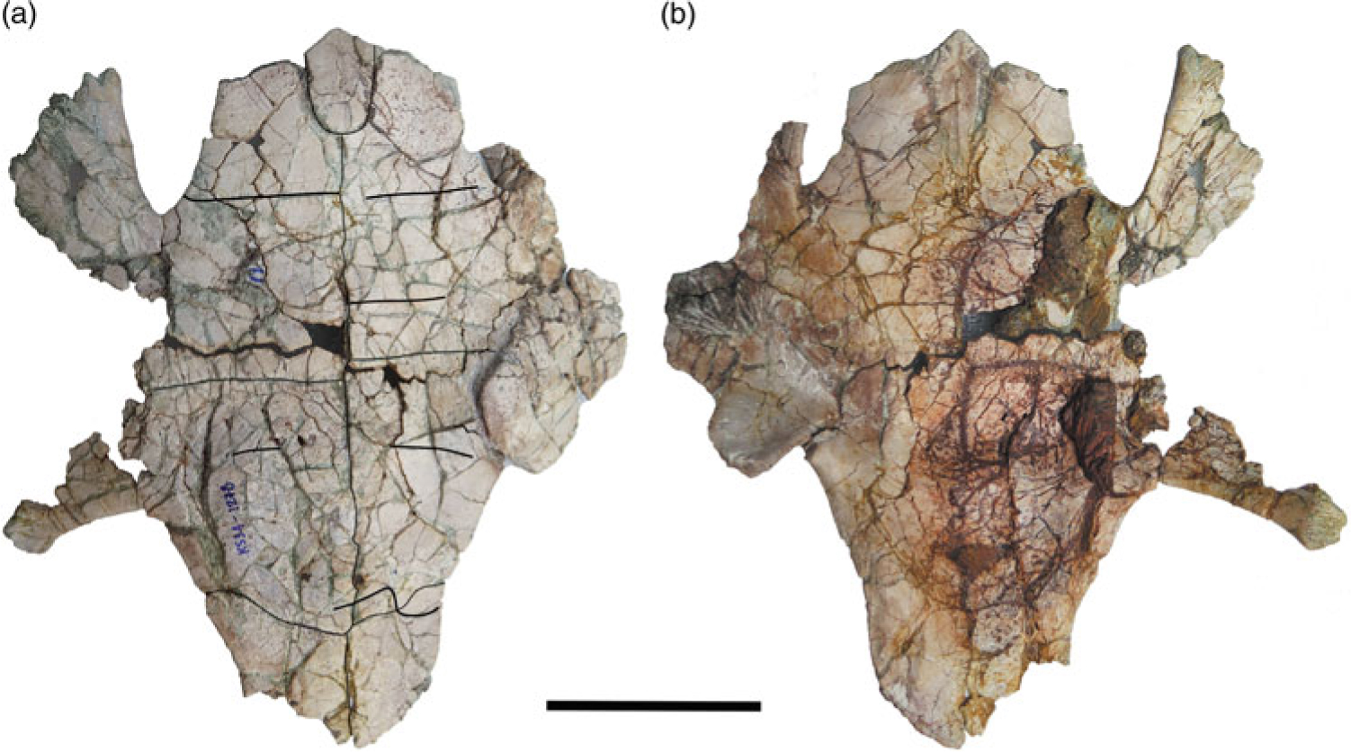
Kalasinemys prasarttongosothi n. gen. n. sp. (KS34-1278) from the Phu Kradung Formation, Phu Noi locality, Kalasin Province, Thailand. Plastron in ventral (a) and dorsal (b) views. Scale bar = 5 cm.

Etymology: The genus name is from Kalasin Province, where the Phu Noi site is located. The species name is in honour of Dr Prasert Prasarttong-Osoth who supports palaeontological research and education in Thailand.
Holotype: KS34-190, a skull and a shell with articulated carapace and plastron. The skull, which is disarticulated and lying on the carapace (see Fig. 1), belongs very likely to the same individual as the shell. The posterior part of the carapace is damaged. The plastron lacks the anterior end and posterolateral part of the right side.
Hypodigm: KS34-1278, a nearly complete plastron, lacking epiplastra.
Type locality and horizon: Phu Noi locality, Ban Dinji, Kham Muang District, Kalasin Province, NE Thailand; lower part of the Phu Kradung Formation, Late Jurassic (see Tong et al. Reference Tong, Naksri, Buffetaut, Suteethorn, Suteethorn, Deesri, Sila, Chanthasit and Claude2015b).
Diagnosis: a xinjiangchelyid turtle characterized by the following combination of features that differs from all other xinjiangchelyids: skull flat without scales on the skull surface; prefrontal with posterior extension, forming most of the medial edge of the orbit; frontal reduced with short main body, long anterior process and small contribution to the orbital margin; basipterygoid process scalene triangular in shape; short basioccipital ventral exposure; shell low and thick, with smooth surface and rounded outline that is expanded posteriorly; cervical notch present; short and wide nuchal; lateral marginals extending onto costals 2 to 4; first vertebral scute narrower than the nuchal and oval entoplastron that is longer than wide. In addition, it differs from Annemys spp. in having a wider triturating surface, a smaller foramen palatinum posterius and a larger incisura columellae auris; and differs from Xinjiangchelys radiplicatoides in having a flat skull and a remnant of an interpterygoid vacuity.
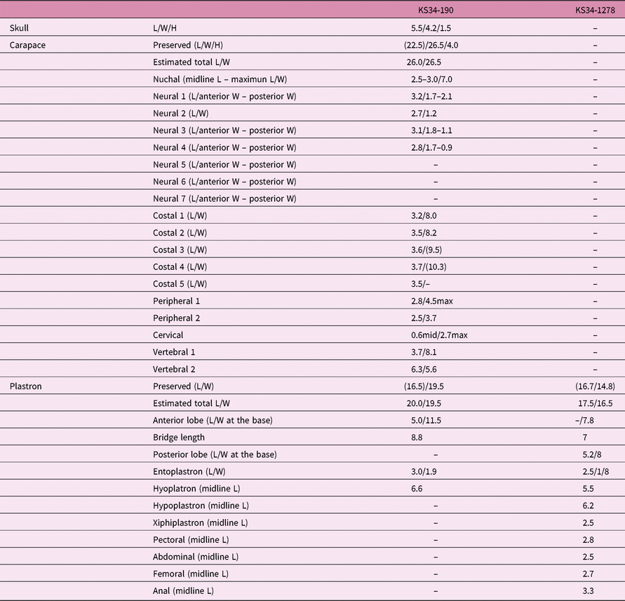
Measurements: see Table 1.
Measurements Kalasinemys prasarttongosothi n. gen. n. sp. (L = length, W = width, H = height) (in cm; incomplete value in parenthesis)

2.a. Description and comparisons
Preservation. The skull, initially stuck onto the shell, was removed during the preparation and fully prepared. It is almost complete, except the tip of the snout that is damaged. It has undergone a dorsoventrolateral crushing and is slightly distorted, with the midline of the dorsal surface shifted towards the left.
General aspect. In dorsal view, the skull has an oblong shape. The lateral margins are nearly parallel to each other; as a result, the anterior end of the skull is wider than in Annemys levensis (Rabi et al. Reference Rabi, Sukhanov, Egorova, Danilov and Joyce2014). Despite deformation, the skull was originally low; comparable to that of A. levensis and Annemys sp. from the Junggar Basin, Xinjiang, China (Brinkman et al. Reference Brinkman, Eberth, Xu, Clark and Wu2013b), but different from the tall skull of Xinjiangchelys radiplicatoides (Brinkman et al. Reference Brinkman, Eberth, Xu, Clark and Wu2013b). The snout is slightly protruding. The external naris, damaged on its margins, is single and appears to face anterodorsally but the precise shape of that opening is unclear. The orbits are large and facing dorsolaterally. The interorbital bar is relatively narrow, with parallel lateral borders as in X. wusu (Rabi et al. Reference Rabi, Zhou, Wings, Ge and Joyce2013) and A. latiens (Rabi et al. Reference Rabi, Sukhanov, Egorova, Danilov and Joyce2014). The temporal emargination is deep, allowing the foramen stapedio-temporale to be exposed. The shape of this emargination is comparable to that of Annemys spp., but shallower. The crista supraoccipitalis extends slightly beyond the posterior end of the squamosal as in A. levensis and X. wusu. In lateral view, the cheek emargination is large and deep, with its upper margin higher than the lower rim of the orbit. The skull roof surface is rough; anteroposteriorly directed parallel ridges are apparent on the posterior end of the parietals. There is no trace of scale sulci on the skull roof.
Nasal. The upper margin of the external narial opening is damaged; the nasals are apparently completely missing.
Prefrontal. The prefrontal is an elongate bone forming almost the entire upper orbital margin. Anteriorly, the prefrontals meet one another along the midline, while the posterior portion of the right and left prefrontals is separated by the anterior process of the frontals. The prefrontal is surrounded by the frontal medially and posteriorly.
Frontal. The frontals are reduced. The right and left frontals form together an inverse T-shaped exposure on the skull roof, that can be divided into an anterior process and a transverse main body. The anterior process is long and lies between the prefrontals. The main body is short, with a narrow lateral lappet that turned anterolaterally to contribute to the posteromedial corner of the orbital margin. The frontal contacts the parietal posteriorly with a straight transverse suture. Laterally, the frontal has a short contact with the postorbital.
Parietal. The parietal is the largest element on the skull roof and forms the medial half of the temporal emargination. In addition to the anterior contact with the frontal, the parietal has a long contact with the postorbital laterally; and posteriorly it has a short contact with the supraoccipital. As preserved on the left side, the parietal sends a posterolateral process along the temporal emargination margin to meet the squamosal.
Postorbital. The postorbital is an anteroposteriorly elongate flat bone that forms the posterior margin of the orbit. Ventrolaterally, the postorbital sends a small process between the jugal and the quadratojugal that barely reaches the cheek margin. Anteromedially, the postorbital has a short contact with the frontal, and medially a long contact with the parietal. In addition, the postorbital contacts the jugal anterolaterally, the quadratojugal posterolaterally and the squamosal posteriorly. The postorbital does not reach the temporal emargination margin, prevented by the parietal/squamosal contact.
Jugal. The jugal is a small bone that forms the anterior half of the cheek emargination. It has also a small contribution to the posteroventral corner of the orbital margin. Anterolaterally the jugal contacts the maxilla; and medially and posteriorly, it is surrounded by the postorbital. The jugal fails to contact the quadratojugal posteriorly, prevented by a small ventral process of the postorbital.
Quadratojugal. The quadratojugal is a triradiate bone in lateral view. The anterior and the ventral branches make up together the posterior half of the cheek emargination. The posterior and the ventral branches frame posteriorly the cavun tympani. The quadratojugal contacts the postorbital anteriorly and dorsomedially, and the quadrate posteriorly. The posterior contact with the squamosal is unclear.
Squamosal: The squamosal is a cone-shaped bone that forms the posterolateral corner of the skull and caps the cavum tympani. Posteriorly, the squamosal extends almost to the level of the posterior end of the crista supraoccipitalis. Medially, it forms the lateral portion of the temporal emargination. The squamosal contacts the parietal anteromedially, the postorbital anteriorly and the quadrate ventrally on the skull roof. Within the temporal fossa, it contacts the opisthotic medially; while the contact with the quadrate is not clearly discernible.
Premaxilla. Both premaxillae are damaged; only their posterior portion is preserved. The premaxillae form the lower edge of the external narial opening. The contact areas with the maxilla laterally are damaged on both sides. In ventral view, the premaxilla contributes to the anteromedial edge of the internal narial opening and contacts the vomer posteriorly.
Maxilla. In lateral view, the maxilla forms the lower rim of the orbit and contacts the jugal posteriorly. The suborbital portion of the maxilla is low as in Annemys spp. Anteriorly the contact areas with the premaxilla, the nasal and the prefrontal are damaged. In ventral view, the maxilla forms most of the triturating surface, which is flat and moderate in width but wider than that of Annemys levensis and A. sp. from China (Wings et al. Reference Wings, Rabi, Schneider, Schwermann, Sun, Zhou and Joyce2012; Brinkman et al. Reference Brinkman, Eberth, Xu, Clark and Wu2013b). The labial ridge is almost straight and sharp; while the lingual edge, without a distinct ridge, is curved and convergent anteriorly towards the labial ridge. The maxilla forms the lateral edge of the internal narial opening and the anterolateral margin of the foramen palatinum posterius; the latter being smaller than that of Annemys spp. Medially the maxilla contacts the palatine, and posteromedially it has a relatively long contact with the pterygoid lateral to the foramen palatinum posterius.
Vomer. The vomer is almost complete, with the surface of the anterior end damaged. The vomer forms a narrow bar between the internal narial openings, which has a slightly concave ventral surface and a widened anterior portion. Anteriorly, the vomer contacts the premaxilla, and posteriorly it extends between the palatines to reach or almost reach the pterygoids.
Palatine. The palatine forms the anterior and medial edge of the foramen palatinum posterius. It contacts the vomer medially, the maxilla anterolaterally and the pterygoid posteriorly.
Quadrate. The left quadrate is intact while the right one is crushed. In lateral view, the cavum tympani, better preserved on the left side, is large, kidney-shaped and deep, with a large antrum postoticum. The incisura columellae auris is a large fissure that opens ventrally. In comparison, Annemys levensis and A. sp. have a very narrow incisura. Inside the cavum tympani, the quadrate bears a process that rises dorsally, ventrolateral to the incisura columellae auris. The quadrate contacts the quadratojugal anteriorly and anterodorsally, and the squamosal posterodorsally. Dorsally within the temporal fossa, the quadrate contributes to the lateral edge of the foramen stapedio-temporale and contacts the prootic anteromedially, the opisthotic posteromedially and the squamosal posteriorly. On the ventral surface, the quadrate forms a short processus articularis and contacts the pterygoid medially. The condylus mandibularis is short anteroposteriorly.
Pterygoid and epipterygoid: The pterygoids form the ventral surface of the palate posterior to the palatines. Anteriorly the pterygoid contributes to the posterior margin of the foramen palatinum posterius, and contacts the palatine anteromedially and the maxilla lateral to that foramen. The processus pterygoideus externus is a large process that is directed posteriorly. A pair of deep triangular depressions is present on the anterior end of the pterygoids, just posterior to the pterygoid/palatine suture. The anterior portion of the pterygoid has a long contact with its counterpart. Anterior to the basisphenoid, there is a slit-like opening along the midline between the pterygoids that we interpret as the remnant of the interpterygoid vacuity. Posteriorly, the pterygoid has a curved contact with the basisphenoid. The foramen posterius canalis carotici palatinum is located along the pterygoids/basisphenoid suture on each side of the basisphenoid tip, facing posteriorly.
Posteriorly, the pterygoids extend between the quadrate and the basisphenoid to the level of the basisphenoid/basiocciptal contact, but fail to reach the basioccipital as in other xinjiangchelyids. The ventral surface of the posterolateral branch of the pterygoid bears a large triangular depression. The depression is bordered medially by a distinct ridge that follows the posterolateral edge of the sulcus caroticus. Laterally, the depression is bordered by a high ridge formed by the pterygoid and the quadrate that extends to the processus articularis of the quadrate. The pterygoid is sutured to the basisphenoid posterior to the sulcus caroticus to enclose the canalis caroticus inernus. The foramen posterius canalis carotici interni is located dorsolateral to the medial ridge of the depression, close to its posterior end.
The region around the foramen trigemini and epipterygoid is deformed on both sides. The epipterygoid borders the foramen nervi trigemini ventrally, and contacts the quadrate posteroventrally and the pterygoid ventrally. The contact with the parietal anterior to the foramen trigemini is visible on the left side.
Basisphenoid. The basisphenoid has an elongate triangular ventral exposure, with an uneven surface. The sulcus caroticus is an anteromedially directed narrow groove that is entirely formed by the basisphenoid. The foramen posterius canalis carotici cerebralis lies at its anterior end, and the foramen anterior canalis carotici interni is located at its posterior end. The diameter of these foramina is smaller than that of the foramen posterius canalis carotici palatinum. On the anterior half of the basisphenoid, a pair of large basipterygoid processes of scalene triangular shape extends from the main body of the basisphenoid laterally. Anteriorly and laterally the basisphenoid is surrounded by the pterygoids, and posteriorly it contacts the basioccipital. The ventral surface of the basisphenoid also bears a pair of pits close to its posterior border.
Basioccipital and exoccipital. The basioccipital and exoccipitals are likely fused; no suture between these elements can be traced. The occipital condyle is deformed. In ventral view, the basioccipital is short and wide, with a shallow concave surface. The lateral margins of the basioccipital are markedly convergent backward; a pair of prominent tuberculae basioccipitale with rounded lateral border is developed at the posterolateral end, as in Annemys levensis and A. sp. In posterior view, the basioccipital/exoccipital complex, together with the supraoccipital, form the foramen magnum. One large foramen nervi hypoglossi that pierces the bone lateral to the condylus occipitalis is visible on the left side. The exoccipital contacts the supraoccipital dorsally and the opisthotic laterally.
Supraoccipital. The supraoccipital forms the dorsal edge of the foramen magnum and the crista supraoccipitalis, a vertical blade extending posteriorly to the level of the posterior end of the squamosal. The supraoccipital contacts the opisthotic and the prootic laterally, and the exoccipital ventrally. The contact with the parietal is not visible.
Prootic. The dorsal surface of the right otic capsule is exposed, but damaged, the sutures are partly discernible; the left side is covered by matrix. The temporal emargination is deep, allowing the foramen stapedio-temporale and part of the prootic to be exposed. The foramen stapedio-temporale is located on the junction of the prootic, opisthotic and the quadrate. From that foramen a groove extends medially. The prootic forms the anterior margin of the foramen and the groove, and contacts the quadrate laterally, the opisthotic posteriorly and the supraoccipital medially. The anterior extent of the bone (including the processus trochlearis oticum) and the contact with the parietal are not visible.
Opisthotic. On the dorsal surface within the temporal fossa, the opisthotic contacts the prootic anteriorly, the supraoccipital medially, the exoccipital ventromedially, and the quadrate and the squamosal laterally. The opisthotic contributes also to the posterior edge of the foramen stapedio-temporale. Ventrally, the opisthotic roofs the cavum acustico-jugulare. The fenestra postotica faces posteroventrally, allowing the processus interfenestralis of the opisthotic to be exposed ventrally. This is a robust process extending ventrally from the opisthotic to reach or almost reach the posterior end of the pterygoid. Medially, the processus borders the fenestra perylymphatica and laterally it frames the fenestra ovalis; both openings communicate with the cavum labyrinthicum. The entrance for the canalis cavernosus is visible lateral to the fenestra ovalis.
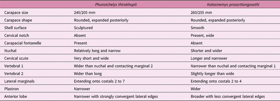
Shell surface ornamentation. Both carapace and plastron surfaces are almost smooth, although light parallel striations are visible on the costal margins, perpendicular to the intercostal sutures.
Carapace. The shell is low. The carapace is wide and expanded posteriorly. There is a wide cervical notch formed by the nuchal and first peripherals, unlike Phunoichelys thirakhupti which has a straight anterior carapace margin. The anterior carapace rim is smooth and blunt, unlike the holotype of P. thirakhupti which has a sharp carapace margin. The anterolateral border of the carapace is slightly upturned, but not forming a clear gutter. The shell seems to be more heavily built when compared to P. thirakhupti.
The nuchal is a short and wide trapezoidal element, with the width being more than twice the length. The first to fourth neurals are discernible in KS34-190, all longer than wide. The first neural is hexagonal with short posterolateral sides. The second neural is rectangular. The third to fourth neurals are hexagonal with short anterolatetral sides. The first costal is relatively short as in other primitive turtles; it is as long as the second costal and bears a sharp ridge on its inner surface, extending up to the lateral end of the plate. The third costal, as shown in KS34-190, is slightly enlarged laterally, with its lateral end longer than its medial end. The peripheral series is not complete. The first peripheral is trapezoidal and slightly wider than long. The second peripheral is roughly square.
The scute sulci are poorly preserved in KS34-190. The cervical scute is about three times wider than long, and clearly narrower and longer than that of P. thirakhupti. The first vertebral scute is wider than long, with convex lateral margins. Anteriorly, it is slightly narrower than the nuchal and contacts the lateral end of the first marginal scute. The second vertebral scute is slightly longer than wide. The third and fourth vertebral scutes are incomplete. The first to third marginal scutes are restricted to the peripherals, while the lateral marginals extend onto the second to fourth costals.
Plastron: The plastron is relatively wide. The anterior end of the plastron is missing in KS34-190, so the position of the front margin of the plastron relative to that of the carapace is unclear. Although none of the plastra has the anterior end preserved, the anterior lobe is apparently shorter than the bridge, with the lateral borders convergent gently forward. The bridge is long. The posterior lobe is shorter than the bridge without an anal notch. The plastron/carapace connection seems to be ligamentous since some pegs are visible on the anterior and posterior ends of the bridge. The axillary and inguinal buttresses are long. The axillary buttresses are directed anteriorly, extending beyond the epi/hyoplastral suture and reaching the anterior third of the second peripheral in KS34-190. The inguinal buttress does not contact the costal plate in KS34-190, but its relationship with the peripheral is not clear since this part of the carapace is much fractured. The inguinal notch is wider than the axillary notch.
The epiplastra are not preserved in either specimen. The entoplastron is oval and clearly longer than wide in ventral view as in other xinjiangchelyids. Although the epiplastra are missing, the complete entoplastron with preserved anterior sutural margin in KS34-1278 suggests that this plate does not reach the front edge of the plastron. In dorsal view, the entoplastron has a long posterior process that extends almost up to the base of the anterior lobe. The hyoplastron is longer than the hypoplastron in the bridge region. The hypo/xiphiplastron suture is strongly convex posteriorly.
The scute sulci are better preserved in KS34-1278. The humeropectoral sulcus lies far posterior to the entoplastron, at the base of the anterior lobe as in other xinjiangchelyids. The pectoral is slightly shorter than the abdominal. The femoroanal sulcus is omega-shaped, extending onto the hypoplastron as in most xinjiangcheyids. No inframarginals are visible in either specimen.
3. Discussion
3.a. Systematic assignment and relationships
Based on the general shell morphology, KS34-190 and KS34-1278 are considered as belonging to the same taxon. They are different from Phunoichelys thirakhupti from the same locality in the following features: the shell is more heavily built, with thicker shell plates; the shell surface is smooth, devoid of ornamentation; a cervical notch is present; the nuchal plate is shorter and wider; the cervical scute is narrower and longer; the lateral marginales extend onto costals 2 to 4 (instead of costals 2 to 7 in Phunoichelys); the first vertebral scute is narrower than the nuchal, contacting marginal 1; the plastron is wider and the anterior lobe is broader with less convergent lateral borders. The detailed comparisons of the shell between Kalasinemys and Phunoichelys are shown in Table 2.
Comparisons between Kalasinemys prasarttongosothi n. gen. et n. sp. and Phunoichelys thirakhupti

The shell of Kalasinemys lacks a pair of mesoplastra. As in Phunoichelys, the morphology of the shell of Kalasinemys agrees with that of the basal eucryptodiran Xinjiangchelyidae. The xinjiangchelyid characters include the lateral marginals extending onto costals; a relatively broad plastron with a long and narrow bridge; a large entoplastron of oval shape and longer than wide; and the femoroanal sulcus omega-shaped, extending onto the hypoplastron. In Macrobaenidae, the lateral marginals are restricted to the peripherals; the plastron is more reduced, with a shorter and wider bridge; the entoplastron is narrower and leaf-shaped and the femoroanal sulcus is restricted to the xiphiplastron.
Known since the Middle Jurassic, xinjiangchelyids are a group of basal eucryptodiran turtles that was dominant in the freshwater ecosystems during the Late Jurassic in Asia. The family Xinjiangchelyidae includes the following genera in Asia: Xinjiangchelys, Annemys, Chengyuchelys, Tienfuchelys, Yanduchelys and Protoxinjiangchelys (Ye, Reference Ye1986; Nessov, Reference Nessov1995; Rabi et al. Reference Rabi, Joyce and Wings2010, Reference Rabi, Zhou, Wings, Ge and Joyce2013, Reference Rabi, Sukhanov, Egorova, Danilov and Joyce2014; Tong et al. Reference Tong, Danilov, Ye, Ouyang, Peng and Li2012a, b, Reference Tong, Dong and Wang2015a;Wings et al. Reference Wings, Rabi, Schneider, Schwermann, Sun, Zhou and Joyce2012; Brinkman et al. Reference Brinkman, Eberth, Xu, Clark and Wu2013b). The last four genera are endemic to the Sichuan Basin, and the skull is only known in Xinjiangchelys and Annemys. Among Xinjiangchelyidae, the shell of Kalasinemys is more derived than those of Chengyuchelys, Tienfuchelys and Yanduchelys from the Late Jurassic of Sichuan Basin in the ligamentous plastron/carapace attachment. It more resembles that of Annemys in the narrow vertebrals 2–3. In Xinjiangchelys, vertebrals 2–3 are wider.
KS34-190 represents the first xinjiangchelyid skull from SE Asia. The ventral aspect of the skull reveals important features of the arterial system and related structures. Sterli & Fuente (Reference Sterli and Fuente2010) studied the evolution of the arterial system in turtles and recognized two groups and six patterns. The more recent studies of the xinjiangchelyid skulls from Asia provided additional information on that matter for the basal eucryptodiran turtles (Brinkman et al. Reference Brinkman, Eberth, Xu, Clark and Wu2013b; Rabi et al. Reference Rabi, Zhou, Wings, Ge and Joyce2013, Reference Rabi, Sukhanov, Egorova, Danilov and Joyce2014). In Kalasinemys, the internal carotid artery enters the skull via the foramen posterius canalis carotici interni that lies at the posterior end of the pterygoid and runs anteriorly within an enclosed canalis caroticus internus. The internal carotid artery exits the skull via the foramen anterius canalis carotici interni, then branches off on the skull surface into two main arteries. The medial one, the cerebral artery, is nested in an open groove, the sulcus caroticus, and enters the skull via the foramen posterius canalis carotici cerebralis that is situated at the anterior end of the sulcus. The lateral branch, the palatine artery, runs further anteriorly outside, and enters the skull via the slit-like foramen posterius canalis carotici palatinum that lies at the contact between the pterygoid and the anterior edge of the basipterygoid process of the basisphenoid. A remnant of the interpterygoid vacuity is present between the pterygoids, anterior to the basisphenoid and there is a pair of basipterygoid processes. This morphology corresponds well to that of xinjiangchelyids as seen in Annemys spp. from the Late Jurassic of Mongolia; and Xinjiangchelys radiplicatoides, X. wusu and A. sp. from the Late Jurassic of Junggar and Turpan basins, China (Rabi et al. Reference Rabi, Joyce and Wings2010, Reference Rabi, Sukhanov, Egorova, Danilov and Joyce2014; Wings et al. Reference Wings, Rabi, Schneider, Schwermann, Sun, Zhou and Joyce2012; Brinkman et al. Reference Brinkman, Eberth, Xu, Clark and Wu2013b). The remnant of the interpterygoid vacuity is no longer functional as the entrance of the palatine artery, in contrast to the more basal turtles, such as Proganochelys quenstedti, Heckerochelys romani, Condorchelys antiqua and Kayentachelys aprix (Sukhanov, Reference Sukhanov2006; Sterli & Joyce, Reference Sterli and Joyce2007; Gaffney & Jenkins, Reference Gaffney and Jenkins2010; Sterli & Fuente, Reference Sterli and Fuente2010; Sterli et al. Reference Sterli, Müller, Anquetin and Hilger2010).
In Kalasinemys as in other xinjiangchelyids, the sulcus caroticus is located about the mid-length of the basisphenoid. The canalis caroticus internus is relatively short and the foramen posterius canalis carotici cerebralis lies posteriorly, distant from the foramen posterius canalis carotici palatinum. The families Sinemydidae and Macrobaenidae present a more derived condition. In these groups, the interpterygoid vacuity is completely closed. In most macrobaenids with the well-preserved relevant region, the foramen posterius canalis cartici cerebralis lies more anteriorly, close to the foramen posterius canalis carotici palatinum (e.g. Ordosemys liaoxiensis, O. sp., Kirgizemys (Hangaiemys) hoburensis (Brinkman & Wu, Reference Brinkman and Wu1999; Li & Liu, Reference Li, Liu, Chen and Jin1999; Sukhanov, Reference Sukhanov2000)). In Judithemys and Kirgizemys dmitrievi, these two foramina are no longer distinct on the skull surface (Parham & Hutchison, Reference Parham and Hutchison2003; Danilov, Reference Danilov2006). This suggests that in macrobaenids the branches of the internal carotid artery are shifted forward relative to the xinjiangchelyid condition and the embedding of the internal carotid system in bone is more accomplished. A similar morphology is also seen in the trionychoids Basilochelys and Adocus (Meylan & Gaffney, Reference Meylan and Gaffney1989; Tong et al. Reference Tong, Claude, Naksri, Suteethorn, Buffetaut, Khansubha, Wongko, Yuangdetkla, Buffetaut, Cuny, Le Loeuff and Suteehorn2009a). Among marcrobaenids, Dracochelys which is similar to xinjiangchelyids in this respect seems to present the most primitive condition in that group (Gaffney & Ye, Reference Gaffney and Ye1992).
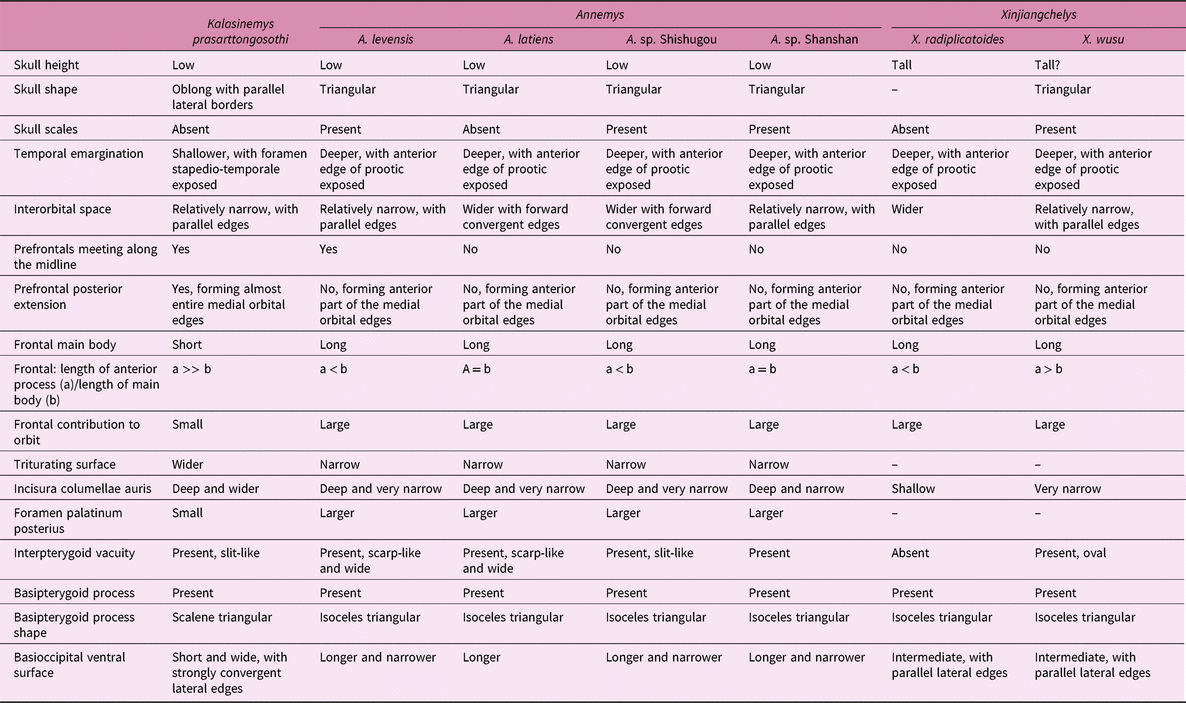
Rabi et al. (Reference Rabi, Sukhanov, Egorova, Danilov and Joyce2014) recognized three morphotypes in the skull of xinjiangchelyids: an inflated and relatively high skull with relatively shallow temporal emargination in Xinjiangchelys radiplicatoides; a small, flat and triangular skull with deeper temporal emargination in Annemys levensis and A. sp.; and an elongate and narrower variant of the second type seen in Annemys latiens which probably corresponds to different feeding niches and strategies. The skull of Kalasinemys closely resembles that of Annemys in being flat, but the shape of the skull that has more parallel lateral edges is different from the triangular skull as seen from above in Annemys.
The prefrontals of Kalasinemys extend posteriorly and form most of the medial edge of the orbit. In other turtles, the prefrontals are shorter and form only the anterior part of the orbit margin. In relation to the posterior extension of the prefrontal, the frontal of Kalasinemys is reduced, with a long anterior process, short main body and small contribution to the orbital edge. In Xinjiangchelys spp., Annemys spp. and A. sp., the frontal is larger with a shorter anterior process; the main body is as long as, or even longer than, the anterior process; and the contribution to the orbital margin is greater. The basipterygoid process in Kalasinemys skull is scalene triangular in shape, while in Annemys and Xinjiangchelys it is isosceles triangular. In addition, the skull of Kalasinemys has a wider triturating surface, smaller foramen palatinum posterius and shorter basioccipital when compared with Annemys. The detailed comparison of Kalasinemys with other xinjiangchelyid taxa is shown in Table 3.
Comparisons between skull of Kalasinemys prasarttongosothi and other xinjiangchelyid taxa

3.b. Relationships of Basilochelys macrobios
The remains of Basilochelys macrobios Tong, Claude, Naksri, Suteethorn, Buffetaut, Khansubha, Wongko and Yuangdetkla, 2009, a large eucryptodiran turtle, are common in the upper part of the Phu Kradung Formation, stratigraphically some 500 m above the Phu Noi locality (Tong et al. Reference Tong, Claude, Naksri, Suteethorn, Buffetaut, Khansubha, Wongko, Yuangdetkla, Buffetaut, Cuny, Le Loeuff and Suteehorn2009a, b, Reference Tong, Naksri, Buffetaut, Suteethorn, Suteethorn, Deesri, Sila, Chanthasit and Claude2015b). Originally Basilochelys was considered as a basal trionychoid (Tong et al. Reference Tong, Claude, Naksri, Suteethorn, Buffetaut, Khansubha, Wongko, Yuangdetkla, Buffetaut, Cuny, Le Loeuff and Suteehorn2009a), which was confirmed by more recent phylogenetic analyses (Joyce et al. Reference Joyce, Rabi, Clark and Xu2016), but an alternative analysis placed it at a more basal position, outside of crown Testudines, between xinjiangchelyids and sinemydids/macrobaenids (Rabi et al. Reference Rabi, Zhou, Wings, Ge and Joyce2013). The primitive feature that is responsible for its more basal position is the presence of the basipterygoid process. We re-examined the skull and confirm that a pair of large basipterygoid processes are present in Basilochelys. Although the distinct entrance of the cerebral and palatine arteries is still exposed on the skull surface as in xinjiangchelyids, the skull of Basilochelys is clearly more derived than Kalasinemys and other xinjiangchelyids. In Basilochelys, the interpterygoid vacuity is completely closed. The sulcus caroticus is shorter and lies close to the foramen posterius canalis palatinum that is placed on the pterygoid/basisphenoid suture. The reduced and more anteriorly placed sulcus caroticus in Basilochelys is related to a longer canalis caroticus internus, which indicates that the division of the internal carotid artery into the cerebral and palatine arteries is more anterior relative to the condition seen in xinjiangchelyids. The position of the sulcus caroticus in Basilochelys is comparable to that of Adocus sp. (Meylan & Gaffney, Reference Meylan and Gaffney1989), even though the foramen posterius canalis palatinum is no longer exposed in the latter. The shell of Basilochelys also shows some derived characters such as the sculptured shell surface, the wide and large entoplastron that is located more posteriorly, the pectoroabdominal sulcus cutting the entoplastron, the relatively long pygal, the suprapygal contacting the peripheral 10 and the marginals 11–12 extending onto the suprapygal. All these are shared with the basal trionychoid Adocidae and Nanhsiungchelyidae, thus supporting the trionychoid affinities of the Basilochelys in spite of some primitive features of the skull mentioned above.
3.c. The turtle assemblage from Phu Noi site and its relationships with other Late Jurassic / Early Cretaceous turtle assemblages from Asia
The Phu Kradung Formation, to which the Phu Noi site belongs, is the basal unit of the Khorat Group in NE Thailand (Racey et al. Reference Racey, Love, Canham, Goodall, Polachan and Jones1996; Racey, Reference Racey, Buffetaut, Cuny, Le Loeuff and Suteehorn2009). It unconformably overlies the Late Triassic Nam Phong Formation. The successive formations of the Khorat Group above the Phu Kradung Formation (from bottom to top: Phra Wihan, Sao Khua, Phu Phan and Khok Kruat formations) are considered as Early Cretaceous (Berriasian to Aptian) in age, while the age of the Phu Kradung Formation is still uncertain. Evidence from vertebrate remains, notably dinosaurs, supports a Late Jurassic age for that formation (Buffetaut et al. Reference Buffetaut, Suteethorn and Tong2001, 2006; Buffetaut & Suteethorn, Reference Buffetaut and Suteethorn2007), whereas based on detrital zircon thermochronology and palynology, it is dated as Early Cretaceous (Carter & Bristow, Reference Carter and Bristow2003; Racey & Goodall, Reference Racey, Goodall, Buffetaut, Cuny, Le Loeuff and Suteehorn2009). However, a Late Jurassic age cannot be excluded for the lowermost part of the Phu Kradung Formation because of the absence of the key Early Cretaceous marker pollen taxon (Racey & Goodall, Reference Racey, Goodall, Buffetaut, Cuny, Le Loeuff and Suteehorn2009). Stratigraphically, the Phu Noi site is located in the lower part of the Phu Kradung Formation (Cuny et al. Reference Cuny, Liard, Deesri, Liard, Khamha and Suteethorn2014).
The turtle assemblage from Phu Noi consists of basal eucryptodiran xinjiangchelyids which can be correlated with those from the Late Jurassic of mainland Asia; while the remains of more advanced turtles, the trionychoid Basilochelys, are common in the upper part of the Phu Kradung Formation. In China, the turtle faunas from the Late Jurassic Shangshaximiao Formation of Sichuan Basin, the Middle–Late Jurassic of the Junggar and Turpan basins (Shishugou, Toutunhe and Qigu formations) and the Middle Jurassic Chuanjie Formation in Yunnan are composed almost exclusively of xinjiangchelyids (Peng & Brinkman, Reference Peng and Brinkman1993; Matzke et al. Reference Matzke, Maisch, Ge, Pfretzschner and Stöhr2004, Reference Matzke, Maisch, Sun, Pfretzschner and Stöhr2005; Rabi et al. Reference Rabi, Joyce and Wings2010; Tong et al. Reference Tong, Danilov, Ye, Ouyang, Peng and Li2012a, 2015a; Wings et al. Reference Wings, Rabi, Schneider, Schwermann, Sun, Zhou and Joyce2012; Brinkman et al. Reference Brinkman, Eberth, Xu, Clark and Wu2013b). This is also the case in Central Asia (Mongolia, Russia and Kirghizia (Nessov, Reference Nessov1995; Sukhanov, Reference Sukhanov2000; Rabi et al. Reference Rabi, Sukhanov, Egorova, Danilov and Joyce2014)). Only a few shells of a basal trionychoid, Sinaspideretes wimani, have been recorded from the Late Jurassic Shangshaximiao Formation in the Sichuan Basin (Tong et al. Reference Tong, Li and Ouyang2014). In addition, the turtle assemblage from Phu Noi is distinct from those from the Early Cretaceous of inland Asia (Central Asia and China) which are generally composed of more advanced eucryptodiran Sinemydidae/Macrobaenidae (Brinkman & Peng, Reference Brinkman and Peng1993a, b; Sukhanov, Reference Sukhanov2000; Tong et al. Reference Tong, Ji and Ji2004; Danilov, Reference Danilov2006; Danilov & Parham, Reference Danilov and Parham2007; Zhou, Reference Zhou2010a, b; Brinkman et al. Reference Brinkman, Yuan, Ji, Li and You2013a; Zhou et al. Reference Zhou, Rabi and Joyce2014; Zhou & Rabi, Reference Zhou and Rabi2015). Danilov & Parham (Reference Danilov and Parham2007) have assigned the holotype of ‘Sinemys’ wuerhoensis Ye, Reference Ye1973 from the Early Cretaceous of the Tugulu Group, Xinjiang, China, to Xinjiangchelyidae. This is the only specimen of xinjiangchelyid turtle that is hitherto known from the Early Cretaceous of mainland Asia. Trionychoids from the Early Cretaceous deposits are rare in these regions, known by isolated records (Danilov & Parham, Reference Danilov and Parham2007; Li et al. Reference Li, Joyce and Liu2015a, b; Brinkman et al. Reference Brinkman, Rabi and Zhao2017).
Xinjiangchelyids are reported from the Early Cretaceous Tetori Group of Japan (Hirayama & Sonoda, Reference Hirayama and Sonoda2015). The turtle assemblage from the Okurodani Formation (Berriasian?) comprises xinjiangchelyids, trionychoids, sinemydids and sinochelyids (Hirayama, Reference Hirayama2006a). This assemblage has not yet been described in detail. Hirayama (Reference Hirayama2004) figured two shells, one of them associated with a skull, from the Okurodani Formation and referred them to Testudinoidea. These specimens were later assigned to Xinjiangchelyidae (Hirayama, Reference Hirayama2006a). Although these shells share some features with xinjiangchelyids (such as the lateral marginals extending onto the costal 2, the suprapygal 1–2 similar in size, expanded peripherals 8–11 and a wider than long cervical scute; reduced but relatively wide plastron relative to sinemydids/macrobaenids and presence of intergular scutes), other characters seen in these specimens appear to be more derived than in xinjiangchelyids from China. These include a narrower and longer cervical scute; a longer pygal (in two figured specimens, this plate is clearly longer than wide in one specimen, but shorter in the second specimen. In xinjiangchelyids the pygal is wider than long); a relatively wide and diamond-shaped entoplastron (instead of longer than wide oval-shaped in xinjiangchelyids); reduced gular scutes which are smaller than intergulars (in xinjiangchelyids they are similar in size); and the presence of a shallow anal notch (the anal notch is absent in xinjiangchelyids). The turtle fauna from the Kuwajima Formation (Valanginian) consists of trionychoids, xinjiangchelyids and sinemydids (Hirayama, Reference Hirayama2006a). Abundant isolated shell plates from that formation have been previously referred to Testudinoidea (Hirayama, Reference Hirayama2000), but now assigned to Xinjiangchelyidae (Hirayama, Reference Hirayama2006a; Hirayama & Sonoda, Reference Hirayama and Sonoda2012). The xinjiangchelyid remains from the Kuwajima Formation are morphologically close to those from the Okurodani Formation, but appear more derived relative to the latter in the reduced cervical scute that is roughly as long as wide and a reduced suprapygal 1. In the overlying Akaiwa Formation, the trionychoid Kappachelys okurai and fragmentary shells with adocid-like sculpture have been reported (Hirayama et al. Reference Hirayama, Isaji and Hibino2013; Hirayama, pers. comm. 2017). The youngest formation of the Tetori Group, the Kitadani Formation (Barremian–Aptian), has yielded four taxa of trionychoids (a trionychid (Nakajima et al. Reference Nakajima, Danilov, Hirayama, Sonoda and Scheyer2017), two taxa of adocids and a nanhsiungchelyid) and two other eucryptodires (xinjiangchelyids and sinemydids, represented by large peripherals; Hirayama, pers. comm. 2017). Trionychoids are by far the dominant group in this formation, representing about 80 % of the turtle assemblage (Sonoda et al. Reference Sonoda, Azuma, Hirayama and Ando2015).
In Europe, several basal eucryptodiran turtles from the Early Cretaceous have been referred to Xinjiangchelyidae. Based on some shell features, Hirayama et al. (Reference Hirayama, Brinkman and Danilov2000) assigned Brodiechelys brodiei (Lydekker, Reference Lydekker1889) from the Early Cretaceous of the Isle of Wight, southern England, to Xinjiangchelyidae, but noticed that the plastral buttresses are restricted within peripherals in this taxon, not overlapping costals as in xinjiangchelyids. Later, several taxa from the Early Cretaceous of Spain (B. royoi Pérez-García et al. Reference Pérez-García, Gasulla and Ortega2014, Larachelus morla Pérez-García & Murelaga, Reference Pérez-García and Murelaga2012 and Camerochelys vilanovai Pérez-García & Murelaga, Reference Pérez-García and Murelaga2013) have been referred to Xinjiangchelyidae (Pérez-García & Murelaga, Reference Pérez-García and Murelaga2012, Reference Pérez-García and Murelaga2013; Pérez-García et al. Reference Pérez-García, Gasulla and Ortega2014; Pérez-García, Reference Pérez-García2017). As indicated by Hirayama et al. for B. brodiei, these taxa share with xinjiangchelyids from Asia the lateral marginals extending onto the costals, the sinuous midline sulcus of the plastron and the femoroanal sulcus intersecting the hypoplastron (Hirayama, Reference Hirayama2006b), but show a relatively small and short entoplastron as in plesiochelyids.
As mentioned above, the general morphology of the shell of xinjiangchelyids from Phu Noi (Phunoichelys and Kalasinemys) more resembles those from the Jurassic of China and Central Asia than those from the Early Cretaceous of Japan or Europe. The skull of Kalasinemys is close to that of Annemys from the Jurassic of Mongolia and China. To sum up, both the morphology of xinjiangchelyids from Phu Noi and the composition of the turtle fauna of that locality are in favour of a Late Jurassic age for the lower part of the Phu Kradung Formation, to which the Phu Noi locality belongs.
4. Conclusion
A new genus and new species of Xinjiangchelyidae, Kalasinemys pasarttongosothi n. gen. n. sp., is erected on the basis of skull and shell material from the Phu Kradung Formation of the Khorat Plateau, NE Thailand. The skull presents an arterial system characteristic of the Xinjiangchelyidae (basal Eucryptodira), and its outline is similar to that of Annemys spp. However the skull of Kalasinemys is distinct from that of Annemys in having the more parallel lateral edges; the prefrontals extending posteriorly and forming most of the medial edge of the orbit; the reduced frontals with a long anterior process, short main body and small contribution to the orbital margin; the wider triturating surface; smaller foramen palatinum posterius; a scalene triangular-shaped basipterygoid process and a shorter basioccipital. Kalasinemys is the second taxon of Xinjiangchelyidae from the Phu Noi locality. It was a freshwater turtle as indicated by its morphology and the depositional environment.
The turtle assemblage from Phu Noi has close affinities with those from the Middle–Late Jurassic of China and Central Asia. The correlation based on the turtle faunas supports a Late Jurassic age for the lower part of the Phu Kradung Formation to which the Phu Noi locality belongs. From a palaeobiogeographical point of view, it suggests a marked faunal interchange between SE Asia and more northern parts of Asia (China and Central Asia) at the time of deposition of the lower part of the Phu Kradung Formation. This discovery adds a new element to the still poorly known turtle fauna from the lower part of the Phu Kradung Formation in NE Thailand, and provides new insight on the evolution of the basal eucryptodiran turtles.
Acknowledgements
We thank students and members of the Palaeontological Research and Education Centre (PRC) of Mahasahakham University and Sirindhorn Museum, Department of Mineral Resources (DMR) of Thailand who took part in the field work at Phu Noi, and two anonymous referees for their reviews. This research is supported by the project PalBioDivASE under a ‘Groupe de Recherche International’ (GDRI) grant from CNRS; the Mahasarakham University Development Fund and the National Research Council of Thailand (NRCT) of the fiscal year 2016 and the Department of Mineral Resources, Thailand. Thanks to the Sirindhorn Museum (Kalasin, Thailand) for the facilities to access the fossil collection housed there.